622
https://doi.org/10.1590/0004-282X20180091 VIEW AND REVIEW
Basal cortisol levels and the relationship with
clinical symptoms in multiple sclerosis: a
systematic review
Níveis de cortisol basal e a relação com sintomas clínicos na esclerose múltipla: uma
revisão sistemática
Gabriela Magalhães Pereira1, Nayron Medeiros Soares2, Andreo Rysdyk de Souza1, Jefferson Becker3,
Alessandro Finkelsztejn4, Rosa Maria Martins de Almeida5
1Universidade Federal do Rio Grande do Sul, Instituto de Ciências Básicas da Saúde, Laboratório de Psicologia Experimental, Neurociências e
Comportamento, Porto Alegre RS, Brasil;
2Universidade Federal do Rio Grande do Sul, Faculdade de Medicina, Laboratório de Psicologia Experimental, Neurociências e Comportamento, Porto Alegre
RS, Brasil;
3Pontifícia Universidade Católica do Rio Grande do Sul, Hospital São Lucas, Serviço de Neurologia, Porto Alegre RS, Brasil;
4Hospital de Clínicas de Porto Alegre, Ambulatório de Esclerose Múltipla, Porto Alegre RS, Brasil;
5Universidade Federal do Rio Grande do Sul, Instituto de Psicologia, Laboratório de Psicologia Experimental, Neurociências e Comportamento, Porto Alegre RS, Brasil.
Correspondence: Rosa Maria Martins de Almeida; Instituto de Ciências Básicas da Saúde da UFRGS; Rua Sarmento Leite, 500; 90050-170 Porto Alegre RS, Brasil; E-mail: [email protected]
Conflict of interest: There is no conflict of interest to declare.
Received 15 April 2018; Received in final form 31 May 2018; Accepted 6 June 2018.
ABSTRACT
Multiple sclerosis (MS) is a demyelinating, progressive and neurodegenerative disease. A disturbance on the hypothalamic-pituitary-adrenal axis can be observed in patients with MS, showing altered cortisol levels. We aimed to identify basal cortisol levels and verify the relationship with clinical symptoms in patients with MS. A systematic search was conducted in the databases: Pubmed, Web of Science and SCOPUS. Both higher and lower cortisol levels were associated with MS. Higher cortisol levels were associated with depression and anxiety, while lower levels were associated with depression, fatigue and urinary dysfunction. Higher cortisol levels may be associated with the progression and severity of MS.
Keywords: Multiple sclerosis; hydrocortisone; pituitary-adrenal system; neurologic manifestations; general symptoms.
RESUMO
A esclerose múltipla (EM) é uma doença desmielinizante, progressiva e neurodegenerativa. Um distúrbio no eixo hipotálamo-hipófise-adrenal pode ser observado em pacientes com EM, mostrando níveis alterados de cortisol. Nosso objetivo foi identificar os níveis basais de cortisol e verificar a relação com os sintomas clínicos em pacientes com EM. Uma busca sistemática foi realizada nas bases de dados: Pubmed, Web of Science e SCOPUS. Ambos os níveis de cortisol elevado e baixo foram associados com a EM. Níveis mais elevados de cortisol foram associados à depressão e ansiedade, enquanto níveis mais baixos foram associados à depressão, fadiga e disfunção urinária. Níveis altos de cortisol podem estar associados à progressão e gravidade da EM.
Palavras-chave: Esclerose múltipla; hidrocortisona; sistema hipófise-suprarrenal; manifestações neurológicas; sintomas gerais.
Multiple sclerosis (MS) is a progressive and inflammatory neurodegenerative disease, characterized by demyelinating lesions and atrophy, in the central nervous system (CNS)1,2,3.
It affects 1/1,000 people in the western world and leads to chronic disability in young adults ranging between 20 and 40 years old4. Multiple sclerosis presents an unpredictable and
often progressive course, with many neurological symptoms5,
which include sensory disorders6, visual problems7, fatigue8,
alterations in balance9, dysfunction of the lower urinary
tract10, limitations in walking11 and cognitive dysfunction12.
The etiology of MS is considered multifactorial and involves genetic and environmental mechanisms, which affect the immunological response13. Although the origin
is still unknown, autoimmune mechanisms are considered
central triggers of MS14. A widely-accepted model considers
MS to be an autoimmune chronic inflammation, mediated by T-cells and macrophages infiltrating the CNS through the peripheral immunological system, which is involved in myelin sheath destruction along with microglia10,15. Nowadays B-cells
623
Pereira GM et al. Cortisol levels and clinical symptoms in MS
It has been demonstrated that alterations in the neuro-endocrine system may also be involved in immune suppres-sion or activation, increasing the vulnerability and severity of autoimmune diseases such as MS4,16,17. In this context, sev
-eral studies have indicated that there is a role of the hypotha-lamic-pituitary-adrenal (HPA) axis in the control of MS
pro-gression18. In physiological conditions, the HPA axis releases
glucocorticoids able to mediate the expression of inflam-matory genes of cytokines, action of monocytes and macro-phages, and adhesion and migration molecules, which have immunomodulatory effects19,20. In patients with MS,
activa-tion of the HPA axis appears to be dysregulated, and chronic hyperactivity occurs in about 50% of the patients14,18,21,22. The
reactivity of the HPA axis has been correlated with MS
pro-gression and global increase in the activation of the neurode
-generative process21,23,24.
Cortisol is an end-product glucocorticoid of the HPA axis in humans, considered to be the stress hormone and a powerful natural immunosuppressant, involved in reg-ulatory functions such as in glucose metabolism, insu-lin release, arterial pressure, immune and inflammatory
responses4,25. In MS, as a consequence of the disrupted HPA
axis, cortisol levels become altered. In patients with MS, the cortisol response after intravenous administration of cor-ticotrophin-releasing hormone is higher, when compared with healthy adult subjects26,27,28. This increase of the
cor-tisol response was associated both positively29, and nega
-tively30, with the number of acute lesions due to central
neu-roinflammation in MS.
In several studies, higher levels of basal cortisol were observed in the cerebrospinal fluid (CSF) and blood, as well as an increase in the cortisol awakening response measured
in saliva5,25,30,31,32,33. As well, evidence in postmortem humans
showed that higher levels of cortisol in the CSF are neuro-protective while lower levels are related to a higher number
of lesions30.
Physiologically, cortisol release levels and the acti-vated cerebral regions depend on the type of stress factor, where motor stress has been associated with brainstem activation while psychological stress has been associated with limbic regions34. In MS, some studies have
investi-gated the relationship between cortisol levels and symp-toms that arise during the course of the disease;
how-ever, there is still no consensus about the role of cortisol
as a cause or consequence of the symptoms. Current evi-dence is directed, mainly, to the relationship of cortisol with fatigue and depression, which are the most common symptoms in MS35,36.
In affective symptoms, such as anxiety and depression, significant correlations are found with higher levels of corti-sol, especially in patients suffering from relapsing-remitting multiple sclerosis (RRMS)5,29,37. Furthermore, studies have
shown that cortisol is correlated with fatigue and might have an important role in RRMS25,38. Nevertheless, contradictory
results have also been observed, where MS patients with depression and chronic fatigue showed cortisol levels with no increase or significant correlations39,40,41.
Even though several investigations have considered
the role of the HPA axis in MS, there is still little consensus about the cortisol levels and their role in MS pathophysiol-ogy. In this study, we aimed to investigate basal cortisol lev-els in MS and the methods of cortisol evaluation, though a systematic review. Moreover, we intended to verify whether cortisol levels were related to clinical symptoms observed in multiple sclerosis.
METHODS
The systematic review was performed on the results of the database searches from January to March in 2018, which were conducted according to PRISMA guidelines42. The
arti-cles were selected from three databases: PubMed, Web of Science and SCOPUS. To find representative articles, the fol-lowing keywords were input: [multiple sclerosis AND corti-sol]; [multiple sclerosis AND cortisol AND progression]; [ mul-tiple sclerosis AND stress AND cortisol]; [multiple sclerosis AND cortisol AND progression AND stress].
The selected articles in this review were assessed inde-pendently by two evaluators and met the following crite-ria: 1) the sample comprised only humans ( from 18 years of age); 2) the articles were published between 2006 and 2017; 3) patients had been affected only by MS, with no neuro-logical diseases from other etiologies; 4) cortisol had been assessed from urine, blood, CSF, saliva or hair; 5) there was an assessment of any physical, behavioral and/or cogni-tive symptom; 6) the full text of the article was available in the database; 7) the full article was in English. Articles were excluded from the sample if: 1) they were review arti-cles, book chapters and abstracts published in journals; 2) they used pharmacological, physical or psychological stim-ulation as a stressing agent to observe the HPA axis func-tion. Only articles that fulfilled all the requirements were included in this review.
In the initial assessment, only the abstracts of the arti-cles were read. If the data were not enough, the evaluators assessed the methods and results of the articles. Accepted articles that met all the inclusion criteria were read and ana-lyzed comprehensively.
624 Ar q Neur opsiquia tr . 2018; 76(9):622-634
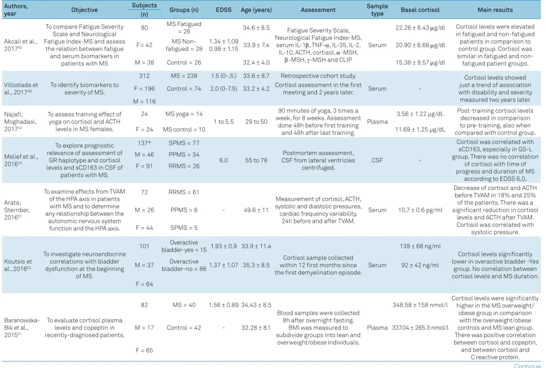
Table 1. Characteristics of the studies that assessed basal cortisol levels and their relationship to symptoms of multiple sclerosis (n = 20).
Authors,
year Objective
Subjects
Groups (n) EDSS Age (years) Assessment Sample
type Basal cortisol Main results
(n)
Akcali et al., 201750
To compare Fatigue Severity Scale and Neurological Fatigue Index-MS and assess
the relation between fatigue and serum biomarkers in
patients with MS
80 MS Fatigued = 26
1.34 ± 1.09 0.98 ± 1.15
34.6 ± 8.5 Fatigue Severity Scale, Neurological Fatigue Index-MS,
serum IL-1β, TNF-α, IL-35, IL-2, IL-10, ACTH, cortisol, α-MSH,
β-MSH, γ-MSH and CLIP
Serum
22.26 ± 8.43 μg/dl Cortisol levels were elevated in fatigued and non-fatigued patients in comparison to control group. Cortisol was similar in fatigued and non-fatigued patient groups. F= 42 MS
Non-fatigued = 28 33.9 ± 7.4 20.90 ± 6.68 μg/dl
M = 38 Control = 26 32.4 ± 4.0 15.38 ± 9.57 μg/dl
Villoslada et al., 201749
To identify biomarkers to severity of MS.
312 MS = 238 1.5 (0-.5.) 33.6 ± 8.7 Retrospective cohort study.
Serum
-Cortisol levels showed just a trend of association with disability and severity measured two years later. F = 196 Control = 74 2.0 (0-7.5) 33.2 ± 4.2 Cortisol assessment in the first
meeting and 2 years later.
M = 116
Najafi; Moghadasi, 201743
To assess training effect of yoga on cortisol and ACTH
levels in MS females.
24 MS yoga = 14
1 to 5.5 29 to 50
90 minutes of yoga, 3 times a week, for 8 weeks. Assessment
done 48h before first training and 48h after last training.
Plasma
3.56 ± 1.22 µg/dL Post-training cortisol levels decreased in comparison to pre-training, also when compared with control group.
F = 24 MS control = 10 11.69 ± 1.25 µg/dL
Melief et al., 201624
To explore prognostic relevance of assessment of
GR haplotype and cortisol levels and sCD163 in CSF of
patients with MS.
137* SPMS = 77
6.0 55 to 78
Postmortem assessment, CSF from lateral ventricles
centrifuged.
CSF
-Cortisol was correlated with sCD163, especially in GS-L group. There was no correlation
of cortisol with time of progress and duration of MS
according to EDSS 6,0. M = 46 PPMS = 34
F = 91 RRMS = 26
Arata; Sternber, 201651
To examine effects from TVAM of the HPA axis in patients with MS and to determine any relationship between the
autonomic nervous system function and the HPA axis.
72 RRMS = 61
- 49.6 ± 11
Measurement of cortisol, ACTH, systolic and diastolic pressures, cardiac frequency variability,
24h before and after TVAM.
Serum 10.7 ± 0.6 pg/ml
Decrease of cortisol and ACTH before TVAM in 18% and 25% of the patients. There was a significant reduction in cortisol
levels and ACTH after TVAM. Cortisol was correlated with
systolic pressure. M = 26 PPMS = 6
F = 44 SPMS = 5
Koutsis et al.,201652
To investigate neuroendocrine correlations with bladder dysfunction at the beginning
of MS.
101 Overactive
bladder-yes = 15 1.93 ± 0.9 33.9 ± 11.4
Cortisol sample collected within 12 first months since the first demyelination episode.
Serum
139 ± 68 ng/ml
Cortisol levels significantly lower in overactive bladder -Yes
group. No correlation between cortisol levels and MS duration. M = 37 Overactive
bladder-no = 861.37 ± 1.07 35.3 ± 8.5 92 ± 42 ng/ml
F = 64
Baranowska-Bik et al., 201531
To evaluate cortisol plasma levels and copeptin in recently-diagnosed patients.
82 MS = 40 1.56 ± 0.89 34.43 ± 8.5
Blood samples were collected 8h after overnight fasting.
BMI was measured to subdivide groups into lean and
overweight/obese individuals.
Plasma
348.58 ± 158 nmol/l
Cortisol levels were significantly higher in the MS overweight/
obese group in comparison with the overweight/obese controls and MS lean group. There was positive correlation between cortisol and copeptin, and between cortisol and
C reactive protein.
M = 17 Control = 42 - 32.28 ± 8.1 337.04 ± 265.3 nmol/l
F = 65
625
P
er
eir
a GM e
t al
. Cor
tisol l
e
vels and clinical s
ymp
toms in MS
Table 1. Characteristics of the studies that assessed basal cortisol levels and their relationship to symptoms of multiple sclerosis (n = 20). Continuation
Powell et al., 201525
To explore the relationship between cortisol and fatigue
in RRMS.
76 RRMS = 38 4.3 5 ±1.40 41.89 ± 7.53 Ecological momentary assessment performed 4
consecutive days in two projects: one based in events (CAR) – collection performed at
awakening; 30 and 45 minutes later; based in time (DCS) — 6 quasi-random samples distributed in 1000h and 2000h.
Saliva
13.57 ± 3.77 nmol/L/min
CAR was higher in RRMS than in the control group. Accumulated fatigue in RRMS
was associated with lower cortisol levels at awakening and higher CAR. The CAR was not associated with fatigue on
the same day. M = 14 Control = 38 - 40.34 ± 8.16
F = 62 11.78 ± 2.95
nmol/L/min
Eftekhari et al., 201444
To determine if a resistance training program and whole body vibration has any effect
on hormone changes in female MS patients.
24 RRMS = 12 2.87 ± 0.82 35.08 ± 6.89 vibration, 3 weekly sessions Resistance training and for 8 weeks. Cortisol was measured before and after intervention, between the 8th
and 10th day of the follicular
phase of the menstrual cycle.
Serum
12.32 ± 4.1 ng/ml
Significant reduction in cortisol concentrations after training
in RRMS compared with control group. F = 24 Control = 12 2.79 ± 0.65 33.75 ± 5.32 9.51 ± 3.4 ng/ml
Melief et al., 201330
To investigate how activity of the HPA axis in MS is related to severity, neurodegeneration,
depression, lesions and genic expression in normal-appearing white
matter.
49
- 3 to 9 32 a 83
Postmortem study. Normal-appearing white matter and hypothalamus were dissected and stored for 30 days. CSF collected to analyze cortisol.
Corticotropin-releasing hormone expressing neurons counted.
CSF 236 nmol/l
Higher cortisol levels are associated with delay in MS progression, above all in women
with SPMS. Lower cortisol was related to higher number
of active lesions and smaller remyelinated plates, and related to quick progression of MS. No differences in lower and higher cortisol levels with
humor disorders. M = 13
F = 36
Kern et al., 201345
To measure cortisol daily release, including CAR under
basal conditions.
111 RRMS = 55 2.71 36.56 EDSS, CES-D and TICS.
Saliva
-Circadian release of cortisol in RRMS was different in
the control group. There was no difference in CAR in
treated and never treated patients. Follow-up groups
with progression of EDSS showed a significant increase
in CAR when compared with the controls. Stress and depression did not correlate
with CAR in RRMS. M = 46 SPMS = 22 4.86 45.91
Cortisol collected 6 times over 24h (on awakening, 20, 45, 60 minutes later, 3 pm and 10pm, on
2 separate days within 2 weeks.
F = 65 Control = 34 35.56
626
Ar
q Neur
opsiquia
tr
. 2018;
76(9):622-634
Table 1. Characteristics of the studies that assessed basal cortisol levels and their relationship to symptoms of multiple sclerosis (n = 20). Continuation
Wipfler et al., 201346
To investigate a potential circadian periodicity of expression levels of several
cytokines relevant to MS, adhesion molecules and
cytokine receptors.
68 RRMS = 34 (12 PAD; 22 PNAD)
1.75
(0.0–5.0) 36.7 ± 7.6
Blood collection at 7am, 11am, 2:30pm, 6pm and 9:30pm.
Cortisol.
Serum
22.00 lg/dl - 7.30am
No significant differences in cortisol levels during the day. There was a decrease in cortisol between 7:30am and 9:30pm in both MS groups. Cortisol was higher in PAD if
compared to PNAD.
M = 16 Control = 34 3.60 lg/dl - 9.30 pm
F = 52 - 36.8 ± 7.2
Kern et al., 20115
To examine circadian function of the HPA axis and CAR in
patients with RRMS, with maximal duration of 36
months.
48 RRMS = 32 - 30.53
CAR assessed on two different days in a week. Sample collected on awakening, 30, 45,
60 minutes later, at 3pm and 10pm. EDSS and BDI.
Saliva
Low BDI 1299.92 ± 394.33 nmol/l
RRMS showed higher CAR levels than the control group.
Only RRMS patients with moderately higher BDI had different levels compared to
controls.
M = 11 Control = 16 - 30.37 High BDI 1486.40 ±
435.29 nmol/l
F = 37
Gold et al., 201137
To examine the role of the HPA axis activity in subpopulations of T-cells from patients with RRMS and
MDD.
44 RRMS = 34 2.2 ± 0.2 35.8 ± 0.7
Circadian profile collected on 2 consecutive days at awakening, 11am, 3pm, 8pm and 10pm.
EDSS and Hospital.
Saliva
-Patients with MDD showed HPA axis hyperactivity, with elevated cortisol levels at night. No differences observed
in CAR between groups. Decrease in cortisol levels was
not associated with CAR but was a significant predictor of
severity in depression. F = 44 RRMS-MDD
= 10 3.3 ± 0.3 37.2 ± 2.2 Anxiety and Depression Scale.
Lombardi et al.,201153
To investigate the correlation between blood hormones and SD in women of reproductive
age with MS.
55 MS-SD = 31 2.9 (1.5-6) 34.7
(26-44) Hormone assessment,
including cortisol on the third day of menstrual cycle. FSFI,
EDSS.
Serum 232.14 (180–366) nmol/l
No alteration in cortisol levels in comparison to standard
laboratory levels and no correlation with the FSFI.
F = 55 EM = 24 -
627
P
er
eir
a GM e
t al
. Cor
tisol l
e
vels and clinical s
ymp
toms in MS
Table 1. Characteristics of the studies that assessed basal cortisol levels and their relationship to symptoms of multiple sclerosis (n = 20). Continuation
Gold et al., 201047
To explore if specific sub-regional volumes of hippocampus can be linked
to alterations in daytime cortisol secretion.
49 RRMS = 29 2.5 ± 0.2 37.5 ± 1.6 BDI-II, BAI, MRI of hippocampus, dentate gyrus, subiculum and entorhinal cortex. Diurnal
cortisol was collected at awakening, 4pm and 9pm on
two consecutive days.
Saliva
-Patients with RRMS and depressive symptoms showed
higher cortisol levels and a smaller CA23DG. The volume
of CA23DG was correlated cortisol levels. M = 6 Control = 20 - 35.1 ± 1.9
F = 43
Heidbrink et al.,201048
To determine DHEA and cortisol levels in CSF and blood of patients with MS,
OIND and NIND.
78 MS = 34 - 41 (16-65)
Blood and CSF collected consecutively between 1pm
and 3pm.
CSF and Serum
-Serum cortisol in MS was just as observed in NIND. CSF
cortisol levels in MS were significantly lower. There was
positive correlation between MS and OIND in paired CSF and
blood analysis. Cortisol levels were lower in CSF and normal in
blood during an acute relapse. M = 33 OIND = 16 - 42 (20-86)
F = 45 NIND = 28 - 49 (22-81)
Mackereth et al.,200954
To compare the effects of muscle relaxing and reflexology training in people
with MS.
50 Group 1 = 25 - 48.1 ± 11.06 Six weekly session of progressive muscular relaxation; followed by 4 weeks
of washout and six-weekly reflexology sessions. SF-36, QHG 28, SAI, cortisol, systolic
and diastolic pressure.
Saliva
7.88 ± 5.35
Diminished cortisol levels after one and six weeks. Reduction
of anxiety symptoms and systolic pressure.
M = 12 Group 2 = 25 - 52.5 ± 11.6 8.49 ± 5.11
F = 38
Ysrraelit et al., 200832
To investigate HPA activity in MS subgroups.
233 PPMS = 40 4.3 ± 1.6 49.5 ± 12.7
EDSS, BDI, HDS, MFIS. Cortisol, ACTH, DHEAS.
Plasma µg/dL*
Statistically higher cortisol levels in all groups with MS patients. RRMS relapse group
showed higher hyperactivity. There was no correlation
between fatigue and depression with cortisol levels.
M = 79 SPMS = 41 5.6 ± 1.4 51.2 ± 10.1 23.0 ± 7
F = 154 RRMS = 58 1.3 ± 1.1 36.8 ± 10 18.4 ± 4.1
RRMS relapse = 34 2 ± 1.4 37.5 ± 8.4 18.4 ± 5.1
Control = 60 - 49.0 ± 5.0 24.6 ± 5.5
Urine µg/24h
348.3 ± 115.6
271.8 ± 65.7
294.1 ± 7
441.5 ± 67.9
Téllez et al., 200640
To test if fatigue in MS is associated to endocrine
biomarkers.
38 MS with
fatigue = 29 6.0 (6.5-4.0) 50.1 ± 8.1
FSS, cortisol, DHEAS
and basal DHEA. Serum
-No differences in cortisol levels between groups. F = 25 MS without
fatigue = 9 6.0 (6.0-3.5) 45.0 ± 7.7
M = 13
628 Arq Neuropsiquiatr. 2018;76(9):622-634
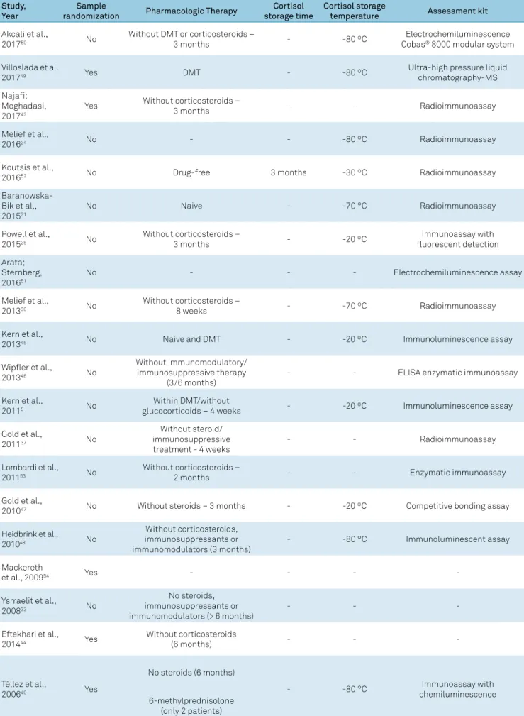
Table 2. Analysis of methodological quality of bias risk in the selected articles (n = 20). Study,
Year
Sample
randomization Pharmacologic Therapy
Cortisol storage time
Cortisol storage
temperature Assessment kit
Akcali et al.,
201750 No
Without DMT or corticosteroids –
3 months - -80 ºC
Electrochemiluminescence Cobas® 8000 modular system
Villoslada et al.
201749 Yes DMT - -80 ºC
Ultra-high pressure liquid chromatography-MS Najafi;
Moghadasi, 201743
Yes Without corticosteroids –
3 months - - Radioimmunoassay
Melief et al.,
201624 No - - -80 ºC Radioimmunoassay
Koutsis et al.,
201652 No Drug-free 3 months -30 ºC Radioimmunoassay
Baranowska-Bik et al., 201531
No Naive - -70 °C Radioimmunoassay
Powell et al.,
201525 No
Without corticosteroids –
3 months - -20 ºC
Immunoassay with fluorescent detection Arata;
Sternberg, 201651
No - - - Electrochemiluminescence assay
Melief et al.,
201330 No
Without corticosteroids –
8 weeks - -70 ºC Radioimmunoassay
Kern et al.,
201345 No Naive and DMT - -20 ºC Immunoluminescence assay
Wipfler et al.,
201346 No
Without immunomodulatory/ immunosuppressive therapy
(3/6 months)
- - ELISA enzymatic immunoassay
Kern et al.,
20115 No
Within DMT/without
glucocorticoids – 4 weeks - -20 ºC Immunoluminescence assay
Gold et al.,
201137 No
Without steroid/ immunosuppressive treatment - 4 weeks
- - Radioimmunoassay
Lombardi et al.,
201153 No
Without corticosteroids –
2 months - - Enzymatic immunoassay
Gold et al.,
201047 No Without steroids – 3 months - -20 ºC Competitive bonding assay
Heidbrink et al.,
201048 No
Without corticosteroids, immunosuppressants or immunomodulators (3 months)
- -80 °C Immunoluminescent assay
Mackereth
et al., 200954 Yes - - -
-Ysrraelit et al.,
200832 No
No steroids, immunosuppressants or immunomodulators (> 6 months)
- -
-Eftekhari et al.,
201444 Yes
Without corticosteroids
(6 months) - -
-Téllez et al.,
200640 Yes
No steroids (6 months)
- -80 °C Immunoassay with
chemiluminescence 6-methylprednisolone
629
Pereira GM et al. Cortisol levels and clinical symptoms in MS RESULTS
In the initial screening, using the above-mentioned key-words, 87 articles were found in PubMed, 149 in Web of
Scienceand 103 in SCOPUS, 339 articles in total. Out of these,
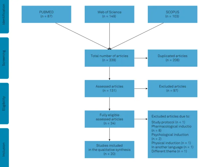
208 articles were excluded as they were in more than one database and 97 articles did not meet the criteria, leaving 34 articles eligible for assessment. The final process of selection resulted in 20 articles being included. All the articles assessed basal cortisol levels in patients with MS, but only 13 studied the relationship of cortisol with any symptom present in the course of MS. The selection process is shown in Figure 1.
Assessment of basal cortisol levels in MS
A total of 20 articles investigated cortisol levels in basal conditions in MS (Table 1). The sample size in MS groups ranged from 2443,44 to 17332 individuals, and some studies
included control groups without MS5,25,31,32,45,46,47,48,49. According
to Kurtzke’s Expanded Disability Status Scale (EDSS), the
severity of disease, when assessed, ranged from 1–6.5 points, in individuals from 29–65 years old. In most of the studies (14/20), the patients had not used glucocorticoids, immuno-modulators or immunosuppressors for at least one month and, in only three articles, the patients either had never been subjected to pharmacological treatment or used to receive the conventional treatment.
Generally, the studies had three types of general objec-tives: to verify the effect of treatment, pretreatment or post-treatment on cortisol levels, excluding articles about phar-macological/psychological induction (n = 4); to describe the relationship between cortisol and MS symptoms (n = 13) or another condition (n = 1); and to observe the HPA axis profile in patients with MS (n = 13).
The results of cortisol levels were divergent, many articles (9/20) found higher cortisol levels in MS groups5,25,30,31,32,37,45,47,50,
while others (4/20) showed lower levels30,48,51,52 or did not
differentiate (3/20) from the control group or laboratory
thresholds24,46,53. Some articles did not classify higher or
PUBMED (n = 87)
Web of Science (n = 149)
Total number of articles (n = 339)
Assessed articles (n = 131)
Fully eligible assessed articles
(n = 34)
Studies included in the qualitative synthesis
(n = 20)
Duplicated articles (n = 208)
Excluded articles (n = 97)
Excluded articles due to: Study protocol (n = 1) Pharmacological inductio (n = 8)
Psychological induction (n = 2)
Physical induction (n = 1) In another language (n = 1) Different theme (n = 1)
SCOPUS (n = 103)
Screening
Eligibility
Identification
Inclusion
630 Arq Neuropsiquiatr. 2018;76(9):622-634
lower cortisol levels (4/20), presenting only the treatment effect43,44,49,54, where there was a reduction in levels after
inter-ventions or just the relationship with the symptom (1/20)40.
Five studies did not show a significant correlation between cortisol levels with the duration, progression or severity of MS5,24,32,47,52. One study showed that the cortisol
awakening response was associated with the progression of RRMS45. There was also a study in which cortisol levels
showed a trend to correlation with severity progress in their
results49. In another study, low cortisol was associated with
fast progression and severity of MS30.
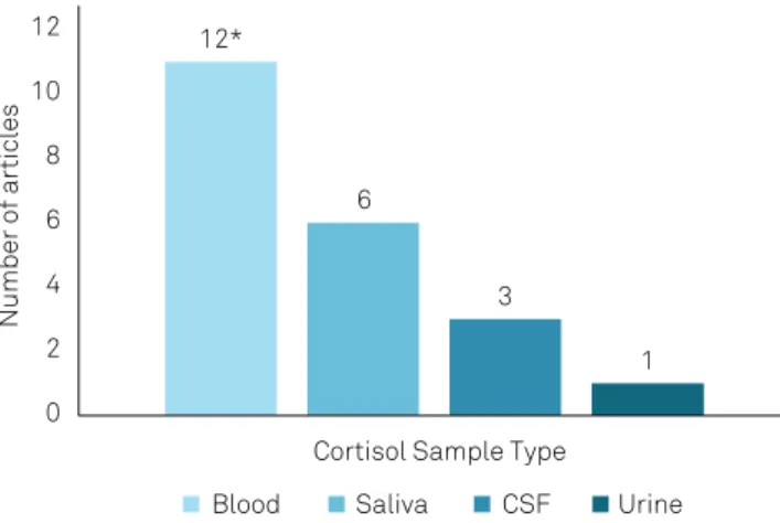
Types of cortisol samples
Cortisol levels in the articles were assessed from blood, saliva, CSF and urine (Figure 2), while most of the studies used serum samples or blood plasma (60%; n = 12). Two articles performed double sampling of cortisol through plasma/urine32 and serum/CSF48, the latter showing a
sig-nificant difference between the sampling types (CSF: p = 0.0256; serum: p = 0.2886) in different stages of the disease.
All the studies that collected saliva investigated the circa
-dian and daytime response of cortisol. No study assessed cortisol from hair samples.
Two postmortem studies included in this review assessed cortisol from CSF and just one in vivo study performed this
type of sampling. Storing temperature, if mentioned, ranged between -30º C and -80° C (Table 2). The most-used method of analysis was radioimmunoassay (31.6%; n = 6) and the least-used methods were the competitive binding assay (5.3%; n = 1), and the ultra-high pressure liquid chromatography with mass spectrometry (5.3%, n = 1).
Relationship between cortisol and MS symptoms
A total of 13 articles verified the relationship of cortisol levels with symptoms or comorbidities present in patients with MS5,25,30,31,32,37,40,45,47,50,52,53,54. The severity of the disease
ranged from 1.3–9 points on the EDSS and the age varied from
32–82 years old. The investigated symptoms were
depres-sion5,30,32,37,45,47, fatigue25,32,40,50, urinary dysfunction52, female
sexual dysfunction31, anxiety54 and obesity31.
The most-assessed symptom was depression and three articles found hyperactivity of the HPA axis, correlating high cortisol levels with depression5,37,47. In contrast, other
studies (4/20) did not find a correlation between lower30
or higher30,32,45 cortisol levels. Next, fatigue was assessed by
four articles, which verified a relationship in patients with a low cortisol awakening response25, high levels of cortisol
in patients with fatigue compared to controls50 or did not
observe any relationship between fatigue and cortisol32,40.
One study found that there may be a reduction of anxiety symptoms associated with lower cortisol levels54. Another
study did not find a correlation between cortisol and female sexual dysfunction53, however, lower cortisol levels were
related to urinary symptoms in both genders52. Finally,
over-weight patients with MS showed a higher release of cortisol when compared with lean patients32.
DISCUSSION
This review included 20 articles, which assessed basal cortisol levels and verified their relationship with any type of symptoms and comorbidities (n = 13) in patients with MS. Those studies assessed cortisol through in vivo (18/20
studies) and postmortem(2/20 studies) samples. The most-common sample type for assessing cortisol was blood (12/20 studies). Hyperactivity of the HPA axis was observed in most studies (9/20). The most-investigated symptom was depres-sion (6/13 studies). Among the assessed articles, only three found any correlation between cortisol and the duration and
progression of MS.
The results found in the various studies in this review sup-port the HPA axis profile in MS, through measuring released cortisol levels. Generally, in most studies, patients with MS showed higher cortisol levels, indicating hyperactivity of the HPA axis, similar to previously-found results26,27. Although
this impairment can be observed in many cases, its cause-and-effect relationship remains unknown. However, some hypotheses point out its role in affective disorders, such as
depression55, which arises during the course of MS. Thus,
depressed patients had higher levels of serum cortisol and this hyperactivity may be related to a decreased response of negative feedback mediated by endogenous glucocorticoids56.
Alternatively, this hyperactivity of the HPA axis may be linked to the inflammatory activity that happens in active MS. During the inflammatory process, release of cytokines, such as IL-1, IL-6 and TNFα, can perform a modulatory role of the HPA axis, increasing the cortisol release4. Alternatively, the
HPA axis hypoactivity found in some studies in this review, and decrease in cortisol levels, may be related to the sup-pression of corticotrophin-releasing hormone neurons that
12*
6
3
1
0 2 4 6 8 10 12
Number of articles
Cortisol Sample Type
Blood Saliva CSF Urine
631
Pereira GM et al. Cortisol levels and clinical symptoms in MS
occurs in active lesion in MS18. Furthermore, applying the
experimental model of autoimmune encephalomyelitis to animal models of MS has demonstrated that the severity and progression of MS is linked to HPA hyporesponsiveness57,58.
Additionally, a postmortem study has shown that lower cortisol levels were associated with larger active lesions and fewer remyelinated plates in humans, while higher cortisol levels were associated with a lower number of active lesions and an increase in plate remyelination30. From this perspective,
hyperresponsive patients showed fewer lesions highlighted by gadolinium, suggesting a neuroprotection of acute lesions59.
Controversially, the HPA axis hyperresponsiveness in MS may be associated with atrophy in the cornu ammonis
and dentate gyrus in the hippocampus47, and its relationship
with symptoms found in MS5,31,37,47. Besides this, patients with
exacerbations of RRMS have been shown to have higher cor-tisol levels compared with healthy subjects45. As well, lower
cortisol levels were found during the acute relapse than in
the stable stage of MS48.
In this systematic review, many of the studies did not find any relationship between cortisol and MS duration, progres-sion or severity; however, a remarkable number of previous studies have found a relationship between HPA dysfunction
and MS progression18,21,29,59. The lower cortisol levels found
in this review were correlated with the progression of MS, contradicting the fact that lower levels were correlated with more brain lesions30.
Although these studies preferentially collected blood serum or plasma, this type of sampling only accounts for
acute levels of cortisol release, relative to urine, saliva and
CSF samples. Moreover, blood and saliva samples provide a momentary profile, while urine samples refer to cortisol lev-els over a 24-hour period60. However, higher cortisol levels
found in studies with CSF samples might have been influ-enced either by the stress generated in response to lumbar puncture or, in postmortem studies, by the response of the HPA axis to the death process33,48.
Cortisol responses found in these articles refer to acute measurements of the hormone. An alternative to minimiz-ing the interference of stress responses generated by some factors in an ambulatory assessment might be a noninvasive gathering of hair samples. This type of sampling has been con-sidered a reliable method of measuring levels months after exposure to cortisol and it is not influenced by acute stress. Also hair samples can be stored at room temperature60.
The difference found between the blood serum sample and the CSF in the results can be explained either by the lower
activation of cortisone through 11β-hydroxysteroid dehydro-genase type 1 or by inactivation via 11β-hydroxysteroid dehy-drogenase type 248, or can be regulated by the efflux of
corti-sol from the brain, which provides a balance to corticorti-sol levels in blood and CSF61. In contrast, the similarity in levels found
between plasma and urine32 might occur as both sample
types provide cortisol levels in the peripherals.
Articles that described assessment characteristics and sample storage did this according to instructions provided by manufacturers of the respective commercial kits. Nevertheless, many studies did not clarify the duration of storage and did not use sample randomization, which made comparison of results among the studies and validity of processing more difficult and increased the bias risk. Similarly, the choice of an accurate and systematic recruitment in case control studies needed to be obtained in the light of data of healthy individuals and patients without MS, who presented for an investigated condition, such as depression. Furthermore, results in some articles were just described as higher or lower, without ever showing mean
val-ues found in each group.
Different symptoms are found in MS and they generally tend to worsen as the disease advances. Fatigue is the most common and debilitating symptom, present in more than 80% of the patients with MS62. Although it is regarded as a
residual symptom of depression63, the involvement of cortisol
levels remains unclear25. The main results encountered in this
review on MS did not find any relationship between cortisol
and fatigue, corroborating previous evidence that did not
observe an influence of cortisol on the fatigue experienced by the patients39.
Excessive cortisol in the blood has been related to mood
disorders64, such as anxiety and depression. Cortisol
per-forms a central role at the onset and during the course of major depression disorder, where higher basal cortisol levels may be found65. Patients with MS who had HPA axis
hyperac-tivity may be susceptible to developing depression66. In fact, it
has been observed that symptoms of depression may precede the onset of specific neurological symptoms during the initial process of MS; however, in spite of the involvement of several epigenetic factors, the etiology of the depression is multifac-torial and varies among patients67. In this context, higher cor
-tisol levels found in those studies in patients with MS who had anxiety and depression symptoms may have been related to a hyperresponsiveness of the HPA axis found in mood dis-orders. The lack of correlation with cortisol levels in some studies may have been due to the methodological design and the materials of investigation employed to classify and assess
the depressive disorder.
The lower cortisol levels found in urinary dysfunction may indicate a relationship between the hormones of the HPA axis and the deficit of bladder activity inhibition52. Results on
the relationship between higher cortisol levels and obesity are controversial, and may be justified by several confound-ing factors that influence cortisol concentration, such as the increase of ACTH release due to copeptin production or the metabolic activity due to the increase of adipose tissue31.
632 Arq Neuropsiquiatr. 2018;76(9):622-634
sample types may have distorted the interpretation and involvement of cortisol with symptoms in MS. Finally, this review did not include a cohort study that evaluated long-term cortisol in patients with MS, for further understanding of its involvement in the progression of the disease.
In conclusion, this systematic review included an over-view of studies that investigated basal cortisol levels and symptoms in MS. The results found pointed to a cortisol level dysfunction and some involvement with symptoms, mainly depression, present in MS. Although there was a satisfac-tory number of studies and promising investigations on the subject, the results still did not present a consensus on the activity of the HPA axis and cortisol release in patients with MS. However, the majority of studies indicated higher corti-sol levels associated with the progression and severity of MS.
Differences related to the type of sample were found among both peripheral and central samples, though the number of studies was not enough to clarify the validity and differences among the sample types. The divergences found were limited to lack of methodological consistency, sample size and stan-dardization, such as, for example, the duration of the disease and type of MS, as well as the evaluation types used in some studies. Because of this, further investigations are necessary to better understand the role of cortisol in MS, such as: (1)
observation of the cortisol release in peripheral and central
samples; (2) verification of the role of cortisol as a trigger for relapses and several motor, cognitive and behavioral symp-toms that arise with the disease; and (3) elaboration on stan-dardized methods that control the influence of the circadian cycle on this hormone.
References
1. Altowaijri G, Fryman A, Yadav V. Dietary interventions and multiple sclerosis. Curr Neurol Neurosci Rep. 2017 Mar;17(3):28. https://doi.org/10.1007/s11910-017-0732-3
2. Lassmann H, van Horssen J, Mahad D. Progressive multiple sclerosis: pathology and pathogenesis. Nat Rev Neurol. 2012 Nov;8(11):647-56. https://doi.org/10.1038/nrneurol.2012.168
3. Loma I, Heyman R. Multiple sclerosis: pathogenesis and treatment. Curr Neuropharmacol. 2011 Sep;9(3):409-16. https://doi.org/10.2174/157015911796557911
4. Deckx N, Lee WP, Berneman ZN, Cools N. Neuroendocrine immunoregulation in multiple sclerosis. Clin Dev Immunol. 2013;2013:705232. https://doi.org/10.1155/2013/705232
5. Kern S, Schultheiss T, Schneider H, Schrempf W, Reichmann H, Ziemssen T. Circadian cortisol, depressive symptoms and neurological impairment in early multiple sclerosis. Psychoneuroendocrinology. 2011 Nov;36(10):1505-12. https://doi.org/10.1016/j.psyneuen.2011.04.004
6. Ortiz P, Bareno J, Cabrera L, Rueda K, Rovira A. [Magnetic resonance imaging with gadolinium in the acute phase of relapses in multiple sclerosis]. Rev Neurol. 2017 Mar;64(6):241-6. Spanish.
7. Sakai RE, Feller DJ, Galetta KM, Galetta SL, Balcer LJ. Vision in multiple sclerosis: the story, structure-function correlations, and models for neuroprotection. J Neuroophthalmol. 2011 Dec;31(4):362-73. https://doi.org/10.1097/WNO.0b013e318238937f
8. Pilutti LA, Greenlee TA, Motl RW, Nickrent MS, Petruzzello SJ. Effects of exercise training on fatigue in multiple sclerosis: a meta-analysis. Psychosom Med. 2013 Jul-Aug;75(6):575-80. https://doi.org/10.1097/PSY.0b013e31829b4525
9. Gunn H, Markevics S, Haas B, Marsden J, Freeman J. Systematic review: the effectiveness of interventions to reduce falls and improve balance in adults with multiple sclerosis. Arch Phys Med Rehabil. 2015 Oct;96(10):1898-912. https://doi.org/10.1016/j.apmr.2015.05.018
10. Phé V, Chartier-Kastler E, Panicker JN. Management of neurogenic bladder in patients with multiple sclerosis. Nat Rev Urol. 2016 May;13(5):275-88. https://doi.org/10.1038/nrurol.2016.53
11. Pearson M, Dieberg G, Smart N. Exercise as a therapy for improvement of walking ability in adults with multiple sclerosis: a meta-analysis. Arch Phys Med Rehabil. 2015 Jul;96(7):1339-1348.e7. https://doi.org/10.1016/j.apmr.2015.02.011
12. Coric D, Balk LJ, Verrijp M, Eijlers A, Schoonheim MM, Killestein J, et al. Cognitive impairment in patients with multiple sclerosis is associated with atrophy of the inner retinal layers. Mult Scler. 2018 Feb;24(2):158-66. https://doi.org/10.1177/1352458517694090
13. Ebers GC. Environmental factors and multiple sclerosis. Lancet Neurol. 2008 Mar;7(3):268-77. https://doi.org/10.1016/S1474-4422(08)70042-5
14. Kümpfel T, Schwan M, Weber F, Holsboer F, Trenkwalder C, Then Bergh F. Hypothalamo-pituitary-adrenal axis activity evolves differentially in untreated versus treated multiple sclerosis. Psychoneuroendocrinology. 2014 Jul;45:87-95. https://doi.org/10.1016/j.psyneuen.2014.03.012
15. Lassmann H, Brück W, Lucchinetti C. Heterogeneity of multiple sclerosis pathogenesis: implications for diagnosis and therapy. Trends Mol Med. 2001 Mar;7(3):115-21. https://doi.org/10.1016/S1471-4914(00)01909-2
16. Taub DD. Neuroendocrine interactions in the immune system. Cell Immunol. 2008 Mar-Apr;252(1-2):1-6. https://doi.org/10.1016/j.cellimm.2008.05.006
17. Eskandari F, Webster JI, Sternberg EM. Neural immune pathways and their connection to inflammatory diseases. Arthritis Res Ther. 2003;5(6):251-65. https://doi.org/10.1186/ar1002
18. Huitinga I, Erkut ZA, Beurden D, Swaab DF. Impaired hypothalamus-pituitary-adrenal axis activity and more severe multiple sclerosis with hypothalamic lesions. Ann Neurol. 2004 Jan;55(1):37-45. https://doi.org/10.1002/ana.10766
19. Barnes PJ. Anti-inflammatory actions of glucocorticoids: molecular mechanisms. Clin Sci (Lond). 1998 Jun;94(6):557-72. https://doi.org/10.1042/cs0940557
20. Bellavance MA, Rivest S. The HPA: immune Axis and the immunomodulatory actions of glucocorticoids in the brain. Front Immunol. 2014 Mar;5:136. https://doi.org/10.3389/fimmu.2014.00136
21. Gold SM, Raji A, Huitinga I, Wiedemann K, Schulz KH, Heesen C. Hypothalamo-pituitary-adrenal axis activity predicts disease progression in multiple sclerosis. J Neuroimmunol. 2005 Aug;165 (1-2): 186-91. https://doi.org/10.1016/j.jneuroim.2005.04.014
22. Heesen C, Gold SM, Huitinga I, Reul JM. Stress and
hypothalamic-pituitary-adrenal axis function in experimental autoimmune encephalomyelitis and multiple sclerosis - a review. Psychoneuroendocrinology. 2007 Jul;32(6):604-18. https://doi.org/10.1016/j.psyneuen.2007.05.002
23. Gold SM, Heesen C. Stress and disease progression in multiple sclerosis and its animal models. Neuroimmunomodulation. 2006;13(5-6):318-26. https://doi.org/10.1159/000104860
633
Pereira GM et al. Cortisol levels and clinical symptoms in MS 25. Powell DJ, Moss-Morris R, Liossi C, Schlotz W. Circadian
cortisol and fatigue severity in relapsing-remitting multiple sclerosis. Psychoneuroendocrinology. 2015 Jun;56:120-31. https://doi.org/10.1016/j.psyneuen.2015.03.010
26. Grasser A, Möller A, Backmund H, Yassouridis A, Holsboer F. Heterogeneity of hypothalamic-pituitary-adrenal system response to a combined dexamethasone-CRH test in multiple sclerosis. Exp Clin Endocrinol Diabetes. 1996;104(1):31-7. https://doi.org/10.1055/s-0029-1211419
27. Then Bergh F, Kümpfel T, Trenkwalder C, Rupprecht R, Holsboer F. Dysregulation of the hypothalamo-pituitary-adrenal axis is related to the clinical course of MS. Neurology. 1999 Sep;53(4):772-7. https://doi.org/10.1212/WNL.53.4.772
28. Heesen C, Gold SM, Raji A, Wiedemann K, Schulz KH. Cognitive impairment correlates with hypothalamo-pituitary-adrenal axis dysregulation in multiple sclerosis. Psychoneuroendocrinology. 2002 May;27(4):505-17. https://doi.org/10.1016/S0306-4530(01)00071-3
29. Fassbender K, Schmidt R, Mössner R, Kischka U, Kühnen J, Schwartz A et al. Mood disorders and dysfunction of the hypothalamic-pituitary-adrenal axis in multiple sclerosis: association with cerebral inflammation. Arch Neurol. 1998 Jan;55(1):66-72. https://doi.org/10.1001/archneur.55.1.66
30. Melief J, Wit SJ, Eden CG, Teunissen C, Hamann J, Uitdehaag BM et al. HPA axis activity in multiple sclerosis correlates with disease severity, lesion type and gene expression in normal-appearing white matter. Acta Neuropathol. 2013 Aug;126(2):237-49. https://doi.org/10.1007/s00401-013-1140-7
31. Baranowska-Bik A, Kochanowski J, Uchman D, Litwiniuk A, Kalisz M, Martynska L et al. Association of copeptin and cortisol in newly diagnosed multiple sclerosis patients. J Neuroimmunol. 2015 May;282:21-4. https://doi.org/10.1016/j.jneuroim.2015.03.011
32. Ysrraelit MC, Gaitán MI, Lopez AS, Correale J. Impaired hypothalamic-pituitary-adrenal axis activity in patients with multiple sclerosis. Neurology. 2008 Dec;71(24):1948-54. https://doi.org/10.1212/01.wnl.0000336918.32695.6b
33. Erkut ZA, Endert E, Huitinga I, Swaab DF. Cortisol is increased in postmortem cerebrospinal fluid of multiple sclerosis patients: relationship with cytokines and sepsis. Mult Scler. 2002 May;8(3):229-36. https://doi.org/10.1191/1352458502ms797oa
34. Thompson SB, Daly S, Le Blanche A, Abidi M, Belkhira C, Marco G. fMRI randomized study of mental and motor task performance and cortisol levels to potentiate cortisol as a new diagnostic biomarker. J Neurol Neurosci. 2016;7(2):92. https://doi.org/10.21767/2171-6625.100092
35. Schapiro R. The pathophysiology of MS-related fatigue: what is the role of wake promotion? Int J MS Care. 2002;(suppl):6-8.
36. Chiaravalloti ND, DeLuca J. Cognitive impairment in multiple sclerosis. Lancet Neurol. 2008 Dec;7(12):1139-51. https://doi.org/10.1016/S1474-4422(08)70259-X
37. Gold SM, Krüger S, Ziegler KJ, Krieger T, Schulz KH, Otte C et al. Endocrine and immune substrates of depressive symptoms and fatigue in multiple sclerosis patients with comorbid major depression. J Neurol Neurosurg Psychiatry. 2011 Jul;82(7):814-8. https://doi.org/10.1136/jnnp.2010.230029
38. Heesen C, Nawrath L, Reich C, Bauer N, Schulz KH, Gold SM. Fatigue in multiple sclerosis: an example of cytokine mediated sickness behaviour? J Neurol Neurosurg Psychiatry. 2006 Jan;77(1):34-9. https://doi.org/10.1136/jnnp.2005.065805
39. Gottschalk M, Kümpfel T, Flachenecker P, Uhr M, Trenkwalder C, Holsboer F et al. Fatigue and regulation of the hypothalamo-pituitary-adrenal axis in multiple sclerosis. Arch Neurol. 2005 Feb;62(2):277-80. https://doi.org/10.1001/archneur.62.2.277
40. Téllez N, Comabella M, Julià E, Río J, Tintoré M, Brieva L, et al. Fatigue in progressive multiple sclerosis is associated with low levels of dehydroepiandrosterone. Mult Scler. 2006;12(4):487-94. https://doi.org/10.1191/135248505ms1322oa
41. Heesen C, Schulz KH, Fiehler J, Von der Mark U, Otte C, Jung R et al. Correlates of cognitive dysfunction in multiple sclerosis. Brain Behav Immun. 2010 Oct;24(7):1148-55. https://doi.org/10.1016/j.bbi.2010.05.006
42. Liberati A, Altman DG, Tetzlaff J, Mulrow C, Gøtzsche PC, Ioannidis JP et al. The PRISMA statement for reporting systematic reviews and meta-analyses of studies that evaluate health care interventions: explanation and elaboration. Ann Intern Med. 2009 Aug;151(4):W65-94. https://doi.org/10.7326/0003-4819-151-4-200908180-00136
43. Najafi P, Moghadasi M. The effect of yoga training on enhancement of Adrenocorticotropic hormone (ACTH) and cortisol levels in female patients with multiple sclerosis. Complement Ther Clin Pract. 2017 Feb;26:21-5. https://doi.org/10.1016/j.ctcp.2016.11.006
44. Eftekhari E, Etemadifar M, Mostahfezian M, Zafari A. Effects of resistance training and vibration on hormonal changes in female patients with multiple sclerosis. Neurol Asia. 2014;19:63-7.
45. Kern S, Krause I, Horntrich A, Thomas K, Aderhold J, Ziemssen T. Cortisol awakening response is linked to disease course and progression in multiple sclerosis. PLoS One. 2013 Apr;8(4):e60647. https://doi.org/10.1371/journal.pone.0060647
46. Wipfler P, Heikkinen A, Harrer A, Pilz G, Kunz A, Golaszewski SM et al. Circadian rhythmicity of inflammatory serum parameters: a neglected issue in the search of biomarkers in multiple sclerosis. J Neurol. 2013 Jan;260(1):221-7. https://doi.org/10.1007/s00415-012-6622-3
47. Gold SM, Kern KC, O’Connor MF, Montag MJ, Kim A, Yoo YS et al. Smaller cornu ammonis 2-3/dentate gyrus volumes and elevated cortisol in multiple sclerosis patients with depressive symptoms. Biol Psychiatry. 2010 Sep;68(6):553-9. https://doi.org/10.1016/j.biopsych.2010.04.025
48. Heidbrink C, Häusler SF, Buttmann M, Ossadnik M, Strik HM, Keller A et al. Reduced cortisol levels in cerebrospinal fluid and differential distribution of 11beta-hydroxysteroid dehydrogenases in multiple sclerosis: implications for lesion pathogenesis. Brain Behav Immun. 2010 Aug;24(6):975-84. https://doi.org/10.1016/j.bbi.2010.04.003
49. Villoslada P, Alonso C, Agirrezabal I, Kotelnikova E, Zubizarreta I, Pulido-Valdeolivas I et al. Metabolomic signatures associated with disease severity in multiple sclerosis. Neurol Neuroimmunol Neuroinflamm. 2017 Jan;4(2):e321. https://doi.org/10.1212/NXI.0000000000000321
50. Akcali A, Zengin F, Aksoy SN, Zengin O. Fatigue in Multiple Sclerosis: is it related to cytokines and hypothalamic-pituitary-adrenal axis? Mult Scler Relat Disord. 2017 Jul;15:37-41. https://doi.org/10.1016/j.msard.2017.03.004
51. Arata M, Sternberg Z. Neuroendocrine responses to transvascular autonomic modulation: a modified balloon angioplasty in multiple sclerosis patients. Horm Metab Res. 2016 Feb;48(2):123-9. https://doi.org/10.1055/s-0035-1547235
52. Koutsis G, Evangelopoulos ME, Sfagos C, Markianos M. Neurochemical and neuroendocrine correlates of overactive bladder at first demyelinating episode. Neurourol Urodyn. 2016 Nov;35(8):955-8. https://doi.org/10.1002/nau.22834
53. Lombardi G, Celso M, Bartelli M, Cilotti A, Del Popolo G. Female sexual dysfunction and hormonal status in multiple sclerosis patients. J Sex Med. 2011 Apr;8(4):1138-46. https://doi.org/10.1111/j.1743-6109.2010.02161.x
54. Mackereth PA, Booth K, Hillier VF, Caress AL. Reflexology and progressive muscle relaxation training for people with multiple sclerosis: a crossover trial. Complement Ther Clin Pract. 2009 Feb;15(1):14-21. https://doi.org/10.1016/j.ctcp.2008.07.002
55. Lobentanz IS, Asenbaum S, Vass K, Sauter C, Klösch G, Kollegger H et al. Factors influencing quality of life in multiple sclerosis patients: disability, depressive mood, fatigue and sleep quality. Acta Neurol Scand. 2004 Jul;110(1):6-13. https://doi.org/10.1111/j.1600-0404.2004.00257.x
634 Arq Neuropsiquiatr. 2018;76(9):622-634
57. Harbuz MS, Leonard JP, Lightman SL, Cuzner ML. Changes in hypothalamic corticotrophin-releasing factor and anterior pituitary pro-opiomelanocortin mRNA during the course of experimental allergic encephalomyelitis. J Neuroimmunol. 1993 Jun;45(1-2):127-32. https://doi.org/10.1016/0165-5728(93)90172-U
58. Stefferl A, Storch MK, Linington C, Stadelmann C, Lassmann H, Pohl T et al. Disease progression in chronic relapsing experimental allergic encephalomyelitis is associated with reduced inflammation-driven production of corticosterone. Endocrinology. 2001
Aug;142(8):3616-24. https://doi.org/10.1210/endo.142.8.8292
59. Schumann EM, Kümpfel T, Then Bergh F, Trenkwalder C, Holsboer F, Auer DP. Activity of the hypothalamic-pituitary-adrenal axis in multiple sclerosis: correlations with gadolinium-enhancing lesions and ventricular volume. Ann Neurol. 2002 Jun;51(6):763-7.
60. Sauvé B, Koren G, Walsh G, Tokmakejian S, Van Uum SH. Measurement of cortisol in human hair as a biomarker of systemic exposure. Clin Invest Med. 2007;30(5):E183-91. https://doi.org/10.25011/cim.v30i5.2894
61. Uhr M, Holsboer F, Müller MB. Penetration of endogenous steroid hormones corticosterone, cortisol, aldosterone and progesterone into the brain is enhanced in mice deficient for both mdr1a and mdr1b P-glycoproteins. J Neuroendocrinol. 2002 Sep;14(9):753-9. https://doi.org/10.1046/j.1365-2826.2002.00836.x
62. Minden SL, Frankel D, Hadden L, Perloffp J, Srinath KP, Hoaglin DC. The Sonya Slifka longitudinal multiple sclerosis study: methods and sample characteristics. Mult Scler. 2006 Feb;12(1):24-38. https://doi.org/10.1191/135248506ms1262oa
63. Targum SD, Fava M. Fatigue as a residual symptom of depression. Innov Clin Neurosci. 2011 Oct;8(10):40-3.
64. Asnis GM, Sachar EJ, Halbreich U, Nathan RS, Ostrow L, Halpern FS. Cortisol secretion and dexamethasone response in depression. Am J Psychiatry. 1981 Sep;138(9):1218-21. https://doi.org/10.1176/ajp.138.9.1218
65. Herbert J. Cortisol and depression: three questions for psychiatry. Psychol Med. 2013 Mar;43(3):449-69. https://doi.org/10.1017/S0033291712000955
66. Pucak ML, Carroll KA, Kerr DA, Kaplin AI. Neuropsychiatric manifestations of depression in multiple sclerosis:
neuroinflammatory, neuroendocrine, and neurotrophic mechanisms in the pathogenesis of immune-mediated depression. Dialogues Clin Neurosci. 2007;9(2):125-39.