Vol.57, n.6: pp. 811-820, November-December 2014 http://dx.doi.org/10.1590/S1516-8913201402852
ISSN 1516-8913 Printed in Brazil
BRAZILIAN ARCHIVES OF BIOLOGY AND TECHNOLOGY
A N I N T E R N A T I O N A L J O U R N A L
Bioremediation of PCP by
Trichoderma
and
Cunninghamella
Strains Isolated from Sawdust
Ngieng Ngui Sing
1, Azham Zulkharnain
1, Hairul Azman Roslan
1, Zani Assim
2and Ahmad
Husaini
1*1Department of Molecular Biology; 2Department of Chemistry; Faculty of Resource Science and Technology;
Universiti Malaysia Sarawak; Kota Samarahan Sarawak - Malaysia
ABSTRACT
Four fungal isolates, SD12, SD14, SD19 and SD20 isolated from the aged sawdust grew on agar plates supplemented with PCP up to a concentration of 100 mg L-1. At high PCP concentration, isolate SD12 showed the highest radial growth rate of 10 mm day-1, followed by SD14 and SD19 both with 4.5 mm day-1 and SD20 with 4.2 mm day-1. Ultrastructural study on the effect of PCP on the PCP tolerant fungi using scanning electron microscope showed that high concentration of PCP caused the collapse of both fungal hyphae and spores. Among the four PCP tolerant fungi examined, isolate SD12 showed the least structural damage at high PCP concentration of 100 mg L-1. This fungal isolate was further characterized and identified as Cunninghamella sp. UMAS SD12. Preliminary PCP biodegradation trial performed in liquid minimal medium supplemented with 20 mg L-1 of PCP using Cunninghamella sp. UMAS SD12 showed that the degradation up to 51.7% of PCP in 15 days under static growth condition.
Key words: Pentachlorophenol, Fungi, Cunninghamella sp., Scanning Electron Microscopy, PCP degradation
*Author for correspondence:haahmad@frst.unimas.my
INTRODUCTION
Pentachlorophenol (PCP) is a widespread, persistent environmental contaminant that has been, and in some developing countries still is one of the extensively used fungicides and pesticides, especially in wood preservation (McAllister et al.
1996;Ge et al. 2007). PCP has been classified as a
priority pollutant by the U.S. Environmental Protection Agency (Keither and Teilard 1979) and has been banned for all usage in certain countries such as India, New Zealand, Sweden and Germany (Ge et al. 2007). Despite being banned and restricted in certain countries due to its toxic effect on health and environment, PCP still remains an
important pesticide from a toxicological
perspective (Proudfoot 2003). PCP is very toxic. Short term exposure to high level of PCP can cause damage to the central nervous system, while
long term exposure, especially for workers in the wood preserving industries can cause serious damage to the liver and kidney (Fisher 1991). PCP is also a possible carcinogen to human (IARC 1999). Dermal exposure to PCP by sawmill workers has been associated with the cases of non-Hodgkin’s lymphoma, multiple myeloma, kidney cancer and also with a number of physical and neuropsychological health effects in former sawmill workers that persisted long after PCP exposure had ceased (Demers et al. 2006; McLean et al. 2009).
freshly sawn lumber, which usually occurs in open-air basins, however, has caused pollution of soil and water in the area (McAllister et al. 1996). Soil samples obtained from the sawmills contained as high as 4500 mg of chlorophenols per kg of dry soil (Persson et al. 2007). Even soil samples obtained from an abandoned sawmill area were
also reported to be contaminated with
chlorophenols that leached deep into the soil (Kitunen and Salkinoja-Salonen 1990).
Fungi are a promising source for the remediation of PCP contaminated site. Many fungi such as
Phanerochaete chrysosporium, Trametes
versicolor and Pleurotus species degrade PCP
(Mileski et al. 1988; Tuomela et al. 1999; Ryu et al. 2000). These basidiomycetes are also known as the white-rot fungi as they are able to decompose lignin in lignocellulosic materials to cause white rotting of wood (Novotny et al. 1999). Due to the non-specific and non-stereoselective free radical mechanism in the decomposition of lignin by these fungi, they can, therefore, be utilized for the degradation of a wide variety of organopollutants, including PCP (Mileski et al. 1988).
Sawdust is the main by-product of wood processing in sawmills that is produced from cutting the wood with a saw (Akpomie et al. 2013). In sawmills, sawdust is generally regarded as a waste that is often heaped or burnt (Lennox et al. 2010). The high recalcitrance lignocellulose content of sawdust has prompted for the isolation
of fungi from sawdust such as Aspergillus niger,
Trichoderma sp. and Penicillium sp. for its
degradation (Nwodo-Chinedu et al. 2005; Lennox et al. 2010). Despite the ability of these fungi to degrade the sawdust, fungi isolated from sawdust, however, have rarely been examined for their ability to degrade PCP, which has been widely applied as a wood preservative in sawmill.
This work aimed in isolation and characterization of fungal isolates from sawdust for PCP degradation and to study the effect of PCP on the morphological structure of the isolated PCP-tolerant fungi using scanning electron microscopy (SEM).
MATERIALS AND METHODS
Collection of samples
Wet and decomposed sawdust samples that were long exposed to the environment were collected
from a sawmill factory in Kota Samarahan, Sarawak.
Isolation of fungi
Fungal isolation were performed using spread plate, pour plate and also by the soil plate method (Giraud et al. 2001). For the soil plate method, 1.0 g of the sample was taken into Petri dishes before sterile agar medium was poured over. Each samples for each isolation method were done in triplicate on Malt Extract Agar (MEA, pH5.6) and
incubated at 280C. Fungal isolates were
distinguished by their colony morphology and sub-cultured periodically for further analysis.
Fungal growth sensitivity towards
pentachlorophenol
The sensitivity of the isolated fungi towards PCP was determined by measuring the rate of mycelial growth of each fungus grown on yeast malt peptone glucose (YMPG) (pH 5.5) agar plates, supplemented with various concentrations of PCP,
ranging from 0 to 20 mg L-1 (Lamar et al. 1990).
The agar plates were inoculated with 5 mm2 plug
cut out of an actively growing fungal mycelium
and incubated at 280C. Three replicates were
prepared for each fungal isolate at each PCP concentration. Mycelial growth was measured daily for 14 days and only the maximum mycelia extension rate observed for these isolates were selected. Based on the preliminary results obtained, fungal isolates that showed high radial growth rate (in the presence of PCP) and high tolerance (in term of the least growth reduction in
the presence of PCP) towards PCP at 20 mg L-1
were further subjected to a higher PCP
concentration of 50 and 100 mg L -1.
Scanning electron microscopy (SEM) analysis
Four fungal isolates capable of growing on agar
plates supplemented with 100 mg L-1 of PCPwere
selected for ultrastructural examination under
SEM. Small pieces of agar blocks (5 x 5 mm2) (pH
5.5) containing 14 days old fungal mycelium and spores were cut out from PCP supplemented agar
plates (0 mg L-1 and 100 mg L-1) and fixed
washing and dehydration were all performed in 1.5 mL microcentrifuge tubes. Using acetone as the intermediate fluid, the dehydrated specimens were critical point dried (Bal-Tec CPD 030) with liquid carbon dioxide. The pieces of agar blocks with mycelium and fungal spores facing up were then placed onto aluminium stub using carbon tape and sputter coated with palladium (Jeol: JFC-1600 Auto Fine Coater) before the fungal hyphae and spores were examined under SEM (Jeol: JSM-6390LA Analytical SEM) (Bianchi et al. 1997; Manoch et al. 2008).
Fungal characterization and identification
Fungal isolates that tolerated up 100 mg L-1 of
PCP were characterized morphologically and molecularly. The morphological appearances of the selected fungal isolates grown on Malt Extract Agar (MEA) plates were characterized by visual
observation and by micro-morphological
techniques. As for molecular identification, fungal DNA was extracted according to the method of
Cubero et al. (1999) and amplified using a
universal primer pair ITS1
TCCGTAGGTGAACCTGCGG-3’) and ITS4 (5’-TCCTCCGCTTATTGATATGC-3’) (White et al. 1990) under the following conditions: initial
denaturation at 950C for 5 min, 30 cycles of
denaturation at 950C for 1 min, annealing at 550C
for 1 min, extension at 720C for 1 min and final
extension at 720C for 7 min. PCR products of 650
bp in molecular weight sizes were then purified and sequenced. Closely related sequences of the isolates were retrieved from the NCBI GenBank
database. A neighbour-joining tree was
constructed and the distances between the sequences were calculated from the models of Jukes and Cantor (1969).
Trial PCP biodegradation in liquid medium
The best PCP-tolerant fungal isolate that showed high growth rate and high tolerance towards PCP was selected for PCP degradation in glucose minimal (GM) liquid medium (Ryu et al. 2000).
The GM medium used contained (g L-1): 1.0
K2HPO4, 0.01 ZnSO4.7H2O, 0.005 CuSO4.5H2O,
0.5 MgSO4.7H2O, 0.01 FeSO4.7H2O, 0.5 KCl, 10
glucose and 3.0 NaNO3 as the sole source of
nitrogen. The pH of the liquid medium was
adjusted to 5.5 before autoclaving at 1210C for 15
min. Two pieces of agar plugs (5 mm in diameter)
of a four days old culture grown on MEA at 280C
were used as inoculums in each of the 20 mL
liquid medium in 100 mL flasks. The cultures were grown under static condition for a period of 24 h at room temperature before PCP dissolved in acetone was added to the culture medium (pH
5.13±0.09) to a final concentration of 20 mg L-1.
Flask containing PCP but with no fungal inoculums was prepared as control. All the experiments were done in triplicate. The biodegradation experiment was done for a period of 15 days at room temperature with sampling at a three days time interval. The entire content of the culture flask was harvested during sampling in order to determine the residual PCP concentration.
Residual PCP extraction and quantification
Residual PCP in the GM liquid medium was extracted using n-hexane. The entire culture was centrifuged at 6000 rpm for 10 min to separate the fungal mycelium from the liquid culture. The pH of the supernatant was brought to pH 2.0 by the addition of 3M HCL. Using a separating funnel, PCP in the acidified supernatant was then extracted three times with 10 mL of hexane. Sufficient amount of anhydrous sodium sulfate was then added to the hexane organic phase to dry the hexane of any aqueous solution. The hexane was left to dry in the fume hood at room temperature. Residual PCP was then re-dissolved in 3.0 mL of 0.5 M NaOH and quantified using spectrophotometer with a detection limit for PCP of 0.1µg/ml at an absorbance of 320 nm (Mueller et al. 1991; Tam et al. 1999). PCP adsorbed on the fungal biomass was also extracted according to the method of Tomasini et al. (2001) and quantified similarly using spectrophotometer. Briefly, the fungal mycelium was sonicated 5.0 mL of 0.2 M carbonate buffer (pH 10.7) for 10 min. The fungal mycelium was then separated from the buffer by centrifugation at 6000 rpm for 10 min. The collected buffer was acidified to pH 2.0 with 3 M HCl and PCP was extracted three times each with 4.0 mL of hexane. Total residual PCP was reported as the total amount of PCP in the supernatant plus the total amount adsorbed on the mycelia (in relative to the maximum that was recovered using the stated extraction method with an extraction efficiency of 84.4%). The percentage of PCP degradation was calculated using the formula:
PCP degradation (%) = − ×100%
t t t
Where tct = Total residual PCP in PCP abiotic
control flask (with no fungal inoculum) at time t
ttt = Total residual PCP in PCP treatment
flask added with fungal inoculum at time t
RESULTS AND DISCUSSION
Fungal isolation
A total number of 33 fungal isolates were isolated from the aging sawdust sample. Only fungal isolates that showed high tolerance towards PCP were further characterized and identified.
PCP tolerance of the isolated fungi
All the 33 fungal isolates were screened for their ability to grow and tolerate PCP at concentrations
ranging from 0 to 20 mg L-1 on solid agar plate.
Results showed that all of the fungal isolates exhibited different degrees of sensitivity towards PCP. Due to the toxic effect of PCP, the maximum mycelial growth rate recorded for all of the isolates were greatly reduced in the presence of PCP and further reduced with increasing PCP concentrations. The fungal isolates were able to
grow in the presence of 5 mg L-1 of PCP and up to
27 isolates were able to grow at 20 mg L-1 of PCP
(data not shown). Among these 33 isolates, isolate SD12 even though not the fastest growing isolate, showed the highest mycelial growth rate on all the different PCP concentration tested.
Seven fungal isolates that showed high growth rate and tolerance towards PCP were selected and tested at higher PCP concentration of 50 and 100
mg L-1. From the results obtained, four isolates
(SD12, SD14, SD19 and SD20) grew tolerating up
to 100 mg L-1 of PCP (Table 1, Fig. 1).
Table 1 -The maximum mycelia extension rate recorded of the seven selected fungal isolates from sawdust within 14 days at higher PCP concentration.
Fungal Isolates
Maximum mycelial extension rate (mm day-1) at higher concentration of PCP (mg L-1)
0 50 100
SD11 50.5 ± 0.6 1.70 ± 0.5 0.0
SD12 34.3 ± 0.5 11.3 ± 2.0 10.0 ± 0.8 SD14 50.9 ± 1.0 6.8 ± 0.0 4.50 ±1.4 SD19 43.8 ± 0.4 8.3 ± 0.8 4.50 ± 0.8 SD20 41.0 ± 0.9 6.5 ± 0.5 4.2 ± 0.5
SD30 40.3 ± 0.8 0.0 0.0
SD32 43.0 ± 0.9 2.0 ± 0.0 0.0
Results are represented as means ± standard deviation of three replica cultures
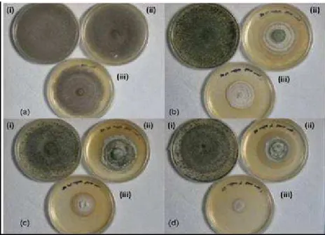
Figure 1 - Growth of fungal isolates (a) SD12 (b) SD14 (c) SD19 and (d) SD20 from sawdust on YMPG agar containing (i) 0 mg L-1 (ii) 50 mg L-1 and (iii) 100 mg L-1 of PCP concentration after 14 days.
Among these four isolates, isolate SD12 showed the highest mycelial extension rate, which was 10
mm day-1 at 100 mg L-1 of PCP. This was followed
SD20 with 4.2 mm day-1. Besides showing high growth rate in the presence of PCP, isolate SD12 was also the most tolerant towards PCP among the four fungal isolates. The increase in PCP
concentration from 50 to 100 mg L-1 only caused a
minor reduction to the growth rate of isolate SD12. The maximum mycelial extension rate obtained
for the isolate SD12 at 100 mg L-1 of PCP was also
higher as compared to the zygomycete, Rhizopus
nigricans that grew at a rate of 6.6 mm day-1 at
100 mg L-1 of PCP (Tomasini et al. 2001).
According to Lamar and co-workers (1990), having a microorganism that is capable of tolerating high contaminant concentrations would be a distinct advantage for the bioremediation of highly contaminated media. Fungal isolates SD12, SD14, SD19 and SD20 showed an interesting potential for PCP bioremediation. Okeke and
co-worker (1994) studied the growth of fungus L.
edodes that was repressed by PCP at only 15 mg
L-1 in agar but was able to tolerate up to 200 mg
kg-1 of PCP in the soil. P. chrysosporium and P.
sordida that were able to grow at 25 ppm of PCP
(Lamar et al. 1990) were able to grow and deplete 88 to 91% of PCP in a field soil with initial
concentrations from 250 to 400 mg kg-1 of PCP
(Lamar and Dietrich 1990). Fungal isolate, especially SD12, which grew and tolerated up to
100 mg L-1 of PCP in agar might be able to
tolerate an even higher concentration of PCP in the soil when applied for bioremediation.
Scanning electron microscopy (SEM) of PCP tolerant fungal isolates
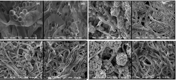
Isolate SD12, SD14, SD19 and SD20 were selected for further examination under SEM to see how well these isolate could tolerate the biocidal effect of PCP. SEM results showed that the
presence of high PCP concentration (100 mg L-1)
caused various degree of morphological changes to the hyphae structure of the tested fungal isolates. At high PCP concentration, the hyphae of all the four isolates, except for isolate SD12 appeared to collapse and had a wrinkles type of appearance as compared to the control (Fig. 2). Not many changes, however, were observed for the hyphae of isolate SD12, which appeared to be fine and smooth. Only minor collapse to the sporangiophores and sporangiola of isolate SD12
was observed at 100 mg L-1 of PCP.
Figure 2 - SEM micrographs of isolate (a) SD12, (b) SD14, (c) SD19 and (d) SD20 grown for 14 days on agar medium containing (i) 0 mg L-1 of PCP as control and (ii) 100 mg L-1 of PCP.
Besides altering the structure of fungal hyphae, PCP also causes the collapse of fungal spores. From the four fungal isolates examined using SEM, isolate SD12 was the only isolate that still
produced sporangiospores at 100 mg L-1 of PCP.
Among the produced sporangiola, however, some
of the sporangiola were collapsed and wrinkled (Fig. 2ii). PCP has been known to strongly inhibit
spore germination at only just 4.0 mg L-1 even in
the cultures of highly efficient PCP degrading
white-rot fungus P. chrysosporium (Mileski et al.
collapse as observed among some of the sporangiola produced by the isolate SD12 might be one of the mechanisms of PCP to inhibit the fungal spore germination. PCP has been shown to prevent the spores from germinating (Carvalho et al. 2009), which, based on the present data, might be related to the collapse of the spores caused by PCP.
Among the four fungal isolates capable of
tolerating up to 100 mg L-1 of PCP, isolate SD12
showed the least structural damage at high PCP concentration. Not only it tolerated the high concentration of PCP, it was also the fastest growing isolate in the presence of PCP. This,
therefore, showed that isolate SD12 was a strong PCP tolerating fungal isolate. The other three
fungal isolates were also strong PCP tolerating
fungi as well. Even with the damage done by the PCP, these fungal isolates were still able to grow at high PCP concentration.
Fungal characterisation and identification
Morphological characterisation of the four PCP tolerant isolates showed that isolate SD12 was a
species of the genera Cunninghamella
(Zygomycetes) while isolate SD14, SD19 and
SD20 belonged to Trichoderma sp (Ascomycetes)
(Table 2).
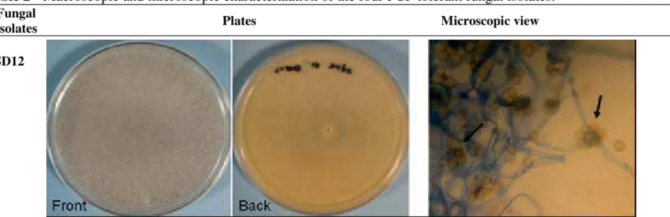
Table 2 - Macroscopic and microscopic characterization of the four PCP tolerant fungal isolates.
Fungal
Isolates Plates Microscopic view
SD12
Fast growing colony that fill up 90 mm Petri dish in 4-5 days. Woolly or cottony mycelial mat formed were white at first but becoming dark grey with sporangiola development. Aerial mycelium form strands that reached the cover of the petri dish. No pigment production and underside of colony is slightly yellowish (color of growth medium)
Presumed as Cunninghamella spp.
Magnification: 600x
Apex of mature sporangiophores with clusters of attached globose to ovoid sporangiola (Arrow). Coenocytic hyphae.
SD14 SD19 SD20
Colony was fast growing and the mycelium covered the whole petri dish in 4 days. Aerial mycelium was first white but become light green upon conidiation. Conidiation causes the white fluffy mycelium to clump, forming thick fluffy tufts which cover mostly the margin of the plate. Mycelium was also slightly raised as it spread from the centre. No pigment production and the underside of the colony is the colour of the growth medium.
Presumed as Trichoderma spp.
Molecular identification of the four PCP tolerant fungal isolates further confirmed that these isolates
were Cunninghamella and Trichoderma species.
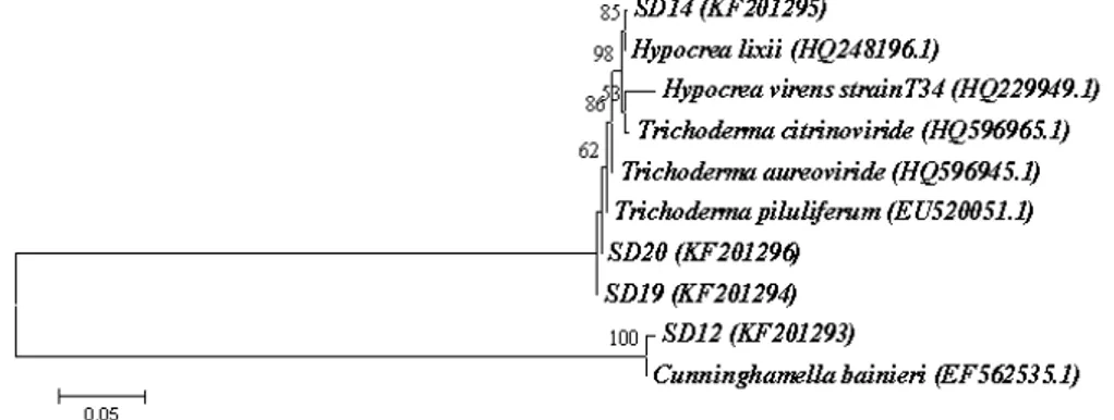
The phylogenetic tree constructed based on the ITS sequences of the four isolates indicated that
isolate SD14 was close to Hypocrea lixii,
teleomorph of Trichoderma harzianum, with up to
99% sequence similarity. SD19 and SD20 showed
99% sequence similarity with Trichoderma
piluliferum and were closely related with
Trichoderma aureoviride, also with 99% sequence
similarity. Isolate SD12 used as out group showed
98% sequence similarity with Cunninghamella
bainieri (Fig. 3).
Figure 3 - Neighbour-joining tree from ITS sequences showing the relationship between the four PCP tolerance fungi from sawdust and closely related sequences of the isolates retrieved from the GenBank (accession number). Bootstrap value >50% (1000 replicates) are shown at the branches. Bar = 5 nucleotide substitutions per 100 nucleotides.
Trichoderma species are free living fungi that are
commonly found in soil, root and foliar environments. These fungi also colonize woody and herbaceous plant materials, in which the sexual teleomorph has most often been found
(Harman et al. 2004). Trichoderma species,
especially T. harzianum and T. virens have been
well known for their application as an effective biological control agent against several soil-borne phytopathogenic fungi and for the commercial
production of cellulase by T. reesei (Samuels
1996; Verma et al. 2007). Studies have also shown the applicability of the soil borne ascomycetes
such as T. viride and T. harzianum for the
degradation of various xenobiotics, including phenanthrene, pyrene, endosulfan, cyanide and pentachlorophenol (Cserjesi 1967; Katayama and Matsumura 1993; Ravelet et al. 2000; Chavez-Gomez et al. 2003; Ezzi and Lynch 2005).
Trichoderma virgatum has been reported to reduce
up to 95% of PCP by methylation of PCP to a less toxic compound (Cserjesi and Johnson 1972). Duncan and Deverall (1964) have also reported on
a less recovery of PCP from Trichoderma treated
wood and found a decreasing activity of PCP against this fungus. They concluded that
Trichoderma was able to degrade
pentachlorophenol.
Different from Trichoderma species, however,
Cunninghamella species have been used widely as
a microbial model of mammalian metabolism of various pharmaceutical drugs and xenobiotics due to their ability to metabolise these compounds in a manner similar to that in the mammals (Abourashed et al. 1999; Asha and Vidyavathi 2009). Several species of this zygomycetes,
normally found in the soil such as Cunninghamella
elegans, Cunninghamella blakesleeana and C.
bainieri have been used to oxidize, transform or
metabolize various drugs (propranolol, warfarin, naproxen, verapamil and flutamide), phenylurea herbicide (chlortoluron, diuron and isoproturon) and organopollutants such as poly aromatic hydrocarbons (anthracene, phenanthrene and fluorine) (Ferris et al. 1973; Cerniglia and Yang 1984; Foster et al. 1989; Rizzo and Davis 1989; Vroumsia et al. 1996; Garon et al. 2000; Zhong et al. 2003; Sun et al. 2004; Amadio and Murphy
2011). Cunninghamella species have also been
reported to degrade organochlorine such as PCP (Seigle-Murandi et al. 1992). Seigle-Murandi and
co-workers (1995) reported that C. bainieri was
PCP initial addition (1 g L-1) in glucose containing GS liquid medium.
Trial PCP biodegradation in liquid medium by isolate SD12
Cunninghamella sp. UMAS SD12that showed the
highest growth rate and tolerance towards PCP was tested for its ability to degrade PCP in glucose minimal liquid medium. The fungus was allowed to grow for 24 h before PCP was added to a final
concentration of 20 mg L-1 (Day 0). Results
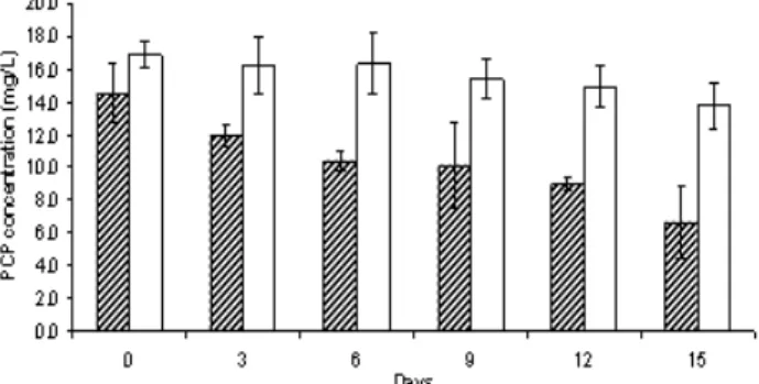
showed that the isolate SD12 removed up to 51.7% of PCP after 15 days (Fig. 4). PCP in the liquid medium was gradually depleted by the
isolate SD12 from 14.5 mg L-1 on day 0 to 10.4
mg L-1 on day 6 and finally to 6.6 mg L-1 on day
15. PCP concentration in the abiotic control flask with no fungal inoculum decreased progressively throughout the 15 days of incubation but the
decrease was only 3.1 mg L-1 of PCP. The ability
of isolate SD12 to tolerate high PCP concentration and at the same time degrade PCP, therefore, highlighted the potential applicability of the fungal isolates from sawdust for bioremediation of PCP contaminated sites.
Figure 4 - Depletion of PCP by fungal isolates SD12 within a period of 15 days in glucose minimal liquid medium supplemented with 20 mg L-1 of PCP. PCP concentration in the abiotic control flask (with no fungal inoculate) is indicated by an empty bar. Each data is represented by the mean of three replicates.
Szewczyk et al (2003) have also previously
reported the isolation of zygomycete, Mucor
ramosissimus from contaminated site that was
capabale of tolerating and transforming 10 mg/L of PCP to tetrachlorohydroquinone (TCHQ). Leon-Santiesteban et al (2008) reported the isolation of tyrosinase and peroxidase producing
zygomycete, Rhizopus oryzae from PCP
contaminated soil that tolerated and removed 90% of initial PCP concentration (12.5%). Carvalho et al (2009) also showed that besides basidiomycetes or white rot fungi, zygomycetes such as
Cunninghamella sp. UMAS SD12 also tolerated
and degraded PCP. Further studies on the metabolites produced by the isolate SD12 during PCP degradation would be crucial in order to facilitate in identifying the possible enzymes and PCP degradation pathway used by the isolates to degrade PCP.
CONCLUSION
The results demonstrated the successful isolation and characterization of four PCP-tolerant fungal isolates. SEM study of the isolates grown on PCP showed that high PCP concentration resulted the collapse of fungal hyphae and spores. Despite that, these fungal isolates were able to grow. The PCP
tolerance fungi were identified as Cunninghamella
and Trichoderma sp. Cunninghamella sp. UMAS
SD12 showed the highest tolerance towards PCP
and its degradation in liquid medium in 15 days.
ACKNOWLEDGEMENTS
This research project was supported by Universiti Malaysia Sarawak and financially funded by Ministry of Higher Education Malaysia (MoHE) under the Fundamental Research Grant Scheme (FRGS) (E14099/F07/69/769/2010(50).
REFERENCES
Abourashed EA, Clark AM, Hufford CD. Microbial models of mammalian metabolism of xenobiotics: An updated review. Curr Med Chem. 1999; 6: 359-374. Akpomie OO, Ubogun E, Ubogun M. Determination of
the cellulolytic activities of microorganisms isolated from poultry litter for sawdust degradation. J Environ Sci Water Resour. 2013; 2: 062-066.
Amadio J, Murphy CD. Production of human metabolites of the anti-cancer drug flutamide via biotransformation in Cunninghamella species.
Biotechnol Lett. 2011; 33: 321-326.
Bianchi A, Zambonelli A, D’Aulerio AZ, Bellesia F. Ultrastructural studies of the effects of Allium sativum on phytopathogenic fungi in vitro. Plant Dis.
1997; 81: 1241-1246.
Carvalho MB, Martins I, Leitao MC, Garcia H, Rodrigues C, Romao, VS, Mclellan I, Hursthouse A, Pereira CS. Screening pentachlorophenol degradation ability by environmental fungal strains belonging to the phyla ascomycota and zygomycota. J Ind Microbiol Biotechnol. 2009; 36: 1249-1256.
Cerniglia CE, Yang SK. Stereoselective metabolism of anthracene and phenanthrene by the fungus
Cunninghamella elegans. Appl Environ Microbiol.
1984; 47: 119-124.
Chavez-Gomez B, Quintero R, Esparza-Garcia F, Mesta-Howard AM, Zavala Diaz de la Serna FJ, Hernandez-Rodriguez CH, Gillen T, Poggi-Varaldo HM, Barrera-Cortes J, Rodriguez-Vazquez R. Removal of phenanthrene from soil by co-cultures of bacteria and fungi pregrown on sugarcane bagasse pith. Bioresour Technol. 2003; 89: 177-183.
Cserjesi AJ. The adaptation of fungi to pentachlorophenol and its biodegradation. Can J Microbiol. 1967; 13: 1243-1249.
Cserjesi AJ, Johnson EL. Methylation of pentachlorophenol by Trichoderma virgatum. Can J Microbiol. 1972; 18: 45-49.
Cubero OF, Crespo A, Fatehi J, Bridge PD. DNA extraction and PCR amplification method suitable for fresh, herbarium-stored, lichenized and other fungi.
Plant Syst Evol. 1999; 216: 243-249.
Demers PA, Davies HW, Friesen MC, Hertzman C, Ostry A, Hershler R, Teschke K. Cancer and occupational exposure to pentachlorophenol and tetrachlorophenol (Canada). CCC. 2006; 17: 749-758. Duncan CG, Deverall, FJ. Degradation of wood preservatives by fungi. App. Environ Microbiol.
1964; 12: 57-62.
Ezzi MI, Lynch JM. Biodegradation of cyanide by
Trichoderma spp. and Fusarium spp. Enzyme Microb Technol. 2005; 36: 849-854.
Ferris JP, Fasco MJ, Stylianopoulou FL, Jerina DM, Daly JW, Jeffrey AM. Monooxygenase activity in
Cunninghamella bainieri: Evidence for a fungal system similar to liver microsomes. Arch Biochem Biophys. 1973; 156: 97-103.
Fisher B. Pentachlorophenol: toxicology and environmental fate. J Pesticide Reform. 1991; 11 (1): 2-5.
Foster BC, Buttar HS, Qureshi SA, McGilveray IJ. Propranolol metabolism by Cunninghamella bainieri.
Xenobiotica. 1989; 19: 539-546.
Garon D, Krivobok S, Seigle-Murandi F. Fungal degradation of fluorene. Chemosphere. 2000; 40: 91-97.
Ge JC, Pan JL, Fei ZL, Wu GH, Giesy JP. Concentrations of pentachlorophenol (PCP) in fish and shrimp in Jiangsu Province, China.
Chemosphere. 2007; 69: 164-169.
Giraud F, Guiraud P, Kadri M, Blake G, Steiman R. Biodegradation of anthracene andfluoranthene by fungi isolated from an experimental constructed wetland for wastewater treatment. Water Res. 2001; 35: 4126-4136.
Harman GE, Howell CR, Viterbo A, Chet I, Lorito M.
Trichoderma species-opportunistic, avirulent plant symbionts. Nature Rev Microbiol. 2004; 2: 43-56. IARC. IARC monographs on the evaluation of
carcinogenic risks to humans 71: Re-evaluation of some organic chemicals, hydrazine and hydrogen peroxide, Lyon, France. World Health Organization, International Agency for Research on Cancer, 1999. Jukes TH, Cantor CR. Evolution of protein molecules.
In: Munro HN (Ed.). Mammalian Protein Metabolism
(Vol. 3). Academic Press, New York, USA, 1969; p.21-132.
Katayama A, Matsumura F. Degradation of organochlorine pesticides, particularly endosulfan, by
Trichoderma harzianum. Environ Toxicol Chem.
1993; 12: 1059-1065.
Keither LH. Teilard WA. Priority pollutants. I. A perspective view. Environ Sci Technol. 1979; 13: 416-423.
Kitunen VH, Salkinoja-Salonen MS. Soil contamination at abandoned sawmill areas. Chemosphere. 1990; 20: 1671-1677.
Lamar RT, Dietrich DM. In situ depletion of pentachlorophenol from contaminated soil by
Phanerochaete spp. Appl Environ Microbiol. 1990; 56: 3093-3100.
Lamar RT, Larsen MJ, Kirk K. Sensitivity to and degradation of pentachlorophenol by Phanerochaete
spp. Appl Environ Microbiol. 1990; 56: 3519-3526. Lennox JA, Abriba C, Alabi BN, Akubuenyi FC.
Comparative degradation of sawdust by microorganisms isolated from it. Afr J Microbiol Res.
2010; 4: 1352-1355.
Leon-Santiesteban H, Bernal R, Fernandez FJ, Tomasini A. Tyrosinase and peroxidase production by Rhizopus oryzae strain ENHE obtained from pentachlorophenol-contaminated soil. J Chem Tech Biot, 2008; 83: 1394-1400
Manoch L, Jeamjitt O, Eamvijarn A, Dethoup T, Kokaew J, Paopun Y, Poochinya P, Umrung P. Light and SEM studies on leaf litter fungi. J. Microsc. Soc. Thailand. 2008; 22: 56-59.
McAllister KA, Lee H, Trevors JT. Microbial degradation of pentachlorophenol. Biodegradation.
McLean D, Eng A, Dryson E, Walls C, Harding E, Wong KC, Cheng S, Mannetje A, Ellison-Loschmann L, Slater T, Shoemack P, Pearce N. Morbidity in former sawmill workers exposed to pentachlorophenol (PCP): A cross-sectional study in New Zealand. Am J Ind Med. 2009; 52: 271-281. Mileski GJ, Bumpus JA, Jurek MA, Aust SD.
Biodegradation of pentachlorophenol by the white rot fungus Phanerochaete chrysosporium. Appl Environ Microbiol. 1988; 54: 2885-2889.
Mueller JG, Middaugh DP, Lantz SE, Chapman PJ. Biodegradation of creosote and pentachlorophenol in contaminated groundwater: chemical and biological assessment. Appl Environ Microbiol. 1991; 57: 1277-1285.
Novotny C, Erbanova P, Sasek V, Kubatova A, Cajthaml T, Lang E, Krahl J, Zadrazil F. Extracellular oxidative enzyme production and PAH removal in soil by exploratory mycelium of white rot fungi. Biodegradation. 1999; 10: 159-168.
Nwodo-Chinedu S, Okochi VI, Smith HA, Omidiji O. Isolation of cellulolytic microfungi involved in wood-waste decomposition: prospects for enzymatic hydrolysis of cellulosic wastes. Int J Biomed Health Sci. 2005; 1(2): 2): 41-51.
Okeke BC, Paterson A, Smith JE, Watson-Craik IA. Relationships between ligninolytic activities of
Lentinula spp. and biotransformation of pentachlorophenol in sterile soil. Lett Appl Microbiol.
1994; 19: 284-287.
Persson Y, Lundstedt S, Oberg L, Tysklind M. Levels of chlorinated compounds (CPs. PCPPs,PCDEs, PCDFs and PCDDs) in soils at contaminated sawmill sites in Sweden. Chemosphere. 2007; 66: 234-242. Proudfoot AT. Pentachlorophenol poisoning. Toxicol
Rev. 2003; 22: 3-11.
Ravelet C, Krivobok S, Sage L, Steiman R. Biodegradation of pyrene by sediment fungi.
Chemosphere 2000; 40: 557-563.
Rizzo JD, Davis PJ. Microbial models of mammalian metabolism: Conversion of warfarin to 4’- hydroxywarfarin using Cunninghamella bainieri. J Pharm Sci. 1989; 78: 183-189.
Ryu WR, Shim SH, Jang MY, Jeon YJ, Oh KK, Cho MH. Biodegradation of white rot fungi under ligninolytic and nonligninolytic conditions.
Biotechnol Bioproc. E. 2000; 5: 211-214.
Samuels GJ. Trichoderma: a review of biology and systematics of the genus. Mycol Res.1996; 100: 923-935.
Seigle-Murandi F, Steiman R, Benoit-Guyod JL, Guiraud P. Biodegradation of pentachlorophenol by micromycetes. 1. Zygomycetes. Environ Toxicol Water Qual. 1992; 7: 125-139.
Seigle-Murandi F, Toe A, Benoit-Guyod JL, Steiman R, Kadri M. Depletion of pentachlorophenol by deuteromycetes isolated from soil. Chemosphere.
1995; 31: 2677-2686.
Sun L, Huang HH, Liu L, Zhong DF. Transformation of verapamil by Cunninghamella blakesleeana. Appl Environ Microbiol. 2004; 70: 2722-2727.
Szewczyk R, Bernat p, Milczarek K, Dlugonski J. Application of microscopic fungi isolated from polluted industrial areas for polycyclic aromatic hydrocarbons and pentachlorophenol reduction.
Biodegradation, 2003; 14: 1-8.
Tam S, Johnson SA, Graham A. The effect of organic structures on pentachlorophenol adsorption on soil.
Water Air Soil Pollut. 1999; 115: 337-346.
Tomasini A, Flores V, Cortes D, Barrios-Gonzalez J. An isolate of Rhizopus nigricans capable of tolerating and removing pentachlorophenol. World J Microbiol Biotechn. 2001; 17: 201-205.
Tuomela M, Lyytikainen M, Oivanen P, Hatakka A. Mineralization and conversion of pentachlorophenol (PCP) in soil inoculated with the white-rot fungus
Trametes versicolor. Soil Biol Biochem. 1999; 31: 65-74.
Verma M, Brar SK, Tyagi RD, Surampalli RY, Valero JR. Antagonistic fungi, Trichoderma spp.: Panoply of biological control. Biochem Eng J. 2007; 37: 1-20. Vroumsia T, Steiman R, Seigle-Murandi F,
Benoit-Guyod JL, Khadrani A. Biodegradation of three substituted phenylurea herbicides (chlortoluron, diuron, and isoproturon) by soil fungi. A comparative study. Chemosphere. 1996; 33: 2045-2056.
White TJ, Bruns T, Lee S, Taylor J. Amplification and direct sequencing of fungal ribosomal RNA genes for phylogenetics. In: Innis MA, Gelfand DH, Sninsky JJ, White TJ, (eds.) PCR Protocols: A Guide to Methods and Applications, Academic Press, New York, USA, 1990; p.315-322.
Zhong DF, Sun L, Liu L, Huang HH. Microbial transformation of naproxen by Cunninghamella
species. Acta Pharmacol Sini. 2003; 24: 442-447.