RESUMO.- [Administração parenteral das vitaminas A, D e E no metabolismo oxidativo e sobre a função de leucócitos polimorfonucleares em suínos.] O período de desmame nos leitões é caracterizado por alterações
isio-Parenteral administration of vitamins A, D and E on the
oxidative metabolism and function of polymorphonuclear
leukocytes in swine
1Alessandra S. Lima2, Rebeca A. Weigel2, Aline A. Morgado2, Giovanna R. Nunes2, Fernando N.
Souza2, Andrea M. Moreno2, Alice Maria M.P. Della Libera2 and Maria Claudia A. Sucupira2*
ABSTRACT.- Lima A.S., Weigel R.A., Morgado A.A., Nunes G.R., Souza F.N., Moreno A.M., Della Libera A.M.M.P. & Sucupira M.C.A. 2012. Parenteral administration of vitamins A, D and E on the oxidative metabolism and function of polymorphonuclear leukocytes in swine.Pesquisa Veterinária Brasileira 32(8):727-734. Departamento de Clínica Médica, Faculdade de Medicina Veterinária e Zootecnia, Universidade de São Paulo, Av. Prof. Dr. Or-lando M. de Paiva 87, São Paulo, SP 05508 270, Brazil. E-mail: [email protected]
The weaning period of piglets is characterized by physiological alterations, such as de-creased weight gain, inde-creased reactive oxygen species (ROS) and inde-creased serum cortisol levels with possible effects on the immune response. The effect of parenteral administra-tion of vitamins A, D and E on producadministra-tion performance, oxidative metabolism, and the func-tion of polymorphonuclear leukocytes (PMNLs) was assessed in piglets during the weaning period. The sample was comprised of 20 male piglets that were given an injectable ADE vitamin combination (135,000 IU vitamin A, 40,000 IU vitamin D and 40mg vitamin E/ animal) at 20 and 40 days of age. Weight gain, concentration of reduced glutathione (GSH), malondialdehyde (MDA), superoxide dismutase (SOD) and the microbicidal and phagocytic activity of PMNLs were assessed. No difference was observed in the average piglet weight during the study; however, a greater percentage of weight gain was observed after weaning in the treated group. The concentrations of GSH and SOD did not differ between groups, although lipid peroxidation was greater in the control group at 60 days of age. The inves-tigated variables of oxidative metabolism were correlated as follows: -0.41 for GSH and MDA, -0.54 for GSH and SOD and 0.34 for MDA and SOD. The intensity of intracellular ROS production, the percentage of ROS-producing PMNLs and the intensity of phagocytosis by PMNLs did not differ between treatment groups. Administration of the injectable ADE com-bination improved the percentage of weight gain between 20 and 40 days of age, decreased oxidative stress at 60 days of age and did not inluence the function of PMNLs in piglets.
INDEX TERMS: Immune response, weaning, piglets, GSH, MSD and SOD.
lógicas como menor ganho de peso, aumento na produção de espécies reativas de oxigênio (EROs) e aumento na con-centração plasmática de cortisol com possíveis implicações para a resposta imune. Foi avaliado o efeito da administra-ção parenteral das vitaminas A, D e E sobre o desempenho produtivo, o metabolismo oxidativo e a função de leucócitos polimorfonucleares (PMNLs) em suínos durante esta fase de crescimento. Foram utilizados 20 leitões, machos, com 20 dias de idade que receberam ADE injetável (135.000 UI vitamina A, 40.000 UI vitamina D e 40mg vitamina E/ani-mal), aos 20 e 40 dias de idade. Foi determinado o ganho de peso e as concentrações de glutationa reduzida (GSH), malondialdeído (MDA) e superóxido dismutase (SOD) e a
1 Received on March 7, 2012.
Accepted for publication on April 10, 2012.
2 Departamento de Clínica Médica, Faculdade de Medicina Veterinária
capacidade microbicida e fagocítica dos PMNLs. Não houve diferença entre o peso vivo médio durante o experimento, porém maior ganho de peso percentual foi observado 20 dias após o desmame para o grupo tratado. As concentra-ções de GSH e SOD não diferiram entre os grupos, porém a lipoperoxidação foi maior no grupo controle aos 60 dias de idade. As correlações entre as variáveis do metabolismo oxidativo foram -0,41 para GSH e o MDA, -0,54 para GSH e SOD e 0,34 para MDA e SOD. A intensidade da produção intracelular de EROs, a porcentagem de PMNLs que produ-ziram EROs e a intensidade de fagocitose dos PMNLs não diferiram entre os tratamentos. A administração de ADE injetável melhorou o ganho de peso percentual no período de 20 a 40 dias de idade, diminuiu o estresse oxidativo aos 60 dias de idade e não inluenciou função dos PMNLs dos leitões.
TERMOS DE INDEXAÇÃO: Resposta imune, desmame, leitões, GSH, MDA e SOD.
INTRODUCTION
Maternal separation, relocation, introduction to new social groups, the establishment of social dominance and a chan-ge in diet to a higher dry mass content induce physiologi-cal changes in recently weaned swine, with a consequent reduction in weight gain being observed (Sutherland et al. 2006, Kojima et al. 2008). These alterations lead to incre-ased cortisol serum levels (Heo et al. 2003) and possible effects on the immune response of animals (Bonnette et al. 1990), such as a reduction in neutrophil microbicidal ca-pacity and reduced function of natural killer cells and lym-phocytes (Sutherland et al. 2006, Da Costa et al. 2008).
Metz and Gonyou (1990) observed that the immune system of swine weaned at four weeks of age is not yet fully developed. Therefore, these animals are more susceptible to disease. Animals weaned at four weeks began to eat food only after 2 or 3 days post-weaning. In response to stress and exposure to antigens, the immune system stimulates the production of proinlammatory cytokines in the brain that reduce the motivation to eat (Kent et al. 1996) and in-teract with growth hormone and insulin-like growth factor 1 (IGF-1). This interaction can decrease cellular growth (Kelly 2004).
During normal aerobic metabolism, cells maintain a ba-lance between the production and elimination of reactive oxygen species (ROS). When the occurrence of oxidative events increases, the system tends toward the pro-oxidative side, resulting in “oxidative stress” (Lykkesfeldt & Svendsen 2007). An increase in oxidative stress might affect the con-centration of antioxidants, such as glutathione and vitamin E, and inally leads to oxidative damage of lipids, proteins, carbohydrates and nucleic acids. This process might be so severe as to eventually cause cell death. The eficacy of the antioxidant system largely depends on the type of molecu-le triggering oxidative stress and its intra- or extracellular location. Damage to the cell membrane might be avoided more effectively by vitamin E, which reacts with peroxyl and hydroxyl radicals, compared to carotenoids, which act by reacting with singlet oxygen (Jordão et al. 1998).
ROS is the main oxidant substance. The physiological action of ROS is directly related to the antimicrobial protec-tion mediated by polymorphonuclear leukocytes (PMNLs). PMNLs usually circulate in the bloodstream in an inactive state during which they may surround or phagocytize bac-teria but do not cause damage (Winterbourn & Assman 1990). Upon exposure to bacteria coated by immunoglobu-lins, immunocomplexes, complement, or leukotrienes, the enzyme NAPDH-oxidase is activated in PMNLs, which trig-gers the production of ROS and facilitates the microbicidal action of PMNLs.
Antioxidants protect biological systems against the da-mage caused by processes that might lead to high levels of oxidation (Krinski 1992). Antioxidants are classiied as endogenous, i.e., synthesized inside the organism, and exogenous, i.e., supplied by the diet. Vitamin E is one of the best-known exogenous antioxidants. Since the 1990s, vita-min E has been investigated as a treatment for vita-minimizing the adverse effects associated with the transitional stage of development, which is represented in swine by weaning (Bonnette et al. 1990). Vitamin A is another exogenous antioxidant able to prevent the deleterious action of ROS released during inlammation, infection and stress or even during resting metabolism (Gomes et al. 2005). Vitamin D is comprised of two major groups: vitamin D3 (cholecalci-ferol, synthesized by animals) and D2 (ergocalci(cholecalci-ferol, syn-thesized by plants); the production of both groups requires sunlight. Animals synthesize approximately 10 different provitamins that form vitamin D upon being irradiated. Thus, special attention must be paid to supplemental vi-tamin D in animals raised in coninement. Lauridsen et al. (2010) concluded that independent of the dose or the form of vitamin D supplied to sows, only a small amount of vi-tamin D is transferred to the offspring. Thus, nursing pigs not exposed to sunlight might require supplemental vita-min D. Indeed, injected supplements of combined A, D and E vitamins are widely used in Brazilian systems of animal production.
Because animals kept in intensive production systems exhibit higher requirements of antioxidant agents, this stu-dy sought to investigate the effect of parenteral administra-tion of vitamins A, D and E on the producadministra-tion performance, oxidative metabolism and PMNL function of swine during their growth phase.
MATERIALS AND METHODS
The sample group consisted of 20 male piglets at 20 days old with a 7.1 kg average live weight. The piglets were hybrids betwe-en commercial gbetwe-enetic lineages cb22 and ag337 Agroceres. Ani-mals were allocated to a commercial farm in the town of Capivari, SP, Brazil. Weaning was performed at 21 days of age, and the pi-glets were subsequently housed in the nursery, where they remai-ned until the end of the study at 60 days of age. Diet was based on corn, soy, bran, dairy products, vitamin and mineral supplements appropriately balanced to promote weight gain compatible with the growth curve of this lineage (Table 1).
vita-min combination (135,000 IU vitavita-min A, 40,000 IU vitavita-min D and 40mg vitamin E/animal/dose) via deep intramuscular injection at 20 and 40 days old. The control group was given the same vo-lume (0.5mL/animal/dose) of sterile physiological saline solution at the same ages and by the same route to mimic the stress effects caused by handling and injection of the animals.
At 20 (M0), 40 (M1) and 60 (M2) days of age, animals were weighed to assess the production performance, and blood was collected to assess oxidative metabolism and PMNL function.
The oxidative metabolism was assessed by means of indirect markers, including a reduction in the endogenous antioxidant en-zymes, glutathione (GSH) and superoxide dismutase (SOD), and a reduction in malondialdehyde (MDA), one of the 2-thiobarbituric acid reactive substances (TBARS), which are themselves products of oxidative reactions. The GSH content was measured in whole blood by means of the colorimetric method described by Beutler et al. (1963). The index of lipid peroxidation was indirectly esta-blished based on the MDA content in heparinized serum as mea-sured by the thiobarbituric acid method described by Esterbauer & Cheeseman (1990). The activity of SOD was measured using the colorimetric method described by Woolliams & Wiener (1983), as adjusted for the LABMAX 240 automatic biochemical analyzer using the RANDOX SD 125 commercial kit (Ransel® Laboratories,
Randox, Crumlin, UK).
Immune function was assessed in circulating PMNLs by mea-suring intracellular ROS produced in vitro following stimulation with phorbol 12-myristate 13-acetate (PMA), Escherichia coli li-popolysaccharides (LPS), and after phagocytosis of Staphylococ-cus aureus conjugated to propidium iodide (Sa-PI). PMNL func-tion, as measured by phagocytosis of S. aureus (American Type Culture Collection (ATCC) 25923), as conjugated to propidium iodide and intracellular ROS production, were performed as su-ggested by Hasui et al. (1989). The samples were analyzed by low cytometry on a FACSCaliburTM cytometer (Becton Dickinson
Immunocytometry SystemTM, San Diego, USA). Twenty thousand
PMNLs were acquired, being identiied by their size and granula-rity (Fig.1 and 2) as described by Busque et al. (1998).
The normal distribution of data was veriied by the Kolmogo-rov-Smirnov test. To assess the differences between the means of results, an analysis of variance (one-way ANOVA) and the Tukey’s test were performed for data exhibiting parametric distribution. The Mann-Whitney test was applied to data exhibiting nonpara-metric distribution. Signiicance was established when P<0.05 in all tests. Data exhibiting a normal distribution are expressed as the mean (± standard deviation); data exhibiting nonparametric distribution are expressed as the median followed by the maxi-mum and minimaxi-mum values. The correlation among variables was investigated by means of the Pearson correlation coeficient. Cor-relation was rated high when r > 0.6, intermediate when r ranged from 0.3 to 0.6 and low when r<0.3. MINITAB® software version
14.1 (GlobalTech Informática™, Belo Horizonte, Minas Gerais,
Bra-zil) was used for statistical analysis.
The present study was approved by the Bioethics Committee of the School of Veterinary Medicine and Zootechnics of the Uni-versity of São Paulo (Faculdade de Medicina Veterinária e Zoo-tecnia da Universidade de São Paulo (FMVZ-USP) under protocol number 1703/2009.
RESULTS
At 60 days of age, the average live weight was 27.6 kg and 30.6 kg in piglets belonging to the control and treated groups, respectively. No difference was found in the ave-rage live weight throughout the study (Table 2). However, a greater percentage of weight gain was observed in vita-min treated animals by 20 days after weaning (P=0.20) (Fig.3).
Due to previous processing, the heparinized blood samples collected at M0 exhibited intense hemolysis. Thus, it was not possible to measure the concentration of SOD or the activity of PMNLs from these samples. Table 2 shows the results obtained for the variables of oxidative metabolism.
Table 1. Diet Composition of pigs in the nursery. São Paulo, 2010
Diet Composition
Sodium chloride 0,45
Limestone 39% 0,26
Corn Grain 8,0 62,2
Rice Bran 5,00
Soybean Meal 46,0 21,0
Yeastof sugar cane 37% 5,00
Meat and bone meal 44% 3,80
Fat lard 1,40
MineralSuplement1 0,10
Vitamin Suplement2 0,10
Phytase3 0,01
Copper Sulp Pent. 25% 0,08
Choline chloride 60% 0,04
DL- methionine 99% 0,08
Lysina HCl 78,8% 0,38
L-Threonina 98% 0,11
Total (%) 100
1 Roligomix Suínos (DSM): Fe, 90g; Cu, 10g; Co, 2g; Mn, 40g; Zn, 2g e
Exci-pient q.s.p. 500g.
2 Premix Inicial (MCassab): Vit. A 8.00.000UI; Vit. D3 2.000.000UI; Vit E
10.000mg; Vit. K3 500mg; Vit. B1 1.500mg; Vit. B2 5.000mg; Vit. B6 2000mg; Vit. B12 20.000mcg; Niacin 25.000mg; Calcium Pantothenate 12.000mg; Folic Acid 800mg; Biotin 50mg, Selenium 280mg e Excipient q.s.p. 1000g.
3 Ronozyme P5000G (DSM).
Fig.2. Cytogram of swine blood polymorphonuclear leukocytes (PMNLs) and subsequent analysis of PMNL function. São Paulo, 2010. (A) Population of PMNLs that produced reactive oxygen species (ROS). (B) Control sample of intracellular ROS production. (C) Popula-tion of PMNLs that performed phagocytosis. (D) Phagocytosis control samples.
Table 2. Mean and corresponding standard deviation of pig live weight following treatment with ADE vitamin combination or an equivalent
volume of saline. São Paulo, 2010
Live Weight (kg)
Treated Control
M0 7.15 a 7.1 a
(2.00) (1.35)
M1 15.4 b 13.15 b
(4.05) (1.86)
M2 30.6 c 27.6 c
(8.24) (2.91)
Note: M0 = weaning and irst dose; M1 = 20 days after M0 and second dose; M2 = 40 days after MO. Different lo-wercase letters in columns indicate differences between time points (P<0.05)
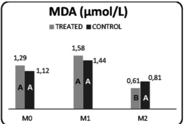
The concentrations of GSH and the enzyme SOD did not differ between the control and treated groups throughout the study (Table 3). Lipid peroxidation was higher in the control group (P=0.012) at M0 (Fig.4).
The correlations between the variables of oxidative me-tabolism exhibited intermediate intensity. There was a ne-gative correlation between GSH and MDA -0.41 (P=0.001) and between GSH and SOD -0.54 (P=0.001); the correlation between MDA and SOD was 0.34 (P=0.04).
The intensity of intracellular ROS production by PMNLs did not differ between treatments, but it was higher (P<0.01) at M2 compared to M1 (Table 4) in both groups. No difference was observed in the percentage of ROS pro-ducing PMNLs between groups, although higher percen-tages (P<0.02) were observed at 60 days of age compared to 40 days of age in both groups (Table 5). The intensity of phagocytosis by PMNLs did not differ between groups or between time-points. In spite of this lack of difference, the treated group exhibited a lower percentage (P=0.03) of phagocytosing PMNLs at M2 compared to M1 (Table 6).
Table 3. Mean and standard deviation of the activity of reduced glutathione (mg/dL), superoxide dismutase (U/g Hb), and malondialdehyde (μmol/L) in pigs treated
or not with ADE vitamin combination. São Paulo, 2010
Reduced Gluta- Superoxide Dismutase (U/g Hb) Malondialdehyde thione (mg/dL) Treated Control (μmol/L)
Treated Control Treated Control
M0 20.76 a 19.29 a * * 1.29 a 1.12 ab
(5.48) (7.92) (0.89) (0.75)
M1 8.78 b 11.50 b 2041.5 a 2111.2 a 1.58 a 1.44 a
(2.24) (5.79) (1284.4/2102.5) (1808.7/2188.1) (0.70) (0.65) M2 21.56 a 23.9 a 1855.2 b 1730.6 b 0.61 B b 0.81 A b
(5.44) (6.91) (1359.9/2016.4) (1618.1/ 2100.8) (0.16) (0.17) Note: M0 = weaning and irst dose; M1 = 20 days after M0 and second
dose; M2 40 days after M0.
* Insuficient sample. Different uppercase letters in rows indicate diffe-rences between treatments (P<0.05). Different lowercase letters in co-lumns indicate differences between time points (P<0.05).
Table 4. Mean staining intensity of intracellular reactive oxygen species in stimulated (i.e., cultured with
Staphylococ-cus aureus and Escherichia coli-derived lipopolysaccharide) and non-stimulated (i.e., basal) blood polymorphonuclear leukocytes from piglets treated or not treated with the ADE
vitamin combination. São Paulo, 2010
Treated Control
Basal Staphylococcus LPS Basal Staphylococcus LPS
aureus aureus
M1 73.76 a 90.39 a 65.11 a 55.36 a 79.98 a 62.66 a
(± 42.24) (± 35.54) (± 41.93) (±25.79) (± 37.26) (± 29.05) M2 143.70 b 264.80 b 138.70 b 164.60 b 172 b 169.10 b
(± 26.34) (± 317.3) (± 17.45) (± 71.15) (± 95.52) (± 67.44)
Different lowercase letters in columns indicate P<0.01. Basal = non-sti-mulated. LPS = lipopolysaccharide from Escherichia coli (strain 055:b5).
Table 5. The percentage (%) of stimulated (i.e., cultured with Staphylococcus aureus and Escherichia coli–derived lipopolysaccharide) and non-stimulated (i.e., basal) blood polymorphonuclear leukocytes (PMNLs) that stain positive for reactive oxygen species; PMNLs were obtained from
piglets treated or not treated with the ADE vitamin combination. São Paulo, 2010
Treated Control
Basal Staphylococcus LPS Basal Staphylococcus LPS
aureus aureus
M1 83.92 a 87.09 a 86.38 a 84.21 a 84.92 a 87.39 a
(± 12.33) (± 5.62) (±5.89) (± 10.34) (± 8.53) (± 9.46) M2 97.09b 93.56 b 97.5 b 96.70 b 96.70 b 96.71 b
(± 1.46) (± 4.54) (±1.20) (± 1.82) (± 1.82) (± 2.60)
Different lowercase letters in columns indicate P<0.02. Basal = non-sti-mulated. LPS = lipopolysaccharides of Escherichia coli (strain 055:b5).
Table 6. In vitro phagocytosis of propidium iodide (PI)-con-jugated Staphylococcus aureus was assessed by the percent of PI positive and mean staining intensity of blood polymor-phonuclear leukocytes from pigs treated or not treated with
ADE vitamin combination. São Paulo, 2010
Treated Control
Percentage Intensity Percentage Intensity
M1 63.18 a 16.99 68.14 13.36
(± 19.40) (± 19.89) (± 17.67) (± 7.75) M2 48.42 b 9.55 44.98 8.48
(± 20.69) (± 7.56) (± 18.31) (± 7.98)
Different lowercase letters in columns indicate differences between time points (P=0.03).
Fig.4. Mean and standard deviation of malondialdehyde concen-tration (μmol/L) in pigs treated with the ADE vitamin combi-nation or saline only. São Paulo, 2010.
Note: M0 = weaning and irst dose; M1 = 20 days after M0, and se-cond dose M2 = 40 days after M0. Different uppercase letters indicate differences between treatments (P<0.05).
DISCUSSION
The percentage of weight gain in piglets shows that the animals in the treated group exhibited better performance at the irst time-point assessed, speciically when animals were transferred from the mother to the nursery.
Few studies have investigated the effects of an injecta-ble combination of vitamins A, D and E in swine produc-tion. Conversely, many studies have assessed the effects of these vitamins separately.
Initially, the serum concentration of vitamin E is high in piglets, probably due to greater bioavailability in milk com-pared to a diet based on concentrates. Immediately after weaning, the serum concentration of vitamin E falls dras-tically (Sivertsen et al. 2007). The importance of vitamin E is related to its solubility in lipids and its localization in the lipid membrane. For these reasons, this vitamin is able to move across the membrane layers, where it performs its main function: to interrupt the chain reaction of lipid pero-xidation, mainly by neutralizing the peroxyl lipid radicals to produce lipid hydroperoxides and tocopheroxyl radicals (Faustman et al. 2009).
The concentration of vitamin A is high in swine colos-trum, and the concentration falls by 25% during the irst ive days of lactation (Heidebrecht et al. 1950). Few stu-dies have investigated the action of vitamin A in piglets, and most of them focused on sows. The effect of vitamin A (450,000 IU of retinol palmitate injected on the day of weaning or mating) on the productive performance was assessed by Silveira et al. (1998): These researchers con-cluded that injectable vitamin A supplements improved the reproductive performance of sows independent of the productive phase, including an increased number of total and live born piglets, and increased litter weight at birth. Thompson (1994) suggested that injectable vitamin A is more effective than dietary supplements because injecta-ble vitamin A might be taken up by cells directly, rather than being stored and released by the liver.
The assessment of oxidative metabolism showed that the concentration of SOD decreased with time during the post-weaning phase, regardless of treatment, which may indicate a higher requirement for antioxidants. SOD, an en-dogenous antioxidant, belongs to the class of metalloenzy-mes that counteract superoxide anion toxicity (Celi 2010). SOD is the most abundant enzymes and is the ifth most abundant protein in quantitative terms. SOD is present in all aerobic organisms and is necessary for the clearance of a wide variety of superoxide radicals.
The concentration of GSH and MDA changed in dispa-rate ways in our study. GSH acts directly or indirectly on important biological processes, and it is needed to pre-vent the peroxidation of unsaturated fatty acids in the cell membrane. For instance, GSH acts to keep the iron atoms of hemoglobin in the ferrous form (Rover Junior et al. 2001). Variations in the glutathione content directly affect the syn-thesis of proteins and deoxyribonucleic acid. GSH oxidation or depletion can decrease protein synthesis. Under condi-tions of intense oxidative stress, GSH can be irreversibly oxidized and thus rendered nonfunctional (Uhlig & Wendel 1992). Dietary supplementation with vitamin E or vitamin D3 can increase the levels of GSH in the liver (Sardar et al. 1996). MDA, the main representative of the thiobarbituric acid-reactive substances that arises during the peroxida-tion of fatty acids by ROS, is considered to be a marker of cellular damage and oxidative stress because its
concentra-tion increases with excess ROS (Celi 2010). In the present study, the lowest post-weaning levels of GSH were found in the treated group. Together with the lower index of lipid peroxidation exhibited by this group at M2 in both series of comparisons (intra- and inter-group), our data indicate that the antioxidant system of the treated group was better prepared to react to the oxidative stress caused by weaning. The two markers of the oxidative metabolism investi-gated in this study, speciically, the decrease in GSH con-centration and the increase in MDA concon-centration, beha-ve in such a way as to counteract oxidatibeha-ve stress. Similar behavior of these metabolites was described by Weigel et al. (2010). These researchers assessed the oxidative meta-bolism of sheep intoxicated with copper.
The assessment of the innate immune response did not reveal beneicial effects of vitamins A, D and E on PMNL function at the time-points and doses assessed in this stu-dy. However, measures of immune function were greatest at 60 days in both groups, including the percentage and in-tensity of ROS production by PMNLs, and phagocytosis of S. aureus conjugates either basally or post-stimulation. Lower microbicidal capacity was observed at 40 days of age com-pared to 60 days, although the percentage of PMNLs that phagocytized Staphylococcus aureus was lower in the con-trol group at age 60 days compared to 40 days. Thus, the lower microbicidal capacity at the age of 40 days might be a function of age (Hoskinson et al. 1990, Butcher et al. 2005) and the consequences of weaning (Sutherland et al. 2006, Kojima et al. 2008). Weaning increases the concentration of cortisol (Heo et al. 2003) and consequently reduces the mi-crobicidal capacity of neutrophils (Hoskinson et al. 1990, Butcher et al. 2005). In agreement with this interpretation, Hoskinson et al. (1990) found lower microbicidal capacity between the third and ifth weeks of life in piglets, and a subsequent increase in this function at sixth weeks and ol-der. These data agree with studies performed in other spe-cies (Coignoul et al. 1984, Da Costa et al. 2008) and with the indings of the present study. However, the present study falls short of discerning whether the lower microbicidal ca-pacity at 40 days is age-related or is a function of weaning because data from 20-day-old piglets is absent.
S. aureus-phagocytizing PMNLs exhibited by animals in the treated group but not the rate of phagocytosis on a per cell basis (as measured by staining intensity).
Dietary vitamin E supplements can potentially increase the resistance of sows and piglets to enteric diseases, such as those caused by E. coli. Indeed, E. coli infection is one of the most common illnesses among newborns and con-tributes to high mortality during the pre-weaning phase (Pinelli-Saavedra 2003). Vitamin A is also involved in the immune response of swine. As such, several studies report an increased concentration of antibodies against Escheri-chia coli and Salmonella Dublin lipopolysaccharides, as well as total gamma globulins, in piglets receiving vitamin A su-pplements compared to controls (Lüdke et al. 1985). The eficiency of vitamin A in the immune response is related with the maintenance of epithelium integrity, the function of the adrenal gland, which releases corticosteroids, and the antibody response to T-cell dependent antigens. Vita-min A plays an important role in maintaining appropria-te numbers of natural killer cells (Zhao & Ross 1995). In addition, Katz et al. (1987) state that retinoic acid contri-butes to increased macrophage phagocytic capacity and a possible role in leukocyte differentiation. Published results suggest that retinoic acid is involved in increased pro-in-lammatory cytokine production, including interleukin 1 (Trechsel et al. 1985).
CONCLUSIONS
The present study shows that administration of an in-jectable ADE combination (135,000 IU vitamin A, 40,000 IU vitamin D and 40mg vitamin E/animal) via deep intramus-cular injection at 20 and 40 days of age can increase the percentage of weight gained at these time points.
Our data also show that reduced oxidative stress and MDA production is observed in treated animals at 60 days of age but that no effect on PMNL function was observed in treated piglets.
REFERENCES
Balteskard L., Unneberg K., Halvorsen D., Hansen J.B. & Revhaug A. 1998. Effects of insulin-like growth factor 1 on neutrophil and monocyte func-tions in normal and septic states. J. Parenteral and Enteral Nutrition 22(3):127-135.
Beutler E., Duron O. & Kelly B.M. 1963. Improved method for the determi-nation of blood glutathione. J. Lab. Clin. Med. 61(5):882-888.
Bonnette E.D., Kornegay E.T., Lindemann M.D. & Hammerberg C. 1990. Hu-moral and cell-mediated immune response and performance of weaned pigs fed supplemental vitamin E levels and housed at two nursery tem-peratures. J. Anim. Sci. 68:1337-1345.
Busque P., Higgins R., Sénéchat S., Marchand R. & Quessy S. 1998. Simul-taneous low cytometric measurement of Streptococcus suis phagocyto-sis by polymorphonuclear and mononuclear leukocytes. Vet. Microbiol. 63:229-238.
Butcher S., Killampalli V., Lascelles D., Wang K., Alpar E.K. & Lord J.M. 2005. Raised cortisol, DHEAs ratios in the elderly after injury: Potential impact upon neutrophil function and immunity. Aging Cell 4(6):319-324. Celi P. 2010. The role of oxidative stress in small ruminants health and
production. Revta. Bras. Zootec. 39(Supl.esp.):348-363.
Coignoul F.L., Bertram T.A., Rorh J.A. & Ghevtlle N.F. 1994. Functional and ultrastructural evaluation of neutrophils from foals and lactating and nonlactating mares. Am. J. Vet. Res. 45:898-902.
Da Costa M.C., Flaiban K.K.M.C., Coneglian M.M., Dognani R., Vettorato E.D., Balarin M.R.S. & Lisboa J.A.N. 2008. Metabolismo oxidativo dos neutrόilos de bezerros das raças Nelore e Limousin nos primeiros quatro meses de vida [Oxidative metabolism of neutrophils of Nelore and Limousin calves during the irst four months of life]. Pesq. Vet. Bras. 28(9):431-436.
Esterbauer H., Dieber-Rotheneder M. & Striegl G. 1991. Role of vitamin E preventing the oxidation of low-density lipoproteins. Am. J. Clin. Nutr. 53(Suppl.1):312S-321S.
Gomes M., Saunders C. & Accioly E. 2005. Papel da vitamina A na preven-ção do estresse oxidativo em recém-nascidos. [Role of vitamin A in the prevention of oxidative stress in newborns]Revta Bras. Saúde Mater. Infant. 5(3):275-282.
Hasui M., Hirabayashi Y. & Kobayashi Y. 1989. Simultaneous measurement by low cytometry of phagocytosis and hydrogen peroxide production of neutrophils in whole blood. J. Immunol. Methods 177(1):53-58. Heo J., Kattesh H.G., Roberts M.P. & Schneider J.F. 2003. Plasma levels of
cortisol and corticosteroid-binding globulin (CBG) and hepatic of CBG--mRNA in pre and postnatal pigs. Domestic Anim. Endocrinol. 25:263-273.
Heidebrecht A.A., Macvicar R., Ross O.B. & Whitehair C.K. 1950. Composi-tion of swine milk: Major constituents and carotene, vitamin A and vita-min C. J. Nutrition 27(Nov.):43-50.
Jordão Jr A.A., Chiarello P.G., Bernardes M.S.M. & Vannucchi H. 1998. Pe-roxidação lipídica e etanol: papel da glutationa reduzida e da vitamina E [Lipid peroxidation and ethanol: The role of reduced glutathione and vitamin E]. Medicina, Ribeirão Preto, 31:434-449, jul./sept.
Katz D., Drzymala M., Turton J.A., Hicks R.M., Hunt R., Palmer L. & Malko-vský M. 1987. Regulation of accessory cell function by retinoids in muri-ne immumuri-ne responses. Brit. J. Exp. Pathol. 38:1-14.
Kelley K.W. 2004.From hormones to immunity: The physiology of immu-nology. Brain, Behavior, and Immunity 18: 95:113.
Kent S., Bret-Dibat V., Kelley K.W. & Dantzer V. 1996. Mechanisms of sickness-induced decreases in food-motivated behavior. Neurosci. Bio-behav. Revs 20: 171-175.
Kojima C.J., Kattesth H.G., Roberts M.P. & Sun T. 2008. Physiological and immunological responses to weaning and transport in the young pig: modulation by administration of porcine somatotropin. J. Anim. Sci. 86:2913-2919.
Hoskinson C.D., Chew B.P. & Wong T.S. 1990. Age-related changes in mito-gen induced lymphocyte proliferation and polymorphonuclear neutro-phil function in the piglet. J. Anim. Sci. 68:2471-2479.
Koskinson C.D., Chew B.P. & Wong T.S. 1992. Effects of injectable beta-caro-tene and vitamin A on lymphocyte proliferation and polymorphonucle-ar neutrophil function in piglets. Biology of the Neonate 62(5):325-336. Krinski N.I. 1992. Mechanism of action of biological antioxidants. Proc.
So-ciety for Experimental Biology and Medicine 200(2):248-254. Lauridsen C., Halekoh U., Larsen T. & Jensen S.K. 2010. Reproductive
per-formance and bone status markers of gilts and lactating sows supple-mented with two different forms of vitamin D. J. Anim. Sci. 88:202-213. Liu Y., Cousin J.M., Hughes J., Van Damme J., Seckl J.R., Haslett C., Dransield
I., Savill J. & Rossi A.G. 1999. Glucocorticoids promote nonphlogistic phagocytosis of apoptotic leukocytes. J. Immunol. 162:3639-3646. Lüdke H., Schöne F., Hennig A., Seffner V. & Steinbach G. 1985. Vitamin
A requirements of growing pigs. 3. Effect of vitamin A supply on the state of health of piglets and fattening swine. Archiv für Tierernährung 35(2):97-108.
Lykkesfeldt J. & Svendsen O. 2007. Oxidants and antioxidants in disease: Oxidative stress in farm animals. Vet. J. 173:502-511.
Matteri R.L., Dyer C.J., Touchette K.J., Carroll J.A. & Allee G.L. 2000. Effects of weaning on somatotrophic gene expression and circulating levels of insulin-like growth factor-l (IGF-I) and IGF-2 in pigs. Domestic Anim. Endocrinol. 19:247-259.
Morris D.D., Gaulin G., Strzemienski P.J. & Spencer P. 1987. Assessment of neutrophil migration, phagocytosis and bactericidal capacity in neona-tal foals. Vet. Immunol. Immunopathol. 16:173-184.
Pinelli-Saavedra A. 2003. Vitamin E in immunity and reproductive perfor-mance in pigs. Reprod. Nutr. Dev. 43:397-408.
Rover Júnior L., Höehr N.F., Vellasco A.P. & Kubota L.T. 2001. Sistema an-tioxidante envolvendo o ciclo metabólico da glutationa associada a mé-todos eletroanalíticos na avaliação do estresse oxidativo. [Antioxidant system involving the metabolic cycle of glutathione associated with electroanalytical methods in the assessment of oxidative stress]. Quími-ca Nova 24(1):112-119.
Sardar S. & Chatterjee M. 1996. Comparative effectiveness of vitamin D3 and dietary vitamin E on peroxidation of lipids and enzymes of the he-patic antioxidant system in Sprague-Dawley rats. Int. J. Vitam. Nutr. Res. 66:39-45.
Silveira P.R., Fernandes L.C.O., Filho J.C.M. & Junior W.B. 1998. Efeito da Vi-tamina A no Desempenho Reprodutivo de Porcas [Effect of vitamin A on the reproductive performance of sows]. Revta. Bras. Zootec. 27(4):743-748.
Sivertsen T., Vie E., Bernhoft A. & Baustad B. 2007. Vitamin E and selenium plasma correlations in weanling pigs under ield conditions in Norwe-gian pig herds. Acta Vet. Scand. 49:1.
Sutherland M.A., Niekamp S.R., Rodriguez-Zaz S.L. & Salakjohnson J.L. 2006. Impacts of chronic stress and social status on various
physiologi-cal and performance measures in pigs of different breeds. J. Anim. Sci. 84:588-596.
Thompson J. 1994. Improving litter size through vitamin A injection. Proc. 25th Annual Meeting of American Association of Swine Practitioners,
Chicago, p.18-22.
Trechsel U., Evequoz V. & Fleisch H. 1985. Simulation of interleukin 1 and 3 production by retinoic acid in vitro. Biochem. J. 230:339-344.
Uhlig S. & Wendel A. 1992. The physiological consequences of glutathione variations. Life Sci. 51:1083-1094.
Weigel R.A., Ortolani E.L. & Sucupira M.C.A 2010. Avaliação do metabolis-mo oxidativo de ovinos intoxicados por cobre e tratados com tetratio-molibdato associado ou não a vitaminas antioxidantes [Assessment of the oxidative metabolism of sheep poisoned with copper and treated with tetrathiomolybdate associated or not with antioxidant vitamins]. Braz. J. Vet. Res. Anim. Sci. 47(6):421-428.
Winterbourn C.C. & Assman W.B. 1990. Neutrophil oxidants: production and reactions, p.31-70. In: Das D.K. & Essman W.B. (Eds), Oxygen radi-cals: systemic events and disease processes. Karger, New York. Woolliams J.A., Wiener G., Anderson P.H. & McMurray C.H. 1983. Improved
method for the determination of blood glutathione. Res. Vet. Sci. 34:253-256.