ISJ 5: 110-123, 2008
ISSN 1824-307X
REVIEW
Oxidative stress and bivalves: a proteomic approach
D Sheehan
1, B McDonagh
21
Proteomics Research Group, Dept. Biochemistry, University College Cork, Lee Maltings, Prospect Row, Mardyke, Cork, Ireland
2
Departamento de Bioquímica y Biología Molecular, University of Cordoba, Campus de Rabanales, Córdoba 14071, Spain
Accepted September 3, 2008
Abstract
Bivalves are of major importance in aquatic ecology, aquaculture, are widely used as sentinel
species in environmental toxicology and show remarkable plasticity to molecular oxygen. Excess
reactive oxygen species (ROS) arising from molecular oxygen can cause oxidative stress and this is
also a consequence of exposure to many common environmental pollutants. Indices of oxidative
stress have therefore found favor as biomarkers of exposure and effect in environmental toxicology.
However, there is a growing body of literature on the use of discovery-led proteomics methods to
detect oxidative stress in bivalves. This is because proteins absorb up to 70 % of ROS leading to
complication of the proteome. This article explores the background to these developments and
assesses the practice and future potential of proteomics in the study of oxidative stress in bivalves.
Key words: bivalve; oxidative stress; mussel; clam; ecotoxicology
Introduction
Molecular oxygen (O2) first accumulated on Earth ~ 2.3 billion years ago due to the appearance of photosynthesis. The redox characteristics of the Earth’s atmosphere fundamentally altered from reducing to strongly oxidizing and living cells, with reducing internal environments, for the first time needed to expend considerable energy to survive the surrounding oxidizing environment. Oxygen is paradoxical in that it is on the one hand essential for the most efficient form of energy metabolism; aerobic metabolism. On the other hand, it is a potential chemical threat because it can lead to formation of reactive oxygen species (ROS) (Halliwell and Gutteridge, 2007; Winterbourn, 2008). These include species such as H2O2, the hydroxyl and superoxide radicals (Fig. 1) which are naturally formed in living cells especially in subcellular organelles such as the mitochondrion and endoplasmic reticulum. Cells developed elaborate
___________________________________________________________________________
Corresponding author: David Sheehan
Proteomics Research Group, Dept. Biochemistry, University College Cork, Lee Maltings, Prospect Row, Mardyke, Cork, Ireland E-mail: [email protected]
strategies to cope with ROS as part of adaptation to their changed redox situation including antioxidant enzyme activities (e.g., catalases), small antioxidant molecules (e.g., glutathione, GSH; vitamin E) and redoxins (e.g., thioredoxins). Under normal circumstances, these defenses cope well with the levels of ROS produced and cells exist in a state of redox homeostasis. However, in certain circumstances, the balancing act between ROS production and antioxidant defences tilts in favor of build-up of ROS in the cell leading to a state of oxidative stress (Halliwell and Gutteridge, 2007).
Fig. 1 Oxidative stress arises when there is an imbalance between production and neutralisation of ROS. These can arise from endogenous enzyme mechanisms or from exogenous sources such as metals and PAHs. Oxidative stress can result in modification of biomolecules, especially proteins and lead to toxicity.
wide range of aquatic environments. They are often the major macrofauna on rocky substrates of littoral, shallow sub-littoral and deep-sea vents (Bayne, 1976; Lutz and Kennish, 1993). Their filter-feeding habit adds greatly to their ecological significance in that bivalves are important calcium and carbon accumulators, they link primary producers (bacteria and phytoplankton) with higher organisms in aquatic food-chains and are responsible in tidal zones for filtration of the water body (Newell, 2004). Since most human interaction with the aquatic environment is concentrated along rivers, coastlines and estuaries (Halpern et al., 2008), bivalves have importance in addition to their evolutionary and ecological significance. They are an important food-source for human populations and can be cultured for food and other reasons (Naylor et al., 2000; Newell, 2004). Bivalves have genomes generally comparable in size to the human genome yet they are poorly represented in DNA sequence databases. For this reason, there is growing interest in bivalve genomics as a means of elucidating evolutionary, genetic and toxicological relationships (McKillen et al., 2005; Tanguy et al., 2008).
Because of their sedentary lifestyle, filter-feeding habit, abundance, ease of identification, tolerance to pollution and wide geographical distribution bivalves have found particular applications as sentinel species in environmental toxicology (ecotoxicology) (Bayne, 1979; Goldberg and Bertine, 2000). This has led to a considerable body of research focusing on using levels of particular biomolecules (biomarkers) to reveal effects of environmental pollutants and give insights
to the pollution status of specific sampling sites (Cajaraville et al., 2000; Handy et al., 2003; Depledge and Galloway, 2005; Galloway, 2006). Since many environmental pollutants such as heavy metals, polyaromatic hydrocarbons (PAHs), polychlorinated biphenyls (PCBs) and nanomaterials are known to be strongly pro-oxidant, much of this research has focused on biomarkers reflecting oxidative stress (Viarengo et al., 1991; López-Barea and Pueyo, 1998; Lesser, 2006; Valavanidis et al., 2006). It has increasingly become clear that such studies also need to allow for effects due to non-pollution variables such as seasonality, nutritional status and the normal redox variations inherent in the bivalve lifestyle (Power and Sheehan, 1996; Manduzio et al., 2004). More recently, interest has grown in extending this essentially hypothesis-driven biomarker approach to a discovery-driven approach in which high-throughput proteomics techniques are applied to bivalves to identify novel biomarkers for exposure to environmental pollution. This review explores how bivalves cope with ROS under normal circumstances, describes the biomarker approach to study of oxidative stress and reviews the technology and opportunities offered by proteomics in this important area of research. Bottlenecks to exploitation of this experimental approach are briefly described.
Reactive oxygen species and oxidative stress
variety of oxygen derivatives, collectively called ROS, are naturally produced. These include the superoxide anion radical (O2•-), hydrogen peroxide (H2O2), peroxyl radicals (ROO•), nitrogen oxide (NO•) and hydroxyl radical (HO•) (Lesser, 2006; Winterbourne, 2008). Some ROS contain unpaired electrons (i.e. they are free radicals) whilst others are non-radical species (e.g., H2O2) (Fig.1). ROS can also be formed in response to a wide range of exogenous agents including radiation (X-ray, gamma, UV, or visible light in the presence of a sensitizer), metal ions, solvents, particulate matter, nitrogen oxides, ozone etc. (Davies, 2005). The HO• is the most important ROS in biology with an oxidation rate constant for protein components comparable to the rate of diffusion ~ 108-10 M-1 s-1 (Davies, 2005; Winterbourn, 2008). Due to its high reactivity HO• is quite non-specific in its targets for oxidation, whereas ROS with lower rate constants react more specifically (Davies, 2005; Winterbourn, 2008). ROS are generally removed rapidly by antioxidant mechanisms as they can affect major cellular components including lipids, proteins, carbohydrates and DNA and can ultimately lead to cell death (Fig. 1). ROS can cause serious toxicity because they are capable of interacting rapidly and efficiently with important biomolecular targets (Davies, 2005; Valavanidis et al., 2006; Winterbourne, 2008). The most toxic ROS is HO• which can attack biological membranes in a diffusion-controlled fashion, initiating free radical-mediated chain reactions (Davies, 2005; Lesser, 2006; Winterbourn, 2008). ROS capable of diffusion across biological membranes (e.g., H2O2) can enter into numerous other reactions and so cause effects at a distance from their site of formation. The extent of damage which ROS can generate is dependent on a number of factors including: the concentration of target, the rate constant for reaction of oxidant with the target, the location of the target when compared to the site of oxidant formation, the occurrence of secondary events, the occurrence of oxidant-scavenging reactions and repair reactions (Davies, 2005; Winterbourn, 2008).
Oxidative stress in bivalves: what is “normal”?
Oxidative stress refers to a state where there is an imbalance between the generation and neutralisation of ROS by antioxidants, caused by excessive production of ROS, loss of antioxidant defenses or both (Halliwell and Gutteridge, 2007). Several aspects of bivalve biology make oxidative stress of especial significance. Bivalves can be exposed to relatively high levels of pro-oxidants as a consequence of their filter-feeding habit. Common environmental pollutants known to be pro-oxidants such as PCBs, PAHs, heavy metals and organochlorines are bioconcentrated within bivalves leading to oxidative stress (Viarengo et al., 1991; Rodríguez-Ariza et al., 1992, 1993, 2003; Cheung et al., 2001; Downs et al., 2002; Rodríguez-Ortega et al., 2002; Valavanidis et al., 2006). Moreover, intertidal animals exposed to air during the tidal cycle face particular challenges. These only survive dehydration because of the integrity of the seal between the two halves of their shell. However, this
seal also cuts off access to oxygen from the animals’ surroundings and oxygen inside the shell is rapidly depleted (Stachowitz et al., 2007). Depending on local conditions, individual animals may be exposed to air for a comparatively short time-period (at the lower end of the intertidal zone) or for up to 7 h in every 12 (at the highest end of the intertidal zone). When exposed to air, animals soon experience hypoxia which can turn to anoxia in the more extreme circumstances of the highest part of the intertidal. This is a significant cause of mortality and individual animals can physiologically adapt to this challenge (Widdows et al., 1979; Altieri, 2006; Stachowitz et al., 2007). Normal aerobic metabolism is very quickly depressed with glycolytic fermentation progressively increasing (Widdows et al., 1979; Widdows and Shick, 1985). On re-immersion, the bivalve opens its shell and experiences a rapid increase in oxygen level. Simultaneously, oxidative metabolism resumes (Widdows and Shick, 1985) resulting in a burst of ROS similar to the reperfusion-ischemia injury of mammals (Grace, 1994). Thus, as a routine part of their life-cycle, bivalves must have sufficient plasticity to cope with relatively large fluctuations in oxygen levels which can be further modulated by factors such as seasonality, pollution exposure and nutritional status (Viarengo et al., 1991; Rodríguez-Ariza et al., 1992, 1993, 2003; Power and Sheehan, 1996; Manduzio et al., 2004; Lesser, 2006). Moreover, recent research (primarily in mammalian systems) suggests that, even in sub-stress scenarios, redox modifications may be an important aspect of normal cell signalling capable of triggering apoptosis (Biswas et al., 2006; Ying et al., 2007; Oktyabrsky and Smirnova, 2007; Poole and Nelson, 2008). While outside the scope of this review, bivalve species will no doubt be a fruitful area for future research into this important role of ROS.
Oxidative stress in bivalves: the biomarker approach
al., 2004). Other important biomarkers include glutathione transferases (Fitzpatrick et al., 1995; Lyons et al., 2003), heat shock proteins (Sanders, 1993; Lyons et al., 2003), DNA damage (Steinert, 1999), ubiquitination (Hofmann and Somero, 1995; Buckley et al., 2001) , acetylcholine esterase, and metallothioneins (Vergani et al., 2005; Lehtonen et al., 2006). Investigation of these biomarkers was largely prompted by analogy with mammalian and/or
in vitro toxicology and was therefore hypothesis-driven in that these biomarkers would be expected a priori to vary in response to chemical pollution. Notwithstanding this expectation, there have been some surprising instances where bivalve biomarkers show significant differences either in biochemical properties or toxicological response when compared to those from higher species (Livingstone, 1998; Vergani et al., 2005; Sole and Livingstone, 2005). A significant body of literature has now grown up on biomarkers of oxidative stress in bivalves as pollution indices.
Proteomics approaches
The biomarker approach depends heavily on study of single variables which could either be serendipitously observed to occur in nature or predicted as likely effects of particular pollutants. Proteomics offers an alternative discovery-based approach for biomarker discovery of targets not necessarily predicted a priori.
Proteomic technologies
Each type of biological sample (organism, tissue, cell) expresses a characteristic subset of the proteins encoded in its genome. This protein population is further complicated by a range of possible post-translational modifications such as oxidation (Mann and Jensen, 2003; Spickett et al., 2006; Sheehan, 2006). The proteome is defined as the complement of proteins expressed in a given biological system under a defined set of conditions. In contrast with the genome, the proteome is highly dynamic, changing with the type of cell and in response to variables such as nutritional or pollution status. Since chemical pollutants can alter the profile of proteins in the sample by altering protein structure (e.g., by post-translational modification; PTM) or by changing expression level of specific proteins (protein expression signature; PES) proteomics provides a potentially highly-sensitive means of detecting effects as well as offering potential insights to toxicity mechanisms (Heijne et al., 2005). This approach has the potential to reveal changes in the level/status of specific proteins against a background of unchanged proteins using high-throughput experimental platforms (reviewed in Sheehan, 2007) such as two dimensional electrophoresis (2DE) (Gőrg et al., 2004) and mass spectrometry (MS) (Aebersold and Mann, 2003). While these approaches will be especially emphasized here, emerging proteomic technologies include protein microarrays (Cretich et al., 2006) and the yeast two hybrid system (Zhu et al., 2003). Sample origin and preparation are key variables in environmental proteomics since, as well as studying whole cell extracts prepared from plants or animals,
selected sub-proteomes can be studied such as those prepared by affinity selection (Lee and Lee, 2004) and subcellular organelles (Kislinger et al., 2006). Moreover, when working with genetically ill-defined animals such as bivalves (as opposed to inbred laboratory strains), it is necessary to have sufficient replicates to allow for inter-animal variation (biological replicates). In addition, multiple analyses are required to allow for experimental variation (analytical replicates). These are important facets of experimental design in environmental toxicology studies with bivalves (Karp et al., 2005; Karp and Lilley, 2006; Monsinjon et al., 2006).
2DE
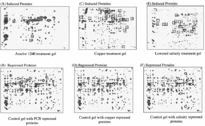
2DE consists of sequential separation of proteins based first on isoelectric focusing (IEF) followed by orthogonal SDS PAGE (O’Farrell, 1975). Originally, a stable pH gradient was established in a polyacrylamide gel by including local buffering agents (ampholytes) when casting the gel. Proteins would migrate along the pH gradient until arriving at their isoelectric point (pI, the pH at which the protein has no net charge) and then stop moving. In this way proteins focus at their pI and the protein population separates in the rod gel into specific bands. In practice, it was technically demanding to achieve reproducible pH gradients in this system and there was a tendency for the entire gradient itself to migrate towards the cathode (“cathodic drift”; Lognonne, 1994). Immobilized pH gradients (IPGs) were later developed in which ampholytes were covalently immobilized along plastic strips (Görg et al., 2000). After IEF, the focused strip is exposed to SDS PAGE sample buffer, laid across the top of an SDS PAGE gel and electrophoresed. When stained with coomassie or silver stain (Rabilloud, 1992), individual proteins become visible as spots which are usually sharply-defined (Fig. 2). Since few proteins have identical pI and relative molecular mass (Mr), especially high-resolution separations are achievable in which pairs of samples (e.g., test and control) can be compared. By increasing the size of the gel, up to thousands of spots may be evident but, even in the smallest gels, several hundred spots may be resolved. Separations are captured and analyzed by image analysis software (Corzett et al., 2006). Independent labeling of lysines in test and control proteomes with up to three different fluorescent labels followed by their mixing and separation on a single gel allows comparison of tests and control samples without gel-to-gel variation, a technique called difference gel electrophoresis (DIGE) (Gade et al., 2003). For the purposes of the present review, it should be noted that 2DE separations can also be interrogated to identify proteins targeted by specific redox lesions, thus revealing redox-related differences not detectable by PES alone (Sheehan, 2006).
Mass spectrometry (MS) methods
Fig. 2 Protein expression signatures (PES) of Mytilus edulis. Whole body proteomes separated by 2DE from animals exposed to (A, B) Arachlor 1248, (C, D) copper or (E, F) lowered salinity. These are composite gels (eight gels, four animals per treatment) and spots highlighted as being up-regulated (relative to controls) in at least 75 % of gels are highlighted with boxes (A, C, E). Spots down-regulated in 75 % of gels are highlighted as circles (B, D, F). From Shepard JL, Olsson B, Tedengren M, Bradely BP. Protein expression signatures identified in Mytilus edulis exposed to PCBs, copper and salinity stress. Mar. Environ. Res. 50: 337-340, 2000.
alone yielding a very accurate mass estimate of the order ± 0.1 %. This allows identification of proteins and peptides which are composed of well-understood structural components of known m. In proteomics, MS analyzes differences between proteomes at the level of individual proteins or is used to identify proteins from tryptic digests of spots in two dimensional gel separations. Protein identification is achieved either by peptide mass mapping (or fingerprinting) (Thiede et al., 2003) or by peptide sequencing (Wu et al., 2006). In both methods proteins are digested into a population of peptides with trypsin (or another protease) and these peptides are identified by comparison with predicted m values from sequence databases. Rapid interrogation/analysis of sequence databases is made possible with powerful bioinformatics programs (Wu et al., 2006; Palagi et al., 2006; Domon and Aebersold, 2006b).
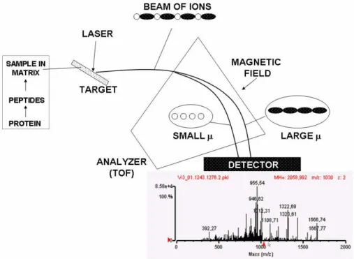
Two common ionization methods are used for MS of proteins/peptides: Matrix-assisted laser desorption ionisation (MALDI) (Tanaka et al., 1988) and electrospray ionization (ESI) (Fenn et al., 1990). MALDI is most commonly used in conjunction with a “time-of-flight” (TOF) detector so this platform is called MALDI-TOF (Fig. 3). Sample is mixed with a “matrix” of a material capable of absorbing some of the laser energy used to ionize the protein or peptide into the gas phase (e.g., sinapinnic acid) and then placed on a target for laser-induced
ionization. Surface enhanced laser desorption ionization (SELDI) is a modification of MALDI increasingly used in biomarker discovery and ecotoxicology in which sub-proteomes are selected for categories of proteins on chips functionalized with selective chemical groups such as ion exchange (Merchant and Weinberger, 2000; Poon, 2007) (Fig. 4). ESI MS achieves ionization of intact proteins by progressively drying droplets containing sample (Fenn et al., 1990). When the droplet becomes sufficiently small, the charges on ions now highly concentrated within the droplet repel each other so strongly that a coulombic explosion occurs ejecting structurally intact ions into the gas phase for separation/detection. This produces extremely accurate m determinations with a minimum of structural breakdown.
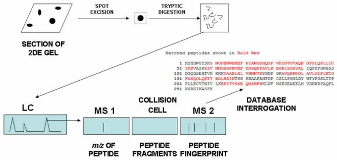
Tandem MS (also called MS/MS or MS2) uses two MS sectors separated by a collision cell (Aebersold and Mann, 2003, 2006a; Wu et al., 2006) (Fig. 5). A tryptic digest is first separated in one MS giving a unique m for each peptide. The peptide then passes through the collision cell where it fragments systematically giving a “ladder” of daughter ions which are separated on the second MS based on m. Since each fragment has a unique
m and is composed of a limited number of chemical building blocks, the ladder can be read as a de novo
Fig. 3 Outline of MALDI-TOF. Sample is mixed with matrix (e.g., sinapinic acid) on a target. A laser beam impacts on this imparting sufficient energy to peptides or proteins to propel them through the TOF analyser. This estimates the time required for each peptide type to reach the detector which depends on momentum (μ). From this time of flight, an accurate m/z value can be calculated. Matching masses of tryptic peptides to masses predicted from sequence databases identifies proteins of origin by PMF.
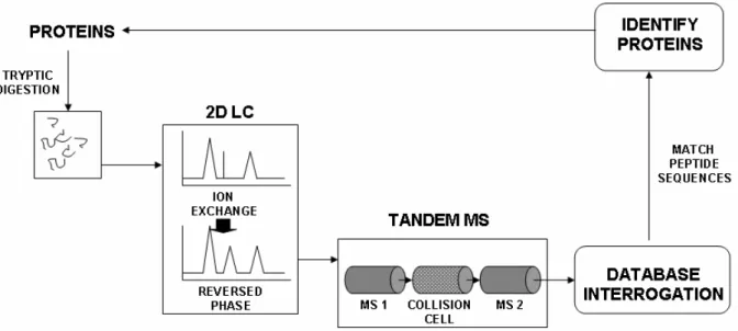
rise to the original digest (Palagi et al., 2006; Aebersold and Mann, 2006b). This dependence on sequence databases means that protein identification by this method in species such as bivalves can be problematic. In such cases, identification may instead need to be based on recognizing sequence homology rather than identity representing a major limitation in identifying target proteins in ecotoxicology (Dowling and Sheehan, 2006; McDonagh and Sheehan, 2007). High-resolution steps such as capillary liquid chromatography (LC) or capillary electrophoresis CE can precede Tandem MS giving “hyphenated” techniques; LC-tandem MS (Powell et al., 2006) and CE-tandem MS (Hu et al., 2005) facilitating extremely high resolution analysis of complex mixtures. In “shotgun sequencing”, a tryptic digest of the entire proteome is separated using two dimensional LC (e.g., ion exchange followed by reversed phase) followed by sequencing in tandem MS (McDonald and Yates, 2002; Peng et al., 2003) (Fig. 6). The very large amounts of MS data generated by such experiments raise important questions for experimental design and statistical analysis which have recently been reviewed (Domon and Aebersold 2006b; Nesvizhskii et al., 2007).
Proteomic studies in bivalves
Environmental toxicology has recently begun to embrace proteomic technologies as a set of techniques suitable for understanding biological
responses to environmental stress (Dowling and Sheehan, 2007; Monsinjon and Knigge, 2007; Nesatyy and Suter, 2007). As is usual with a new field, the approach has been at first tentative with an early emphasis, for example, on identifying PES rather than specific proteins. However, increasingly sophisticated proteomics technologies such as DIGE and SELDI-TOF have gradually been introduced in important landmark studies. This work has revealed a number of points of difficulty some of which are further explored near the end of this article. For example, when studying bivalves significant care must be taken when selecting the study organism with respect to size, sensitivity to stress, availability and previously published data. Moreover, tissues from different organisms may not necessarily be comparable for physiological reasons: mussels are filter feeders while clams are generally sediment feeders. Therefore, proteomic responses in gill from both species might be very different. Notwithstanding these practical limitations, proteomics is now increasingly accepted as a valid means of detecting often subtle effects of environmental pollutants. While not all of the following studies refer explicitly to oxidative stress, they often feature model compounds thought to function as pro-oxidants such as metals and hydrocarbons.
(B)
Fig. 4 (A) Outline of SELDI-TOF. Multiple protein samples can be added to ProteinChip® arrays which select proteins based on ion exchange, reversed phase or affinity interactions with groups immobilised on the array. Use of 8-well or 96-well arrays allows scaling to number of samples required with high throughput. Non-specifically-bound proteins are removed by washing and matrix (e.g., sinapinnic acid) is added. A laser beam propels proteins through a TOF analyser. The output may be represented as a plot of intensity versus m/z or as a synthetic “gel view” (i.e. how the sample might appear if separated by 1D SDS PAGE). (B) Selection of results of SELDI-TOF from Knigge et al. (2004) (reproduced with permission). A protein with m/z = 3268 was up-regulated in M. edulis
Fig. 5 LC-Tandem MS. Proteins in 2DE spots are digested with trypsin to form tryptic peptides. These are separated by capillary HPLC followed by m/z determination in first MS sector. Peptides are then fragmented in a collision cell and fragments are analysed in a second MS sector. Because proteins/peptides fragment in predictable ways, the series of fragments can be interpreted as a sequence. Sequences of several tryptic peptides are searched in sequence databases to give high-confidence identification.
distinction between two samples without any protein identification, it revealed little about underlying mechanisms of toxicity. The first bivalve study combining 2DE with MS identification was in the clam Chamaelea gallina, exposed to four model pollutants (Rodriguez-Ortega et al., 2003). The 15 most dramatically altered protein spots were excised and analysed by MALDI-TOF MS, of which 4 proteins were identified. However, these proteins were cytoskeletal in origin which may point to high relative abundance, prevalence of cytoskeletal proteins in sequence databases or a cytoskeleton-related oxidative stress response (Mirabelli et al., 1989). This approach was later extended to another clam species, Scrobicularia plana, sampled from sites of varying pollution status leading to identification of site-specific PES, identification of sequence tags for 16 proteins of increased abundance and unambiguous identification of more abundant hypoxanthine guanine phosphoribosyl transferase and glyceraldehyde 3-phosphate dehydrogenase (Romero-Ruiz et al., 2006). As a further development Manduzio et al. (2004), identified 19 protein spots in gills of mussels exposed to crude oil. Again, some cytoskeletal proteins were identified but also some antioxidant and metabolic enzymes such as GST, heavy metal binding proteins and aldolase. The authors also reported that, although a number of spots produced good MALDI-TOF spectra, they did not yield a significant score for peptide mapping and hence identification. This highlights the difficulty in identifying bivalve proteins when they are so poorly represented in sequence databases, a recurring theme in bivalve proteomics (Dowling and Sheehan, 2007; Monsinjon and Knigge, 2007).
Knigge et al. (2004) applied a SELDI-TOF approach for biomarker discovery to mussels exposed to PAHs and heavy metals. SELDI-TOF should be capable of giving a proteomic profile of changes in cells under stress conditions as well as being well suited for low abundance and small proteins. However, a major problem encountered in this study was high variability amongst individual samples. These authors went on to combine protein array technology with SELDI-TOF of mussel serum from animals exposed to oil either on its own or spiked with alkylphenols (Bjørnstad et al., 2006). A total of 83 mass peaks were perturbed in the spiked samples while only 49 were altered by oil alone. As with earlier studies, individual proteins were not explicitly identified but the mass patterns could be used as a fingerprint of effect. These studies demonstrate the power of SELDI-TOF for detection of effects when processing relatively large sets of individual complex protein samples although, again, analysis of such datasets is not without its difficulties (Monsinjon et al., 2006).
Fig. 6 Shotgun proteomics. This involves tryptic digestion of whole proteomes with high-resolution separation and identification technologies. In this schematic, tryptic peptides are first passed through 2D LC (ion exchange first with each peptide peak further separated by reversed phase) then each resultant 2D LC peak is passed to tandem MS. High-speed algorithms identify peptide sequences and thus allow identification of proteins in the starting proteome. Up to hundreds of proteins are identifiable in a single experiment.
variation in spot patterns, staining intensity and which have a low linear range (Nesatyy and Suter, 2007).
Oxidative stress can cause a range of reversible and irreversible modifications to proteins and their side chains. Some of these can lead to protein inactivation, some are protective of the protein’s structural integrity and some may be viewed as sensing changes in the cell’s redox status (Davies, 2005; Sheehan, 2006). Protein side chains can be irreversibly modified to aldehyde or ketone groups in a process termed carbonylation. Protein carbonylation can lead to protein aggregation, inactivation and degradation (Levine et al., 1990; Davies, 2005). The 2DE proteomic approach can be combined with immunoblotting to investigate the carbonylation response in individual tissues in response to pollutants known to induce oxidative stress in both mussels and clams (McDonagh et al., 2005; Dowling et al., 2006; McDonagh and Sheehan, 2006). These studies revealed that levels of carbonylated proteins increased in general with oxidative stress although there was a tissue-specific response between organisms and the response was also pollutant-specific. Damaged proteins are removed from cells by proteolysis, mainly via the ubiquitin proteasome pathway (UPP) (Marques et al., 2004). The UPP is responsible for selective digestion of many short lived intracellular regulatory proteins or abnormal cytosolic or nuclear proteins (Marques et al., 2004). Changes in ubiquitination levels have been studied in bivalves by immunoblotting combined with 1DE in response to temperature and season (Hofmann and Somero, 1995; Buckley et al., 2001) and in M. trossulus
exposed to Exxon Valdez oil (Downs et al., 2002). This approach has been extended to 2DE, for
analysing mussels and clams exposed to a variety of oxidative stressors (McDonagh and Sheehan, 2006; Chora et al., 2008; Tedesco et al., 2008). Somewhat surprizingly, the ubiquitination PES is distinct from that for carbonylation suggesting that carbonylated proteins are degraded in a UPP-independent manner.
stress, the method of Baty et al. (2005) was adapted to analyse changes in thiol status within the proteome. In this approach, free thiols are initially blocked with N-ethylmaleimide, disulphides reduced by DTT and resulting free thiols labelled with the thiol- reactive fluorescent compound, 5-iodoacetamidofluorescein (5-IAF). This enabled identification of a number of menadione-responsive proteins in the gills of M. edulis by LC-MS/MS (McDonagh and Sheehan, 2007, 2008). Another technique that uses an affinity column to pre-select for a subproteome in combination with 2DE has also been used in the clam Tapes semidecussatus, where a GSH-agarose column was used and tissue-specific expression of Pi class GST’s was discovered (Dowling et al., 2006). These studies take a sub-proteome approach to oxidative stress in bivalves considerably simplifying subsequent analysis.
Conclusions and perspectives
Recent years have seen development of new proteomic techniques, many of which have yet to be fully embraced by environmental toxicology. Some of these techniques such as “shotgun proteomics” necessitate fully-covered genome sequences which currently precludes many bivalves. Others, such as blue native gel electrophoresis have especial promise for studies of oxidative stress as this technique resolves protein-protein complexes in systems such as mitochondria (Krause, 2006). In the coming years, significantly more sequence data are expected to become available (McKillen et al., 2005; Dupont et al., 2007; Tanguy et al., 2008). This is especially important as “gel-free” techniques overcome many of the well-known shortcomings of 2DE such as the need for detailed image analysis, misidentification due to co-migration of proteins, under/over-selection of some proteins due to their physical properties or position in the cell (e.g., membrane proteins, are not suitable for 2DE separations) (Görg et al., 2004).
One of proteomic’s ultimate goals is to unearth networks of interactions to understand a particular biological state as well as to identify and quantify involvement of individual proteins (Domon and Aebersold, 2006a). Relative quantification between protein samples is a crucial aspect of analysis and 2DE, when combined with image analysis, allows relative spot comparisons to be made between gels. However, numerous gel replicates and careful consideration of total amount of protein on gels is necessary. As the dynamic range of many protein stains is limited, relative quantification can be difficult, although the newer fluorescent stains and dyes have been a significant improvement (Nesatyy and Suter, 2008). Some of the newer approaches for quantification include, isobaric tags for relative and absolute quantification (iTRAQ) allowing relative quantification between tandem mass spectra and use of tags of varying mass that label N-terminus and side-chains of all peptides. Samples are then pooled and analysed by MS/MS. Isotope coded affinity tag (ICAT) reagents that are cysteine-specific offer options especially relevant to redox proteomics. Cysteines are one of the most rarely
used amino acids but are often highly-conserved and involved in the regulation of important cellular processes. ICAT can provide information about the oxidative state of individual proteins (Leichert et al., 2008).
Many proteomic studies of oxidative stress in bivalves have so far been performed under laboratory-controlled conditions, where bivalves are subjected to high levels of contaminants or stressors. This is understandable as proteomics is relatively new to environmental monitoring, and researchers often use artificially high exposures to see if there is measurable response. The next step is to develop proteomic techniques to the point that they can reveal differences in the field at environmentally-relevant pollutant concentrations.
number of proteomic studies including 2DE and SELDI-TOF, and highlighted the need for multivariate statistical methods for interpretation of complex datasets. In general, careful initial planning and outlining of overall objectives can save time over the course of the study. Together with the increasing availability of complete gene and protein databases we should see further developments in the field of environmental monitoring of bivalves as a response to oxidative stress (McKillen et al., 2005; Dupont et al., 2007).
Acknowledgements
Our laboratory is supported by the Irish Research Council for Science Engineering and Technology.
References
Aebersold R, Mann M. Mass spectrometry-based proteomics. Nature 422: 198-207, 2003.
Altieri AH. Inducible variation in hypoxia tolerance across the intertidal subtidal distribution of the blue mussel Mytilus edulis. Mar. Ecol. Prog. Ser. 325: 295-300, 2006.
Apraiz I, Mi J, Cristobal S. Identification of proteomic signatures of exposure to marine pollutants in mussels (Mytilus edulis). Mol. Cell Proteomics 5: 1274-1285, 2006.
Baty JW, Hampton MB, Winterbourn CC. Proteomic detection of hydrogen peroxide-sensitive thiol proteins in Jurkat cells. Biochem. J. 389: 785-95, 2005.
Bayne BL (ed.).Marine Mussels: Their Ecology and Physiology. Cambridge University Press, Cambridge, UK, 1976.
Bayne BL. Assessing effects of marine pollution. Nature 280: 14, 1979.
Biswas S, Chida AS, Rahnam I. Redox modifications of protein thiols: Emerging roles in cell signaling. Biochem. Pharmacol. 71: 551-564, 2006.
Buckley BA, Owen ME, Hofmann, GE. Adjusting the thermostat: the threshold induction temperature for the heat-shock response in intertidal mussels genus (Mytilus) changes as a function of thermal history. J. Exp. Biol. 204: 3571-3579, 2001.
Bjørnstad A, Larse BK, Skadsheim A, Jones MB, Andersen OK. The potential of ecotoxicoproteomics in environmental monitoring: biomarker profiling in mussel plasma using proteinChip array technology. J. Toxicol. Environ. Health A 69: 77-96, 2006.
Cajaraville MP, Bebianno MJ, Blasco J, Porte C, Sarasquete C, Viarengo A. The use of biomarkers to assess the impact of pollution in coastal environments of the Iberian Peninsula: A practical approach. Sci. Total Environ. 247: 295-311, 2000.
Cheung CCC, Zheng GJ, Li AMY, Richardson BJ, Lam PKS. Relationships between tissue concentrations of polycyclic aromatic hydrocarbons and antioxidative responses of marine mussels, Perna viridis. Aquat. Toxicol. 52: 189-203, 2001.
Chora S, McDonagh B, Sheehan D, Starita-Geribaldi M, Roméo M, Bebianno MJ. Ubiquitination and carbonylation as markers of oxidative-stress in Ruditapes decussatus. Mar. Environ. Res. 66: 95-97, 2008.
Corzett TH, Fodor IK, Choi MW, Walsworth VL, Chromy BA, Turteltaub KW, et al. Statistical analysis of the experimental variation in the proteomic characterization of human plasma by two-dimensional difference gel electrophoresis. J. Proteome Res. 5: 2611-2619, 2006.
Cretich M, Damin F, Pirri G, Chiari M. Protein and peptide arrays: Recent trends and new directions. Biomol. Eng. 23: 77-88, 2006.
Davies M J. The oxidative environment and protein damage. Biochim. Biophys. Acta 1703: 93-109, 2005.
Depledge MH, Galloway TS. Healthy animals, healthy ecosystems. Front. Ecol. Environ. 3: 252-258, 2005.
Domon B, Aebersold R. Mass spectrometry and protein analysis. Science 312: 212-217, 2006a Domon B, Aebersold R. Challenges and
opportunities in proteomics data analysis. Mol. Cell Proteomics 5: 1921-1926, 2006b.
Dowling VA, Sheehan D. Proteomics as a route to identification of toxicity targets in ecotoxicology. Proteomics 6: 5597-5604, 2006.
Dowling V, Hoarau PC, Romeo M, O'Halloran J, van Pelt F, O'Brien N, et al. Protein carbonylation and heat shock response in Ruditapes
decussatus following
p,p'-dichlorodiphenyldichloroethylene (DDE) exposure: a proteomic approach reveals that DDE causes oxidative stress. Aquat. Toxicol. 77: 11-18, 2006.
Dowling V, McDonagh B, Cotter EM, O'Brien N, van Pelt F, O'Halloran J, et al. Two-dimensional electrophoresis analysis of glutathione affinity-selected proteins from the clam Tapes semidecussatus: evidence for tissue-specific expression of redox proteins. Comp. Biochem. Physiol. 1D: 267-272, 2006.
Downs CA, Shigenaka G, Fauth JE, Robinson CE, Huang A. Cellular physiological assessment of bivalves after chronic exposure to spilled Exxon Valdez crude oil using a novel molecular diagnostic biotechnology. Environ. Sci. Technol. 36: 2987-2993, 2002.
Dupont S, Wilson K, Obst M, Skold H, Nakano H, Thorndyke MC. Marine ecological genomics: When genomics meets marine ecology. Mar. Ecol. Prog. Ser. 332: 257-273, 2007.
Fenn JB, Mann M, Meng CK, Wong SF, Whitehouse CM. Electrospray ionization - principles and practice. Mass Spectrometry Rev. 9: 37-70, 1990.
Fitzpatrick PJ, Sheehan D, Livingstone, DR. Studies on isoenzymes of glutathione S-transferase in digestive gland of Mytilus galloprovincialis with exposure to pollution. Mar. Environ. Res. 39: 241-244, 1995.
Galloway TS. Biomarkers in environmental and human health risk assessment. Mar. Pollut. Bull. 53: 606-613, 2006.
Goldberg ED, Bertine KK. Beyond the mussel watch - new directions for monitoring marine pollution. Sci. Total Environ. 247: 165-174, 2000.
Görg A, Obermaier C, Boguth G, Harder A, Scheibe B, Wildgruber R, et al. The current state of two-dimensional electrophoresis with immobilized pH gradients. Electrophoresis 21: 1037-1053, 2000.
Görg A, Weiss W, Dunn MJ. Current two-dimensional electrophoresis technology for proteomics. Proteomics 4: 3665-3685, 2004. Gould SC, Calloway SB. Clams and brachiopods;
ships that pass in the night. Palaeobiology 6: 383-396, 1980.
Grace PA. Ischemia-reperfusion injury. Brit. J. Surgery 81: 637-647, 1994.
Halliwell B, Gutteridge JMC. Free Radicals in Biology and Medicine. Fourth Edition, Oxford University Press, Oxford, UK, 2007
Halpern BS, Walbridge S, Selkoe KA, Kappel CV, Micheli F, D’Agrosa C, et al. A global map of human impact on marine ecosystems. Science 319: 946-948, 2008.
Handy RD, Galloway TS, Depledge MH. A proposal for the use of biomarkers for the assessment of chronic pollution and in regulatory toxicology. Ecotoxicology 12 : 331-343, 2003.
Heijne WHM, Kienhuis AS, van Ommen B, Stierum RH, Groten JP. Systems toxicology: Applications of toxicogenomics, transcriptomics, proteomics and metabolomics in toxicology. Expert Rev. Proteomics 2: 767-780, 2005. Hofmann G, Somero G. Evidence for protein
damage at environmental temperatures: seasonal changes in levels of ubiquitin conjugates and hsp70 in the intertidal mussel
Mytilus trossulus. J. Exp. Biol. 198: 1509-1518, 1995.
Hu A, Tsai PJ, Ho YP. Identification of microbial mixtures by capillary electrophoresis/selective tandem mass spectrometry. Anal. Chem. 77: 1488-1497, 2005.
Karp NA, Lilley KS. Design and analysis issues in quantitative proteomics studies. Proteomics S1: 42-50, 2007.
Karp NA, Spencer M, Lindsay H, O'Dell K, Lilley KS. Impact of replicate types on proteomic expression analysis. J. Proteome Res. 4: 1867-1871, 2005.
Kislinger T, Cox B, Kannan A, Chung C, Hu PZ, Ignatchenko A, et al. Global survey of organ and organelle protein expression in mouse; Combined proteomic and transcriptomic profiling. Cell 125: 173-186, 2006.
Knigge T, Monsinjon T, Andersen OK. Surface-enhanced laser desorption/ionization-time of flight-mass spectrometry approach to biomarker discovery in blue mussels (Mytilus edulis) exposed to polyaromatic hydrocarbons and heavy metals under field conditions. Proteomics 4: 2722-2727, 2004.
Krause F. Detection and analysis of protein-protein interactions in organellar and prokaryotic proteomes by native blue electrophoresis: (Membrane) protein complexes and supercomplexes. Electrophoresis 27: 2759-2781, 2006.
Lee WC, Lee KH. Applications of affinity chromatography in proteomics. Anal. Biochem. 324: 1-10, 2004.
Lehtonen KK, Leinio S, Schneider R, Leivuori M. Biomarkers of pollution effects in the bivalves
Mytilus edulis and Macoma balthica collected from the southern coast if Finland (Baltic Sea). Mar. Ecol. Prog. Ser. 322: 155-168, 2006. Leichert LI, Gehrke F, Gudiseva HV, Blackwell T,
Ilbert M, Walker AK, et al. Quantifying changes in the thiol redox proteome upon oxidative stress in vivo. Proc. Natl. Acad. Sci. USA 105: 8197-8202, 2008.
Lesser MP. Oxidative stress in marine environments: Biochemistry and physiological ecology. Ann. Rev. Physiol. 68: 253-278, 2006.
Levine RL, Garland D, Oliver CN, Amici A, Climent I, Lenz AG, et al. Determination of carbonyl content in oxidatively modified proteins. Methods Enzymol. 186: 464-78, 1990.
Livingstone DR. The fate of organic xenobiotics in aquatic ecosystems: quantitative and qualitative differences in biotransformation by invertebrates and fish. Comp. Biochem. Physiol. 120A: 43-49, 1998.
Lognonne JL. 2D-PAGE analysis - a practical guide to principal critical parameters. Cell. Mol. Biol. 40: 41-55, 1994.
López-Barea J, Pueyo C. Mutagen content and metabolic activation of promutagens by mollusks as biomarkers of marine pollution. Mutat. Res. 399: 3-15, 1998.
Lutz RA, Kennish MJ. Ecology of deep-sea hydrothermal vent communities; a review. Rev. Geophys. 31: 211-242, 1993.
Lyons C, Dowling V, Tedengren M, Gardestrőm, J, Hartl MG, O'Brien N, et al. Variablity of heat shock proteins and glutathione S-transferases in gill and digestive gland of blue mussel,
Mytilus edulis. Mar. Environ. Res. 56: 585-597, 2003.
Manduzio H, Monsinjon T, Galap C, Leboulanger F, Rocher W. Seasonal variations in antioxidant defences in blue mussels Mytilus edulis
collected from a polluted area: major contributions in gills of an inducible isoform of Cu/Zn-superoxide dismutase and of glutathione S-transferase. Aquat. Toxicol. 70: 83-93, 2004.
Mann M, Jensen ON. Proteomic analysis of post-translational modifications. Nature Biotechnol. 21: 255-261, 2003.
McDonagh B, Sheehan D. Redox proteomics in the blue mussel Mytilus edulis: carbonylation is not a pre-requisite for ubiquitination in acute free radical-mediated oxidative stress. Aquat. Toxicol. 79: 325-333, 2006.
McDonagh B, Tyther R, Sheehan D. Carbonylation and glutathionylation of proteins in the blue mussel Mytilus edulis detected by proteomic analysis and Western blotting: actin as a target for oxidative stress. Aquat. Toxicol. 73: 315-326, 2005.
McDonagh B, Tyther R, Sheehan D. 2006. Redox proteomics in the mussel, Mytilus edulis. Mar. Environ. Res. 62: S101-S104.
McDonagh B, Sheehan D. Effect of oxidative stress on protein thiols in the blue mussel Mytilus edulis: proteomic identification of target proteins. Proteomics 7: 3395-3403, 2007.
McDonagh B, Sheehan D. Effects of oxidative stress on protein thiols and disulphides in
Mytilus edulis revealed by proteomics: actin and protein disulphide isomerase are redox targets. Mar. Environ. Res. 66: 193-195, 2008. McDonald WH, Yates JR. Proteomics and
biomarker discovery. Dis. Markers 18: 99-105, 2002.
McKillen DJ, Chen YA, Chen CM, Jenny MJ, Trent HF, Robalino J, et al. Marine genomics: a clearing house for genomic and transcriptomic data of marine organisms. BMC Genomics 6: 34-39, 2005.
Merchant M, Weinberger SR. Recent advancements in surface-enhanced laser desorption/ionization-time of flight-mass spectrometry. Electrophoresis 21: 1164-1177, 2000.
Mi J, Apraiz I, Cristobal S. Peroxisomal proteomic approach for protein profiling in blue mussels (Mytilus edulis) exposed to crude oil. Biomarkers 12: 47-60, 2007.
Miller AI. Biotic transitions in global marine diversity. Science 281: 1157-1160, 1998.
Mirabelli F, Salis A, Vairetti M, Bellomo G, Thor H, Orrenius S. Cytoskeletal alterations in human platelets exposed to oxidative stress are mediated by oxidative and Ca2+ -dependent mechanisms. Arch. Biochem. Biophys. 270: 478-488, 1989.
Monsinjon T, Knigge T. Proteomic applications in ecotoxicology. Proteomics 7: 2997-3009, 2007.
Monsinjon T, Andersen OK, Leboulanger F, Knigge T. Data processing and classification analysis of proteomic changes: A case study of oil pollution in the mussel, Mytilus edulis. Proteome Sci. 4: 17, 2006.
Naylor RL, Goldburg RJ, Primavera JH, Kautsky N, Beveridge MCM, Clay J, et al. Effect of aquaculture on world fish supplies. Nature 405: 1017-1024, 2000.
Nesatyy VJ, Suter MJF. Proteomics for the analysis of environmental stress responses in organisms. Environ. Sci. Technol. 41: 6891-6900, 2007.
Nesvizhskii AI, Vitek O, Aebersold R. Analysis and validation of proteomic data generated by tandem mass spectrometry. Nature Methods 4: 787-797, 2007.
Newell, RIE. Ecosystem influences of natural and cultivated populations of suspension-feeding bivalve molluscs: a review. J. Shellfish Res. 23: 51-61, 2004.
O’Farrell PH. High resolution 2-dimensional electrophoresis of proteins. J. Biol.Chem. 250: 4007-4021, 1975.
Oktyabrsky ON, Smirnova GV. Redox regulation of cellular functions. Biochemistry (Moscow) 72: 132-145, 2007.
Palagi PM, Hernandez P, Walther D, Appel RD. Proteome informatics I: bioinformatics tools for processing experimental data. Proteomics 6: 5435-5444, 2006.
Pampanin DM, Viarengo A, Garrigues P, Andersen OK. Background for the BEEP Stavanger workshops: Biological effects on marine organisms in two common, large, laboratory experiments and in a field study. Comparison of the value (sensitivity, specificity, etc.) of core and new biomarkers. Aquat. Toxicol., 78, S1-S4, 2006.
Peng J, Elias JE, Thoreen CC, Licklider LJ, Gygi SP. Evaluation of multidimensional chromatography coupled with tandem mass spectrometry (LC/MS/MS) for large-scale protein analysis: the yeast proteome. J. Proteome Res. 2: 43-50, 2003.
Poole LB, Nelson KJ. Discovering mechanisms of signaling-mediated cysteine oxidation. Curr. Opin. Chem. Biol. 12: 18-24, 2008.
Poon TCW. Opportunities and limitations of SELDI-TOF-MS in biomedical research: practical advices. Expert Rev. Proteomics 4: 51-65, 2007. Powell DW, Merchant ML, Link AJ. Discovery of
regulatory molecular events and biomarkers using 2D capillary chromatography and mass spectrometry. Expert Rev. Proteomics 3: 63-74, 2006.
Power A, Sheehan D. Seasonal variation in the antioxidant defence systems of gill and digestive glands of the blue mussel, Mytilus edulis. Comp. Biochem. Physiol.114C: 99-103, 1996. Prevodnik A, Gardeström J, Lilja K, Elfwing T,
McDonagh B, Petrović N, et al. Oxidative stress in response to xenobiotics in the blue mussel
Mytilus edulis L.: evidence for variation along a natural salinity gradient of the Baltic Sea. Aquat. Toxicol. 82: 63-71, 2007.
Rabilloud T A comparison between low background silver diammine and silver-nitrate protein stains. Electrophoresis 13: 429-439, 1992.
Regoli F, Gorbi S, Frenzilli G, Nigro M, Corsi I, Focardi S, et al. Oxidative stress in ecotoxicology: from the analysis of individual antioxidants to a more integrated approach. Mar. Environ. Res. 54: 419-423, 2002.
Rodríguez-Ariza A, Abril N, Navas JL, Dorado G, López-Barea J, Pueyo C. Metal mutagenicity and biochemical studies on bivalve molluscs from Spanish coasts. Environ. Mol. Mutagen. 19: 112-124, 1992.
Rodríguez-Ariza A, Martínez-Lara E, Pascual P, Pedrajas JR, Abril N, Dorado G, et al.
Rodríguez-Ariza A, Rodriguez-Ortega MJ, Marenco JL, Amezcua O, Alhama J, López-Barea J. Uptake and clearance of PCB congeners in
Chamaelea gallina: Response of oxidative stress biomarkers. Comp. Biochem. Physiol. 134C: 57-67, 2003.
Rodríguez-Ortega MJ, Alhama J, Funes V, Romero-Ruíz A, Rodríguez-Ariza A, López-Barea J. Biochemical biomarkers of pollution in the clam
Chamaelea gallina from the South-Spanish littoral. Environ. Toxicol. Chem. 21: 542-549, 2002.
Rodriguez-Ortega MJ, Grosvik BE, Rodriguez-Ariza A, Goksoyr A, Lopez-Barea J. Changes in protein expression profiles in bivalve molluscs (Chamaelea gallina) exposed to four model environmental pollutants. Proteomics 3: 1535-1543, 2003.
Romero-Ruiz A, Carrascal M, Alhama J, Gómez-Ariza JL, Abian J, López-Barea J. Environmental proteomics studies in clams from the Donaña bank of Guadalquivir Estuary (SW Spain). Proteomics 6 (Suppl 1): S245-S255, 2006.
Sanders BM. Stress proteins in aquatic organisms: an environmental perspective. Crit Rev. Toxicol. 23: 49-75, 1993.
Sheehan, D. Detection of redox-based modification in two dimensional electrophoresis proteomic separations. Biochem. Biophys. Res. Commun. 349: 455-462, 2006.
Sheehan, D. The potential of proteomics for providing new insights into environmental impacts on human health. Rev. Environ. Health 22: 175-194, 2007.
Sheehan D, Power A. Effects of seasonality on xenobiotic and antioxidant defence mechanisms of bivalve molluscs. Comp. Biochem. Physiol. 123C: 193-199, 1999.
Shepard JL, Olsson B, Tedengren M, Bradley BP. Protein expression signatures identified in
Mytilus edulis exposed to PCBs, copper and salinity stress. Mar. Environ. Res. 50: 337-340, 2000.
Sole M, Livingstone DR. Components of the cytochrome P450-dependent monooxygenase system and ‘NADPH-independent benzo[a]pyrene hydroxylase’ activity in a wide range of marine invertebrate species. Comp. Biochem. Physiol. 141C: 20-31, 2005.
Sole M, Porte C, Biosca X, Mitchelmore CL, Chipman JK, Livingstone DR, et al. Effects of the “Aegean Sea” oil spill on biotransformation enzymes, oxidative stress and DNA-adducts in digestive gland of the mussel (Mytilus edulis L). Comp. Biochem. Physiol. 113C: 257-265, 1996. Spickett CM, Pitt AR, Morrice N, Kolch W.
Proteomic analysis of phosphorylation, oxidation and nitrosylation in signal transduction. Biochim. Biophys. Acta 1764: 1823-1841, 2006.
Stachowitz M, Riedel B, Zuschin M, Machan R. Oxygen depletion and benthic mortalities: The
first in situ experimental approach to documenting an elusive phenomenon. Limnol. Oceanogr. Methods 5: 344-352, 2007.
Stadtman ER, Levine RL. Protein oxidation. Ann. NY Acad. Sci. Vol. 899: 191-208, 2000.
Steinert SA. DNA damage as a bivalve biomarker. Biomarkers 4: 492-496, 1999.
Tanaka K, Waki H, Ido Y, Akita S, Yoshida Y, Yoshida T. Protein and polymer analyses up to m/z 100 000 by laser ionization time-of-flight mass spectrometry. Rapid Commun. Mass Sp. 2: 151-153, 1988.
Tanguy A, Bierne N, Saavedra C, Pina B, Bachère E, Kube M, et al. Increasing genomic information in bivalves through new EST collections in four species: development of new genetic markers for environmental studies and genome evolution. Gene 408: 27-36, 2008. Tedesco S, Doyle H, Redmond G, Sheehan D. Gold
nanoparticles and oxidative stress in Mytilus edulis. Mar. Environ. Res. 66: 131-133, 2008. Thiede B, Hohenwarter W, Krah A, Mattow J,
Schmid M, Schmidt F, et al. Peptide mass fingerprinting. Methods 35: 237-247, 2005. Valavanidis A, Vlahogianni T, Dassenakis M,
Scoullos M. Molecular biomarkers of oxidative stress in aquatic organisms in relation to toxic environmental pollutants. Ecotoxicol. Environ. Saf. 64:178-189, 2006.
Vergani L, Grattarola M, Borghi C, Dondero F, Viarengo A. Fish and molluscan metallothioneins - A structural and functional comparison. FEBS J. 272: 6014-6023, 2005. Viarengo A, Canesi L, Pertica M, Livingstone DR.
Seasonal variations in the antioxidant defense systems and lipid peroxidation of the digestive gland of mussels. Comp. Biochem. Physiol. 100C: 187-190, 1991.
Widdows J, Shick JM. Physiological response of
Mytilus edulis and Cardium edule to aerial exposure. Mar. Biol. 85: 278-286, 1985.
Widdows J, Bayne BL, Livingstone DR, Newell RIE, Donkin P. Physiological and biochemical responses of bivalve mollusks to exposure to air. Comp. Biochem. Physiol. 62A: 301-308, 1979.
Winston GW. Oxidants and antioxidants in aquatic animals. Comp. Biochem. Physiol. 100C: 173-176, 1991.
Winterbourn CC. Reconciling the chemistry and biology of reactive oxygen species. Nature Chem. Biol. 4: 278-286, 2008.
Wu X, Edwards N, Tseng CW. Peptide identification via tandem mass spectrometry. Adv. Computers 68: 253-278, 2006.
Ying J, Clavreul N, Sethuraman M, Adachi T, Cohen RA. Thiol oxidation in signalling and response to stress: detection and quantification of physiological and pathophysiological thiol modifications. Free Radic. Biol. Med. 43: 1099-1108, 2007.