RESUMO.- [Efeito do estresse moderado crônico sobre parâmetros de avaliação óssea em ratos adultos ma-chos e fêmeas.]A osteoporose é uma doença multifatorial,
de alta prevalência e que tem um grande impacto na quali-dade de vida, principalmente porque os efeitos sobre a es-trutura do osso aumentam o risco de fraturas, que podem ser muito debilitantes. Com base na observação de que pa-cientes com depressão têm menor densidade mineral óssea que indivíduos saudáveis , muitos estudos têm indicado que o estresse pode ser um fator agravante para a perda óssea. Este estudo avalia o efeito de um protocolo de estresse mo-derado crônico (EMC) em parâmetros de avaliação óssea em ratos machos e fêmeas. Cinco animais de cada sexo, com cinco meses de idade, foram submetidos a um cronograma de aplicação de estressores durante 28 dias. Os estressores incluídos foram: frio, calor, contenção, inclinação da gaiola, isolamento, iluminação durante a noite e privação de água e ração. Cinco animais de cada sexo foram mantidos com um
Effects of chronic mild stress on parameters of bone
assessment in adult male and female rats
1Fabrício L. Valente2, Anna Paula B.R. Ferreira3, Leonardo D. da Costa4, Mário J.Q.
Louzada5, Joaquin H. Patarroyo² and Marlene I. Vargas²*
ABSTRACT.- Valente F.L., Ferreira A.P.B.R., Costa L.D., Louzada M.J.Q., Patarroyo J.H. & Var-gas M.I. 2016. Effects of chronic mild stress on parameters of bone assessment in adult male and female rats. Pesquisa Veterinária Brasileira 36(Supl.1):106-112. Departamento de Veterinária, Universidade Federal de Viçosa, Campus Viçosa, Avenida Peter Henry Rolfs s/n, Viçosa, MG 36570-900, Brazil. E-mail: [email protected]
Osteoporosis is a multifactorial disease of high prevalence and has great impact on qua-lity of life, because the effects on bone structure increase the risk of fractures, what may be very debilitating. Based on the observation that patients with depression have lower bone mineral density than healthy individuals, many studies have indicated that stress could be an aggravating factor for bone loss. This study evaluates the effect of a protocol of chronic mild stress (CMS) on parameters of bone assessment in male and female rats. Five 5-monh--old rats of each sex underwent a schedule of stressor application for 28 days. Stressors included cold, heat, restraint, cage tilt, isolation, overnight illumination, and water and food deprivation. Five rats of each sex were kept under minimum intervention as control group. The animals were weighed at beginning and end of the period, and after euthanasia had their bones harvested. Femur, tibia and lumbar vertebrae were analyzed by bone densi-tometry. Biomechanical tests were performed in femoral head and diaphysis. Trabecular bone volume was obtained from histomorphometric analysis of femoral head and vertebral body, as well as of femoral midshaft cross-sectional measures. Not all parameters analyzed showed effect of CMS. However, tibial and L4 vertebral bone mineral density and cross--sectional cortical/medullar ratio of femoral shaft were lower in female rats submitted to
the CMS protocol. Among male rats, the differences were significant for femoral trabecular bone volume and maximum load obtained by biomechanical test. Thus, it could be confir -med that CMS can affect the balance of bone homeostasis in rats, what may contribute to the establishment of osteopenia or osteoporosis.
INDEX TERMS: Stress, osteoporosis, animal model, bone densitometry, bone histomorphometry, rat.
1 Received on February 16, 2016.
Accepted for publication on May 3, 2016.
2 Departamento de Veterinária, Universidade Federal de Viçosa (UFV),
Campus Viçosa, Avenida Peter Henry Rolfs s/n, Viçosa, MG 36570-900, Brazil. *Correspondig author: [email protected]
3 Centro de Ciência Animal, Universidade Federal de Ouro Preto (UFOP),
Morro do Cruzeiro, Ouro Preto, MG 35400-000, Brazil.
4 Faculdade de Medicina Veterinária e Zootecnia, Universidade Estadual
Paulista (Unesp), Campus Botucatu, Distrito de Rubião Júnior s/n, Botuca-tu, SP 18618-970, Brazil.
5 Faculdade de Medicina Veterinária, Unesp, Campus Araçatuba, Rua
mínimo de intervenção como grupo controle. Os animais
foram pesador no início e no final do período, e após euta -násia tiveram seus ossos coletados. Fêmur, tíbia e vértebra lombar foram analisados por densitometria óssea. Testes
biomecânicos foram realizados na cabeça e na diáfise do fê -mur. Volume trabecular ósseo foi obtido a partir de análise histomorfométricas da cabeça do fêmur e do corpo
verte-bral, bem como medidas da seção transversal diáfise femo -ral. Nem todos os parâmetros avaliados sofreram efeito do protocolo de EMC. No entanto, a densidade mineral óssea da tíbia e da vértebra L4 e a razão osso cortical/medula da
seção transversal da diáfise femoral foram menores nas fê -meas submetidas ao protocolo. Entre os ratos machos, as
diferenças foram significativas no volume trabecular ós -seo da cabeça femoral e na carga máxima obtida no teste
biomecânico. Assim, confirma-se que o protocolo de EMC
pode afetar o equilíbrio da homeostase óssea em ratos, o que pode contribuir para o estabelecimento de osteopenia ou osteoporose.
TERMOS DE INDEXAÇÃO: Estresse, osteoporose, modelo animal, densitometria óssea, histomorfometria óssea, rato.
INTRODUCTION
Osteoporosis is a high prevalence disease that affects pre-dominantly women but also has great importance among men, and develops as the result of an imbalance between bone formation and resorption (WHO 2003), that are re-gulated by a variety of endocrine and immune mediators (Shea & Miller 2005). Among the population aged 50 or
more, one-third of women and one-fifth of men may be at
high risk of fracture (Dawson-Hughes et al. 2012). It is es-timated that the number of high-risk individuals for osteo-porotic fractures will double from 2010 to 2040 worldwide (Odén et al. 2015). The disease is multifactorial and chro-nic exposure to glucocorticoid is considered an important cause of bone loss (Sambrook & Lane 2001). Endogenous glucocorticoid plays a central role in the response to stress (de Kloet et al. 1998, Pacak & McCarty 2000). Endogenous glucocorticoid levels increase with age and may have ne-gative effects on bone metabolism (Weinstein et al. 2010). In humans, depression has been related to reduced bone mineral density (BMD), increasing the risk of osteoporo-sis (Cizza et al. 2009). In the study of stress, the chronic mild stress (CMS) model in rats and mice is widely used with the aim of inducing changes consistent with depres-sion (Gamaro et al. 2003, Detanico et al. 2009, Lucca et al. 2009). However, these studies emphasize the neurological symptoms related to depression. In the CMS model, not only the glucocorticoid produced in response to stress can have an effect on bone metabolism but also norepinephrine also seems to play an important role (Yirmiya et al. 2006), what reiterates the complexity of factors involved. The con-nection between stress and bone metabolism is still poor-ly investigated. To contribute with additional information about this relationship, the present study evaluates bone parameters by histomorphometric, biomechanical and densitometric analyses in adult rats of both sexes submit-ted to a CMS protocol.
MATERIALS AND METHODS
Ethics statement. The ethics committee for animal use of the institution approved the procedures in this experiment (Protocol #52/2009), based on current legislation.
Animals. Twenty 3-month-old Wistar rats (Rattus norvegi-cus) of both sexes were purchased from the Central Animal
Facili-ty of UFV. The animals were kept in plastic cages (five per cage) in
an experimental room used exclusively for this study with cycles of 12h light/dark, 20-24°C. When they were 5-months-old, CMS
protocol was initiated for five males and five females. The remai -ning animals comprised the control group that did not undergo any procedure during the experiment.
Chronic mild stress. The design of CMS protocol used in this
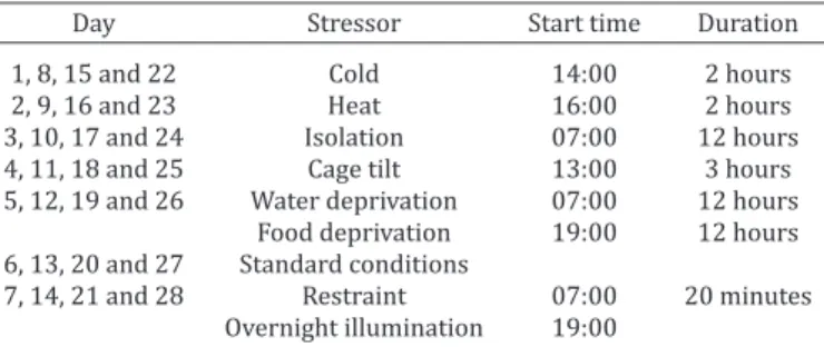
study was based on many sources found in the scientific litera -ture (Gamaro et al. 2003, Dalla et al. 2005, Baker et al. 2006, De-tanico et al. 2009, Lucca et al. 2009) and adapted to the facilities available. Eight stressors were applied, during 28 days (Table 1): (a) 2 hours of lower temperature, when the animals were kept in a plastic cage without wood shavings placed over bags of ice
(temperature next to the floor of the cage was maintained around
10°C); (b) 2 hours of heat, which was produced by an electric heater (1500W) orientated to the open side of a cardboard box containing the cages, for partial thermal insulation so that tem-perature were maintained between 38 and 40°C; (c) 12 hours of isolation, when the animals of each group were transferred to in-dividual cages; (d) 3 hours of 45° cage tilt; (e) 12 hours of water deprivation; (f) 12 hours of food deprivation; (g) 20 minutes of restraint, which was achieved by wrapping the animal in a clo-th wiclo-th hook-and-loop fasteners; and (h) overnight illumination, that was applied by transferring the cages to an adjacent room before the lights of the main room were turned off. On the 29th day from the begging of the experiment, the animals were eutha-nized and had femurs, tibias and lumbar vertebrae harvested and dissected. Body weights were recorded at the beginning and at the end of the experimental period.
Assessment of bone parameters. Right femur and tibia, and
L4 vertebral body were fixed in formalin 10% and then analyzed
in a densitometer DPX-A to obtain bone mineral content (BMC, grams) and bone mineral density (BMD, g/cm²). Next, those same samples were submitted to histological process to obtain
5µm-thick sections of decalcified bone stained by hematoxylin and eo -sin for histomorphometric analysis. The nomenclature used in this
work follows that standardized by Parfitt et al. (1987). Vertebral
bodies were sectioned along the sagittal axis and images of each
slice were captured at a 100x magnification immediately caudal
from the cranial growth plate and cranial from the caudal growth plate. Proximal epiphysis of the femurs were sectioned longitu-dinally and photographed immediately distal to the growth plate of the femoral head. Trabecular bone volume were obtained from micrographs by counting-points method and expressed as bone
volume divided by total volume (BV/TV, %). Femurs were also
cross-sectioned at the middle portion of the diaphysis for: bone
Table 1. Schedule of stressors application during 28 days
Day Stressor Start time Duration
1, 8, 15 and 22 Cold 14:00 2 hours
2, 9, 16 and 23 Heat 16:00 2 hours
3, 10, 17 and 24 Isolation 07:00 12 hours
4, 11, 18 and 25 Cage tilt 13:00 3 hours
5, 12, 19 and 26 Water deprivation 07:00 12 hours Food deprivation 19:00 12 hours 6, 13, 20 and 27 Standard conditions
and medullary cavity diameters (B.Dm and Ma.Dm, µm); cortical thickness (Ct.Wi, µm); bone and medullary cavity area (B.Ar and Ma.Ar, µm²). Moreover, the relationship between cortical bone and medullary cavity was calculated (Ct.Wi/Ma.Dm and Ct.Ar/Ma.Ar).
Mechanical properties of the left femurs were evaluated by flexo --compression to fracture of femoral head and three-point bending test to fracture of femoral diaphysis. The tests were conducted in a Universal Testing Machine (EMIC, DL3000 model), using a load cell at 2000N at a speed of 5mm/minute), as described by Carva-lho et al. (2007). Maximum load to fracture (N) and stiffness (N/ mm) were obtained in each test.
Data analysis. Effects of sex and stress on bone parameters as well as interactions were determined by 2-way ANOVA. In case
of significance, Holm-Sidak test was used as post-hoc test. Initial
and final weights were compared by 2-way repeated measures
ANOVA, considering group (treatment x sex) and time (initial and
final) as factors. Relationship between different parameters were evaluated by Pearson correlation test. A level of significance of 5%
was considered for all tests.
RESULTS Body weight
After 28 days, correlation between initial and final body
weights was 0.934. Nevertheless, male rats subjected to CMS protocol showed a decrease in body weight compared to the beginning of the experiment (Fig.1).
Bone densitometry
Femoral BMC showed effect of sex (p <0.001), but not of CMS or interaction between sex and CMS. Holm-Sidak test pointed the difference between the sexes (Fig.2A), with the highest values among males. For tibia BMC, an interaction effect between sex and CMS was observed (p=0.006). The post-hoc analysis showed that the bone mineral content was higher in males subjected to CMS, while this effect was
Fig.1. Graphical representation of initial and final body weight
means and standard deviations, in grams. MC: male control;
FC: female control; MS: male stress; FS: female stress. Signifi -cant differences in 2-way repeated measures ANOVA between
initial and final body weight: * p<0.001. There were no signifi -cant difference in initial body weight between treatments for each sex.
Fig.2. Graphical representation of bone mineral content (BMC) and bone mineral density (BMD) means and standard devia-tions, according to sex and treatment. Femoral (A), tibia (B) and vertebral (C) BMC. Femoral (D), tibia (E) and vertebral (F) BMD. MC: male control; FC: female control; MS: male stress;
FS: female stress. Significant differences in Holm Sidak-test for
sex within each treatment: * p<0.001 and ** p<0.05; and for treatment within each sex: † p<0.001 and †† p<0.05.
not seen in females (Fig.2B). Vertebral bodies did not show
any significant differences between groups regarding to
mineral content (Fig.2C).
Tibia and vertebral BMDs were affected by CMS (p=0.016 and p=0.032, respectively), with lower values in those groups subjected to the protocol. The p-value for the effect of CMS on femoral BMD was 0.050, and therefore post-hoc test was not performed, since a significance level of 5% was chosen (Fig.2D). The Holm-Sidak test showed
the difference between the sexes within the control groups, as well as the effect of stress on the tibia BMD tibia in fema-les (Fig.2E). The post-hoc test, in the case of the vertebral
body, identified only the effect of CMS in females (Fig.2F).
BMD of any of the three bones showed no correlation with body weight (p<0.005).
Bone histomorphometry
B.Ar, Ma.Ar e Ct.Ar obtained from cross-section femoral analysis showed only effect of sex (p <0.001 for the three measures). The post-hoc test revealed similar results be-tween measurements, with higher values among males compared to females (Fig.3A-C).
able to demonstrate any difference, analysis of the graph indicates that CMS is able to reduce slightly the measure-ments of the cortical bone thickness (Fig.3F).
CMS was able to change the cortical bone thickness/ medullary cavity diameter ratio (p=0.023). It decreased in animals subjected to the protocol, especially in fema-les (Fig.4A). The same happened in the cortical/medulla-ry area ratio, which decreased with CMS (p = 0.036). The post-hoc test also showed only difference between females (Fig.4B).
Correlation between body weight and the cross-sectio-nal histomorphometric parameters is high (ranging from 0.773 to 0.939), except bone cortical/medullary cavity
ra-tios (no correlation) and Ct.Wi (correlation coefficient =
0.594).
In femoral head, CMS and sex showed effect on trabe-cular bone volume, given by BV/TV, with p-values equal to 0.018 and 0.005, respectively. The post-hoc test sho-wed differences between male and female rats from con-trol groups and between CMS and concon-trol rats from male groups (Fig.4C). In the vertebral body, sex and stress did not affect trabecular bone volume (Fig.4D). Femoral BV/TV
correlates to body weight with coefficient of 0.566.
Mechanical testing
The effect of sex was detected in maximum load regis-tered for both head and shaft tests (p=0.002 and p=0.007). CMS affected maximum load only in the former (p=0.035) in which control group showed highest means. Holm-Sidak
test then indicated the differences between sexes among the control groups and between treatments among males (Fig.5A). For the shaft, only the difference between males and females subjected to stress could be noted in the post--hoc test (Fig.5B). CMS-sex interaction showed influence on femoral head stiffness (p=0.021) but only difference be-tween males and females in the groups subjected to CMS protocol was indicated by the Holm-Sidak (Fig.5C). The chart analysis reveals an inverse behavior of means. Males subjected to CMS showed higher stiffness than their con-trol group, while the opposite occurs in females. Stiffness of the femoral shaft showed only the sex effect (p=0.028). The post-hoc test did not identify any difference between the groups (Fig.5D).
Femur BV/TV showed a correlation coefficient of 0.537
with maximum load and 0.492 with the stiffness of the fe-moral head in biomechanical testing.
DISCUSSION
Chronic mild stress applied accordingly to the protocol of this study was able to cause weight loss in male rats. Rats increase their body weight until about 18 months of age
Fig.4. Graphic representation of means and standard deviations of the relationship between cortical bone thickness and mar-row diameter (A) and bone and marmar-row area (B) in femoral midshaft cross-section, and trabecular bone volume (BV/TV) for femoral head (C) and vertebral body (D) according to sex and treatment. MC: male control; FC: female control; MS: male
stress; FS: female stress. Significant differences in Holm Sidak --test for sex within each treatment: * p<0.001 and ** p<0.05; and for treatment within each sex: † p<0.001 and †† p<0.05.
Fig.3. Graphical representation of means and standard devia-tions of femoral midshaft cross-section total area (B.Ar) (A), medullary area (Ma.Ar) (B) cortical area (Ct.Ar) (C), femoral midshaft cross-section total diameter (B.Dm) (D), medullary diameter (Ma.Dm) (E) and cortical width (Ct.Wi) (F), accor-ding to sex and treatment. MC: male control; FC: female
con-trol; MS: male stress; FS: female stress. Significant differences
(Sengupta et al. 2005). Therefore, many authors refer to this effect as a reduction of body weight gain, because they use young animals (Marin et al. 2007, Garcia et al. 2009, Gong et al. 2011). However, not all researchers observe wei-ght loss or reduction of body weiwei-ght gain in rats submitted to CMS (Bekris et al. 2005). The results showed sexual di-morphism in the response of body weight changes to CMS, similar to results reported by García-Cáceres et al. (2010), who observed that rat body weight gain, when subjected to stress, was dimorphic, but apparently not related to sex hormones. Harris et al. (2006) used only immobilization as a chronic stressor in rats and observed that the initial wei-ght loss is related to decreased food intake and increased energy expenditure, although anorexia can also be a symp-tom of depression. The protocol outlined for the present study used collective cages and the animals were subjected to frequent handling and change of cages for the applica-tion of stressors. For isolaapplica-tion, for example, the raapplica-tion was
placed in a pot on the floor of the cage, while in collective
cages the ration was placed on the grid of the lid. Therefo-re, it was not possible to measure accurately the amount of food ingested by the animals throughout the experiment. Moreover, the effects of stress on feeding and body weight may depend on the circadian rhythm (Harris et al. 2002).
Bone densitometry is the standard tool for the
diag-nosis of osteoporosis based on parameters defined by the
World Health Organization (WHO 1994). However, the re-sult may vary depending on the method employed (Faulk-ner et al. 1999, Blake et al. 2002), the part of the skeleton that is analyzed (El Maghraoui et al. 2007, Mounach et al. 2009) and the body composition of the patient (Saarelai-nen et al. 2007). This occurs partly because, although os-teoporosis is a systemic disease, bone loss is not uniform within the skeleton (Shea & Miller 2005), as the activity of bone cells depends on the local blood supply and mechani-cal stimulation (Weinstein & Manolagas 2000, Hazenberg et al. 2006). In medical practice, fracture risk assessment becomes much more reliable when combined with clinical risk factors (Syed & Khan 2002, Leslie et al. 2002). Iwaniec & Turner (2008) recommend that BMC would be used for interpreting densitometry data in animal models, because DMO dilutes the size of the bone, which is a crucial factor that determines bone strength. In the present work, BMC was greater in males subjected to CMS protocol than the control group. This difference was proportional to that ob-tained for bone area, which is assessed for BMD calculation, indicating that the animals selected to compose the group submitted to CMS protocol were bigger than the control group. This could not be detected at the beginning of the study, when the animals were selected accordingly to body
weight, which was homogeneous at that time, as confirmed
by repeated measures ANOVA (Fig.1). Thus, BMD gains im-portance in this work, being evaluated together with the other parameters, which is also recommended by those authors. Ovariectomized rat model is widely used to stu-dy osteoporosis, and Francisco et al. (2011) demonstrated that the age of the animals at the time of surgery, time of
analysis after surgery and the specific site analyzed have influence on results, and changes of BMC are more intense
in the distal femur while changes of BMD are more intense in the proximal portion of tibia.
In this study, some of the parameters evaluated changed due to CMS, mainly between females. According to Willner
(1997), CMS model is difficult to establish, in addition to
being laborious and requiring a lot of space and time. Ho-wever, once established, it provides valuable information
about problems that are extremely difficult to study by
other means. It is important to consider that the type of response to a stressor depends crucially on the phenotypic plasticity, determined by interaction between environment and genome (Anisman & Matheson 2005, Levine 2005). Immunological pathways also have great effect on the ner-vous system and the skeletal system, but there is scant kwledge about the interaction between them. Although no-repinephrine is considered the major mediator of bone loss induced by stress (Yirmiya et al. 2006), the role of glucocor-ticoids is still likely, because they have direct and indirect
Fig.5. Graphical representation of means and standard deviations of femoral head (A) and diaphysis (B) maximum load, and fe-moral head (C) and diaphysis (D) stiffness, according to sex and treatment. MC: male control; FC: female control; MS: male
action on bones, that results in bone loss (Ziegler & Kasperk 1998, Manelli & Giustina 2000, Lafage-Proust et al. 2003). The cardinal feature of glucocorticoid-induced osteoporo-sis is bone formation reduction (McIlwain 2003, Tamura et al 2004), because it affects osteoblast proliferative and metabolic competence (Patschand et al. 2001) and induces osteoblast apoptosis (Lafage-Proust et al. 2003). Glucocor-ticoids also decrease intestinal calcium absorption and re-nal calcium reabsorption (Ziegler & Kasperk 1998, Manelli & Giustina 2000). Stress can also affect sex hormones pro-duction (Epel 2009). These hormones play an important role in bone maintenance (Riggs et al. 2002). However, the effect of stress on these hormones in animal models is con-tradictory (Tsuchiya & Horii 1995, Retana-Márquez 2003, Chichinadze & Chichinadze 2008). The main effect caused by CMS in females is estrous cycles irregularity (Dalla et al. 2005). Variation among data found in literature may occur not only by the variety of protocols used but also by the sexual dimorphism related to each type of stressor. Men have marked response when the stressor involves achie-vement or conquest, while women are more susceptible to stressors that involve social rejection (Stroud et al. 2002). Weinstein et al. (2010) showed that, as well as in humans, circulating glucocorticoid in mice increases with age and this increment is able to impair bone microarchitecture and strength. Osteoporotic fractures do not occur naturally in rats and bone fragility needs to be assessed by
biome-chanical tests (Turner et al. 2001). Many factors influen -ce these tests such as bone type, temperature, degree of hydration and autolysis; therefore, data should always be relative (Turner & Burr 1993). In the present study,
maxi-mum load achieved in flexo-compression test of the femo -ral head of male rats was the only parameter that showed
influence by CMS in statistical analysis. The mild bone loss or sometimes non-significant differences between CMS and
control groups may be due not only to the induction me-chanism but also to the time of observation. El Khassawna et al. (2013), for example, have shown that tibia BV/TV of
ovariectomized rats fed with multideficient diet, assessed by microcomputed tomography, can achieve less than 10%
after three months of induction. Feik et al. (1997) showed that in humans, cortical area of cross-sections from femoral midshaft increases in both sexes until the 7th decade and
then it starts to decline, more pronounced in women. In addition, medullary area expanded over time, about two times in men and three times in women. In the ovariecto-mized rat model of osteoporosis, there is periosteal bone growth and enlargement of medullary cavity due to incre-ment on endosteal bone resorption (Jee & Yao 2001). In the present work, changes on cross-sectional measures of fe-moral shaft caused by CMS could be evident only when the ratio of cortical to medullary cavity.
CONCLUSIONS
The protocol of chronic mild stress used in this study showed negative effects on some of the bone densitome-tric, histomorphometric and biomechanical parameters analyzed, especially in females.
Given the impact that stress takes place in modern life
and hence depression, the results obtained in the present study indicate that chronic mild stress is useful as a compli-mentary model for the study of osteoporosis.
Acknowledgements.- The authors thank CAPES, CNPq e FAPEMIG for fi -nancial support.
Conflict of interest statement.- The authors have no competing interests.
REFERENCES
Anisman H. & Matheson K. 2005. Stress, depression, and anhedonia: ca-veats concerning animal models. Neurosci. Biobehav. Rev. 29:525-546. Baker S.L., Kentner A.C., Konkle A.T.M., Barbagallo L.S.M. & Bielajew C.
2006. Behavioral and physiological effects of chronic mild stress in fe-male rats. Physiol. Behav. 87:314-322.
Bekris S., Antoniou K., Daskas S. & Papadopoulou-Daifoti Z. 2005. Be-havioural and neurochemical effects induced by chronic mild stress ap-plied to two different rat strains. Behav. Brain Res. 161:45-59.
Blake G.M., Knapp K.M. & Fogelman I. 2002. Absolute fracture risk varies with bone densitometry technique used. J. Clin. Densitom. 5:109-116. Carvalho A.A.F., Louzada M.J.Q. & Riso N.D.M. 2007. Hindlimb unloading
producing effects on bone biomechanical properties in mature male rats. Braz. J. Morphol. Sci. 24:175-179.
Chichinadze K. & Chichinadze N. 2008. Stress-induced increase of testos-terone: Contributions of social status and sympathetic reactivity. Physiol. Behav. 94:595-603.
Cizza G., Ravn P., Chrousos G.P. & Gold P.W. 2001. Depression: A major, un-recognized risk factor for osteoporosis? Trends Endocrinol. Metabol. 12:198-203.
Dalla C., Antoniou K., Drossopoulou G., Sagoraris M., Kokras N., Sfikakis
A. & Papadopoulou-Daifoti Z. 2005. Chronic mild stress impact: Are fe-males more vulnerable? Neuroscience 135:703-714.
Dawson-Hughes B., Looker A.C., Tosteson A.N.A., Johansson H., Kanis J.A. & Melton III L.J. 2012. The potential impact of the National Osteoporosis Foundation guidance on treatment eligibility in the USA: an update in NHANES 2005-2008. Osteoporos. Int. 23:811-820.
De Kloet E.R., Vreugdenhil E., Oitzl M.S. & Joël M. 1998. Brain corticoste-roid receptor balance in health and disease. Endocr. Rev. 19:269-301. Detanico B.C., Piato A.L., Freitas J.J., Lhullier F.L., Hidalgo M.P., Caumo W.
& Elisabetsky E. 2009. Antidepressant-like effects of melatonin in the mouse chronic mild stress model. Eur. J. Pharmacol. 607:121-125.
El Khassawna T., Böcker W., Govindarajan P., Schliefke N., Hürter B., Kamp -schulte M., Schlewitz G., Alt V., Lips K.S., Faulenbach M., Möllmann H.,
Zahner D., Dürselen L., Ignatius A., Bauer N., Wenisch S., Langheinrich A.C., Schnettler R. & Heiss C. 2013. Effects of multi-deficiencies-diet on
bone parameters of peripheral bone in ovariectomized mature rat. PLoS One 16: e71665.
El Maghraoui A., Mouinga Abayi D.A., Rkain H. & Mounach A. 2007. Discor-dance in diagnosis of osteoporosis using spine and hip bone densitome-try. J. Clin. Densitom. 10:153-156.
Epel E.S. 2009. Psychological and metabolic stress: a recipe for accelerated cellular aging? Hormones 8:7-22.
Faulkner K.G., Von Stetten E. & Miller P. 1999. Discordance in patient
clas-sification using T-scores. J. Clin. Densitom. 2:343-350.
Feik S.A., Thomas C.D. & Clement J.G. 1997. Age-related changes in cortical porosity of the midshaft of the human femur. J. Anat. 191:407-416. Francisco J.I., Yu Y., Oliver R.A. & Walsh W.R. 2010. Relationship between
age, skeletal site, and time post-ovariectomy on bone mineral and tra-becular microarchitecture in rats. J. Orthop. Res. 29:189-196.
Gamaro G.D., Manoli L.P., Torres I.L.S., Silveira R. & Dalmaz C. 2003. Effects of chronic variate stress on feeding behavior and on monoamine levels in different rat brain structures. Neurochem. Int. 42:107-114.
behavior-al and physiologicbehavior-al behavior-alterations induced by chronic mild stress in rats. Prog. Neuropsychopharmacol. Biol. Psychiatry 33:450-455.
García-Cáceres C., Diz-Chaves Y., Lagunas N., Calmarza-Font I.S., Azoitia I., Garcia-Segura L.M., Frago L.M., Argente J. & Chowen J.A. 2010. The weight gain response to stress during adulthood is conditioned by both sex and prenatal stress exposure. Psychoneuroendocrinology 35:403-413. Gong Y., Chai Y., Ding J.H., Sun X.L. & Hu G. 2011. Chronic mild stress
dam-ages mitochondrial ultrastructure and funtion in mouse brain. Neurosci. Lett. 488:76-80.
Harris R.B.S., Palmondon J., Leshin S., Flatt W.P. & Richard D. 2006. Chronic disruption of body weight but not of stress peptides or receptors in rats exposed to repeated restraint stress. Horm. Behav. 49:615-625. Harris R.B.S., Zhou J., Mitchell T., Hebert S. & Ryan D.H. 2002. Rats fed
only during the light period are resistant to stress-induced weight loss. Physiol. Behav. 76:543-550.
Hazenberg J.G., Lee T.C. & Taylor D. 2006. The role of osteocytes in func-tional bone adaptation. Bonekey Osteovision 3:10-16.
Iwaniec U.T. & Turner R.T. 2008. Animal models for osteoporosis, p.985-1009. In: Marcus R., Feldman D., Nelson D. & Rosen C.J. (Eds), Osteopo-rosis. Academic Press, San Diego.
Jee W.S.S. & Yao W. 2001. Overview: Animal models of osteopenia and os-teoporosis. J. Musculoskel. Neuron. Interact. 1:193-207.
Lafage-Proust M.H., Boudignon B. & Thomas T. 2003. Glucocorticoid-in-duced osteoporosis: Pathophysiology data and recent treatments. Joint Bone Spine 70:109-118.
Leslie W.D., Metge C., Salamon E.A. & Yuen C.K. 2002. Bone mineral density testing in healthy postmenopausal women. J. Clin. Densitom. 5:117-130. Levine S. 2005. Developmental determinants of sensitivity and resistance
to stress. Psychoneuroendocrinology 30:939-946.
Lucca G., Comim C.M., Valvassori S.S., Réus G.Z., Vuolo F., Petronilho F., Dal--Pizzol F., Gavioli E.C. & Quevedo J. 2009. Effects of chronic mild stress on the oxidative parameters in the rat brain. Neurochem. Int. 54:358-362. Manelli F. & Giustina A. 2000. Glucocorticoid-induced osteoporosis.
Trends Endocrinol. Metabol. 11:79-85.
Marin M.T., Cruz F.C. & Planeta C.S. 2007. Chronic restraint or variable stresses differently affect the behavior, corticosterone secretion and body weight in rats. Physiol. Behav. 90:29-35.
McIlwain H.H. 2003. Glucocorticoid-induced osteoporosis: Pathogenesis, diagnosis, and managenment. Prev. Med. 36:243-249.
Mounach A., Abayi M., Ghazi M., Ghozlani I., Nouijai A., Achemlal L., Bezza A. & El Maghraoui A. 2009. Discordance between hip and spine bone mineral density measurement using DXA: Prevalence and risk factors. Semin. Arthritis Rheum. 38:467-471.
Odén A., McCloskey E.V., Kanis J.A., Harvey N.C. & Johansson H. 2015. Burden of high fracture probability worldwide: secular increase 2010-2040. Osteoporos. Int. 26:2243-2248.
Pacak K. & McCarty R. 2000. Acute stress response: experimental, p.7-14. In: Fink G. (Ed.), Encyclopedia of Stress. Academic Press, San Diego.
Parfitt A.M., Drezner M.K., Glorieus F.H., Kanis J.A., Malluche H., Meunier
P.J., Ott S.M. & Recker R.R. 1987. Bone histomorphometry: Standardiza-tion of nomenclature, symbols, and units. J. Bone Miner. Res. 2:595-610. Patschand D., Loddenkemper K. & Buttgereit F. 2001. Molecular
mecha-nisms of glucocorticoid-induced osteoporosis. Bone 29:498-505.
Retana-Márquez S., Bonilla-Jaime H., Vázquez-Palacios G., Martínez-García R. & Velázquez-Moctezuma J. 2003. Changes in masculine sexual beha-vior, corticosterone and testosterone in response to acute and chronic stress in male rats. Horm. Behav. 44:327-337.
Riggs B.L., Khosla S. & Melton L.J. 2002. Sex steroids and the construction and conservation of the adult skeleton. Endocr. Rev. 23:279-302. Saarelainen J., Rikkonen T., Honkanen R., Kröger H. & Tuppurainen M.
2007. Is discordance in bone measurementes affected by body compo-sition or anthopometry?: a comparative study between peripheral and central devices. J. Clin. Densitom. 10:312-318.
Sambrook P. & Lane N.E. 2001. Corticosteroid osteoporosis. Best Pract. Res. Clin. Rheumatol. 15:401-413.
Sengupta S., Ars: had M., Sharma S., Dubey M. & Singh M.M. 2005. Attain-ment of peak bone mass and bone turnover rate in relation to estrous cycle, pregnancy and lactation in colony-bred Sprague-Dawley rats: Suitability for studies on pathophysiology of bone and therapeutic mea-sures for its management. J. Steroid Biochem. Mol. Biol. 94:421-429. Shea J.E. & Miller S.C. 2005. Skeletal function and structure: Implications
for tissue-target therapeutics. Adv. Drug Deliv. Rev. 57:945-957. Stroud L.R., Salovey P. & Epel E.S. 2002. Sex differences in stress responses:
Social rejection versus achievement stress. Biol. Psychiatry 52:318-327. Syed Z. & Khan A. 2002. Bone densitometry: applications and limitations.
J. Obstet. Gynaecol. Can. 24:476-485.
Tamura Y., Okinaga H. & Takami H. 2004. Glucocorticoid-induced osteopo-rosis. Biomed. Pharmacother. 58:500-504.
Tsuchiya T. & Horii I. 1995. Different effects of acute and chronic immobi-lization stress on plasma testosterone levels in male syrian hamsters. Psychoneuroendocrinology 20:95-102.
Turner R.T., Maran A., Lotinun S., Hefferan T., Evans G.L., Zhang M. & Si-bonga J.D. 2001. Animal models for osteoporosis. Rev. Endocr. Metab. Disord. 2:117-127.
Turner C.H. & Burr D.B. 1993. Basic biomechanical measurements of bone: a tutorial. Bone 14:595-608.
Weinstein R.S. & Manolagas S.C. 2000. Apoptosis and osteoporosis. Am. J. Med. 108:153-164.
Weinstein R.S., Wan C., Liu Q., Wang Y., Almeida M., O’Brien C.A., Thosten-son J., RoberThosten-son P.K., Boskey A.L., Clemens T.L. & Manolagas S.C. 2010. Endogenous glucocorticoids decrease skeletal angiogenesis, vascularity, hydration, and strength in aged mice. Aging Cell 9:147-161.
Willner P. 1997. Validity, reliability and utility of the chronic mild stress model of depression: a 10-year review and evaluation. Psychopharma-cology 134:319-329.
World Health Organization 1994. Assessment of fracture risk and its ap-plication to screening for postmenopausal osteoporosis. WHO Technical Report Series 843. 130p.
World Health Organization 2003. Prevention and management of osteo-porosis. WHO Technical Report Series 921. 193p.
Yirmiya R., Goshen I., Bajayo A., Kreisel T., Feldman S., Tam J., Trembovier V., Csernus V., Shohami E. & Bab I. 2006. Depression induces bone loss through stimulation of the sympathetic nervous system. PNAS 103:16876-16881.