eScholarship provides open access, scholarly publishing services to the University of California and delivers a dynamic research platform to scholars worldwide.
Dermatology Online Journal UC Davis
Peer Reviewed Title:
Infantile myofibromatosis – a clinical and pathological diagnostic challenge Journal Issue:
Dermatology Online Journal, 23(4) Author:
Mota, Fernando, Dermatology Department, Centro Hospitalar do Porto, Porto, Portugal
Machado, Susana, Dermatology Department, Centro Hospitalar do Porto, Porto, Portugal Dermatology Research Unit, Centro Hospitalar do Porto, Porto, Portugal Instituto de Ciências Biomédicas Abel Salazar, Universidade do Porto, Porto, Portugal
Moreno, Filipa, Anatomic Pathology Department, Centro Hospitalar do Porto, Porto, Portugal Barbosa, Telma, Pediatrics Department, Centro Hospitalar do Porto, Porto, Portugal
Selores, Manuela, Dermatology Department, Centro Hospitalar do Porto, Porto, Portugal Dermatology Research Unit, Centro Hospitalar do Porto, Porto, Portugal Instituto de Ciências Biomédicas Abel Salazar, Universidade do Porto, Porto, Portugal
Publication Date: 2017
Permalink:
http://escholarship.org/uc/item/4493x33g Keywords:
infantile myofibromatosis, mesenchymal tumor, infancy Local Identifier(s):
doj_34632 Abstract:
Infantile myofibromatosis is a rare disorder offibroblastic/myofibroblastic proliferation andrepresents the most frequent type of mesenchymaltumor in the neonatal period and primary infancy.Three clinical types have been described: solitary,multicentric, and generalized (with visceralinvolvement). A correct characterization of thehistopathology is essential to diagnose theseneoplasias in early infancy. We present a case ofmulticentric infantile myofibromatosis with regressionover time.
eScholarship provides open access, scholarly publishing services to the University of California and delivers a dynamic research platform to scholars worldwide.
Volume 23 Number 4 | April 2017 DOJ 23 (4): 6
- 1 - Dermatology Online Journal || Case Presentation
Fernando Mota1, Susana Machado1,2,3, Filipa Moreno4, Telma Barbosa5, Manuela Selores1,2,3
Affiliations: 1 Dermatology Department, Centro Hospitalar do Porto, Porto, Portugal 2 Dermatology Research Unit, Centro Hospitalar
do Porto, Porto, Portugal 3 Instituto de Ciências Biomédicas Abel Salazar, Universidade do Porto, Porto, Portugal 4 Anatomic Pathology
Department, Centro Hospitalar do Porto, Porto, Portugal 5 Pediatrics Department, Centro Hospitalar do Porto, Porto, Portugal
Corresponding Author: Fernando Jorge Rocha Mota, Rua Central, 180 Branzelo, 4515-498 Melres, Portugal, Tel: 00351914529411, E-mail:
fernandojrmota@gmail.com
Infantile myofibromatosis – a clinical and pathological
diagnostic challenge
Keywords: infantile myofibromatosis; mesenchymal tumor; infancy
Introduction
Infantile myofibromatosis (IM) is considered the most frequent type of mesenchymal tumor in the neonatal period and primary infancy [1]. It represents a rare disorder of fibroblastic/myofibroblastic proliferation with unknown etiology [2]. Three clinical types have been described: solitary (most frequent), multicentric (involving the skin, subcutaneous tissues, muscles and bone), and generalized (with visceral involvement). We present a case of multicentric IM in an infant with spontaneous regression over time.
Case Synopsis
A 2-month-old female infant was referred to our department because of the presence of four congenital erythemato-violaceous cutaneous lesions localized on the temporal, dorsal, and lumbar regions,
Abstract
Infantile myofibromatosis is a rare disorder of fibroblastic/myofibroblastic proliferation and represents the most frequent type of mesenchymal tumor in the neonatal period and primary infancy. Three clinical types have been described: solitary, multicentric, and generalized (with visceral involvement). A correct characterization of the histopathology is essential to diagnose these neoplasias in early infancy. We present a case of multicentric infantile myofibromatosis with regression over time.
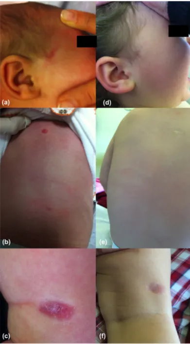
Figure 1. Clinical aspect of the lesions: (A-C) initial lesions; (D-F)
and lower right leg. The patient was otherwise healthy, with normal psychomotor development. On physical examination, the patient had a linear erythemato-violaceous plaque localized to the right temporal region of the head (Figure 1A), two erythematous nodules on the back (Figure 1B), one dorsal and one lumbar, and an erythemato-violaceous ovoid plaque on the popliteal region of the right leg (Figure 1C).
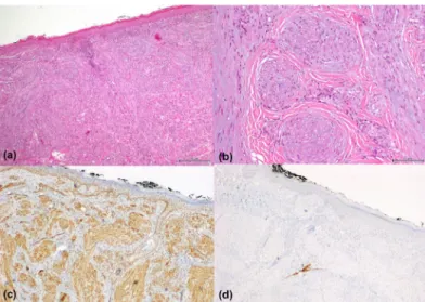
A skin biopsy was performed on both nodules on the back revealing bundles and whorls of cytologically bland myoid spindle cells with tapering nuclei and palely eosinophilic cytoplasm, focally associated with delicate thin-walled vascular channels compatible with myofibroma (Figure 2A, B). Immunostaining was positive for vimentin, smooth muscle actin, calponin, and CD 68 (focally), and negative for desmin, S100 and EMA (Figure 2, C, D).
A diagnosis of IM was made. Chest radiography and abdominal and transfontanelar ultrasound were normal and the patient had no clinical signs of visceral involvement. On follow-up visit after 18 months the lesions had regressed completely leaving residual, slightly atrophic violaceous scars (Figure 1, D-F).
Case Discussion
IM was first described in 1954 by Stout, who named this entity “congenital generalized fibromatosis” [3]. It was only in 1981 that Chung and Enzinger
introduced the term “infantile myofibromatosis” based on histochemical findings that allowed the authors to identify cell lines from which the tumor arises, further dividing this entity into subgroups with distinct prognosis depending on the location and involvement (with or without visceral lesions) [4]. More recently, IM has been considered to be part of a spectrum of tumors with perivascular myoid differentiation [5, 6].
The disorder may be present from birth to two years of age and rarely occurs in older children and adults [7]. Three clinical types have been described. The solitary type (most common) presents as a solitary, firm, well-circumscribed, and painless nodule, usually affecting the skin, muscle, bone and subcutaneous tissue in the head, neck, or trunk. The multicentric form (as in our case), consists of multiple lesions with involvement limited to the skin, subcutaneous tissues, muscles, and bone, without visceral involvement. The generalized form is defined by skin and visceral involvement [8, 9].
The diagnosis must be confirmed by a biopsy of the lesion(s). Histologically, there is a proliferation of spindle-shaped collagen-forming cells, sometimes arranged in whorled or interlacing fascicles. Immunohistochemical staining shows characteristics between fibroblasts and smooth muscle cells, being positive for vimentin and SMA; reactivity for desmin is variable [4]. CD-34 antigen has also been reported to be positive in cases of infantile myofibromatosis with hemangiopericytoma-like pattern [6].
Calponin is considered to be a family of actin filament-associated proteins expressed in both smooth muscle and non-smooth muscle cells [10]. In routine biopsy practice, immunohistochemical detection of calponin mainly serves to confirm smooth muscle and myoepithelial cell differentiation. Monoclonal antihuman calponin antibodies stain positively with differentiated visceral and vascular smooth muscle cells, myoepithelial cells in the lobules, ducts, and galactophorous tissue of normal human breasts, as well as periacinar and periductal myoepithelial cells of the salivary glands. Thus, calponin is expressed not only in some mesenchymal neoplasms, but also in benign or malignant tumors of myoepithelial origin (salivary glands, breast, or skin), [11-14].
Figure 2. Histology and immunohistochemistry: (A) H&E, 100x;
(B) H&E, 200x; (C) positive immunohistochemistry for smooth muscle actin; (D) immunohistochemistry, negative for desmin.
Volume 23 Number 4 | April 2017 DOJ 23 (4): 6
- 3 - Dermatology Online Journal || Case Presentation The prognosis is largely dependent on the presence of visceral involvement. Most cases have a benign, self-limited course, with spontaneous regression being the usual course. However, in cases of visceral involvement the prognosis is poor, with mortality rates of up to 73% [2, 15, 16].
No treatment is usually necessary for the solitary type. Surgical treatment is warranted when vital structures are affected, especially in the generalized variety. Chemotherapy has also been used with some success to treat patients with recurrent or nonresectable tumors [17, 18]. Follow-up is essential in all patients owing to the possibility of recurrence. The diagnosis and treatment of IM is challenging at times. Thorough histopathological evaluation is essential to diagnose these. tumors in early infancy and to avoid unnecessary interventions and morbidity in these patients.
Acknowledgements
We thank Dr. Cristopher D.M. Fletcher, Department of Pathology, Brigham and Women’s Hospital, for the help with the histological diagnosis.
References
1. Scheper MA, DiFabio VE, Sauk JJ, Nikitakis NG. Myofibromatosis: a case report with a unique clinical presentation. Oral Surg Oral Med Oral Pathol Oral Radiol Endod 2005;99:325-330. [PMID: 15716840] 2. Wiswell TE, Davis J, Cunningham BE, Solenberger R, Thomas PJ.
Infantile myofibromatosis: the most common fibrous tumor of infancy. J Pediatr Surg 1988;2:315-8. [PMID: 3385581]
3. Stout AP. Juvenile fibromatosis. Cancer 1954;7:953-978. [PMID: 13199773]
4. Chung EB, Enzinger FM. Infantile myofibromatosis. Cancer
1981;48:1807-1818. [PMID: 7284977]
5. Mentzel T, Dei Tos AP, Sapi Z, Kutzner H. Myopericytoma of skin and soft tissues; clinicopathologic and immunohistochemical study of 54 cases. Am J Surg Pathol 2006;30:104-13. [PMID: 16330949] 6. Kiyohara T, Maruta N, Iino S, Ido H, Tokuriki A, Hasegawa M.
CD34-positive infantile myofibromatosis: Case report and review of hemangiopericytoma-like pattern tumors. J Dermatol. 2016;43:1088-91. [PMID: 27074874]
7. Franzese CB, Carron J. Infantile myofibromatosis: unusual diagnosis in an older child. Int J Pediatr Otorhinolaryngol 2005;69:865-868. [PMID: 15885344]
8. Mashiah J, Hadj-Rabia S, Dompmartin A, Harroche A,
Laloum-Grynberg E, Wolter M, Amoric JC, Hamel-Teillac D, Guero S, Fraitag S, Bodemer C. Infantile myofibromatosis: A series of 28 cases. J Am Acad Dermatol 2014;71:264-70. [PMID: 24894456]
9. Stanford D, Rogers M. Dermatological presentations of infantile myofibromatosis: a review of 27 cases. Austr J Dermatol 2000;41:156-61. [PMID: 10954986]
10. Wu KC, Jin JP. Calponin in non-muscle cells. Cell Biochem Biophys 2008;52:139-48. [PMID: 18946636]
11. Scarpellini F, Marucci G, Foschini MP. Myoepithelial differentiation markers in salivary gland neoplasia. Pathologica 2001;93:662-7. [PMID: 11785118]
12. Furuse C, Sousa SO, Nunes FD, Magalhães MH, Araújo VC. Myoepithelial cell markers in salivary gland neoplasms. Int J Surg Pathol 2005;13:57-65. [PMID: 15735856]
13. Endo Y, Sugiura H, Yamashita H, Magalhães MH, Araújo VC. Myoepithelial carcinoma of the breast treated with surgery and chemotherapy. Case Rep Oncol Med 2013;2013:164761. [PMID: 23984136]
14. Mentzel T, Requena L, Kaddu S, Soares de Aleida LM, Sangueza OP, Kutzner H. Cutaneous myoepithelial neoplasms: clinicopathologic and immunohistochemical study of 20 cases suggesting a continuous spectrum ranging from benign mixed tumor of the skin to cutaneous myoepithelioma and myoepithelial carcinoma. J Cutan Pathol 2003;30:294-302. [PMID: 12753168]
15. Jones VS, Philip C, Harilal KR. Infantile visceral myofibromatosis – a rare cause of neonatal intestinal obstruction. J Pediatr Surg 2007;42:732-4. [PMID: 17448777]
16. Rohrer K, Murphy R, Thresher C, Jacir N, Bergman K. Infantile myofibromatosis: a most unusual case of gastric outlet obstruction. Pediatr Radiol 2005;35:808-811. [PMID: 15841368]
17. Raney B, Evans A, Granowetter L, Schaufer L, Uri A, Littman P. Nonsurgical management of children with recurrent or unresectable fibromatosis. Pediatrics 1987;79:394-398. [PMID: 3103093]
18. Williams W, Craver RD, Correa H, Velez M, Gardner RV. Use of 2-chlorodeoxyadenosine to treat infantile myofibromatosis. J Pediatr Hematol Oncol 2002;24:59-63. [PMID: 11902743]

