317
https://doi.org/10.1590/0004-282X20170046
HISTORICAL NOTE
The relationship between the First World War
and neurology: 100 years of “Shell Shock”
A relação entre a Primeira Guerra Mundial e a neurologia: 100 anos do “Shell Shock”
José Luiz Pedroso1, Stefanie C. Linden2, Orlando G. Barsottini1, Péricles Maranhão Filho3, Andrew J. Lees4,5
1Universidade Federal de São Paulo, Departamento de Neurologia, Divisão de Neurologia Geral e Unidade de Ataxia, São Paulo SP, Brasil;
2Cardiff University, School of Medicine, Division of Psychological Medicine and Clinical Neurosciences, Cardiff, UK;
3Universidade Federal do Rio de Janeiro, Hospital Universitário Clementino Fraga Filho, Departamento de Neurologia, Rio de Janeiro RJ, Brasil;
4University College London, National Hospital for Neurology and Neurosurgery, Institute of Neurology, Queen Square, UK;
5Reta Lila Weston Institute for Neurological Studies, London, UK.
Correspondence: José Luiz Pedroso; Departamento de Neurologia, Divisão de Neurologia Geral e Unidade de Ataxia, UNIFESP; Rua Pedro de Toledo, 650; 04041-002 São Paulo SP, Brasil; E-mail: [email protected]
Conflict of interest: There is no conlict of interest to declare. Received 19 January 2017; Accepted 03 February 2017. ABSTRACT
The First World War was a global war, beginning on 28 July 1914, until 11 November 1918. Soon after the beginning of the war, there was an “epidemic” of neurological conversion symptoms. Soldiers on both sides started to present in large numbers with neurological symptoms, such as dizziness, tremor, paraplegia, tinnitus, amnesia, weakness, headache and mutism of psychosomatic origin. This condition was known as shell shock, or “war neurosis”. Because medically unexplained symptoms remain a major challenge, and considering the close relationship of symptoms described in shell shock with clinical neurology, we should study their history in order to improve future care.
Keywords: First World War; shell shock; combat disorders.
RESUMO
A Primeira Guerra Mundial foi uma guerra global, iniciada em 28 de julho de 1914, até 11 de novembro de 1918. Logo após o início da guerra, exatamente há 100 anos, houve uma “epidemia” de sintomas neurológicos conversivos. Soldados de ambos os lados começaram a apresentar com frequência sintomas neurológicos, tais como: tontura, tremor, paraplegia, zumbido, amnésia, fraqueza, cefaleia e mutismo de origem psicossomática. Esta condição icou conhecida como shell shock, ou “neurose de guerra”. Como muitos sintomas e doenças inexplicadas continuam sendo um grande desaio, e considerando a estreita relação dos sintomas descritos no shell shock com a neurologia clínica, torna-se importante estudar essa parte da história com o objetivo de entendermos e melhorarmos os cuidados aos pacientes.
Palavras-chave: Primeira Guerra Mundial; shell shock; distúrbios de guerra.
he First World War (also known as World War I or the Great War) was a global war, beginning on 28 July 1914, until 11 November 1918. his large conlict involved countries that organized themselves in two opposing lines: the Allies or Triple Entente (including the United Kingdom, France and Russia), versus the Central Powers or Triple Alliance (Germany and Austria-Hungary). Although Italy was a mem-ber of the Triple Alliance, these alliances reorganized and expanded as more nations entered the war: Italy, Japan and the United States joined the Allies, while the Ottoman Empire and Bulgaria joined the Central Powers.
he trigger for the war was the assassination of Archduke Franz Ferdinand of Austria, but other political questions related to the foreign imperialist policies of the great powers of Europe were also at stake. In this Great War, which was one of the largest in history, more than 70 million servicemen,
including 60 million from Europe, were mobilized. With tech-nological advancement and increased lethality of weapons, more than nine million soldiers were killed on battleields.
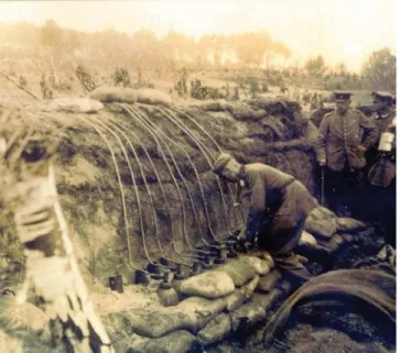
Following an initial period of mobile warfare, trench war-fare became the dominant setting for the WWI battles at the Western front. he armies protected themselves in extensive lines of earth that were dug by the soldiers (Figure 1). his was the most lethal phase of the Great War. Many soldiers fell victim to shell explosions, machine gun ire, and toxic gas. Hygiene was precarious, trenches were infested with para-sites and vermin, and soldiers were constantly exposed to infectious diseases.
318 Arq Neuropsiquiatr 2017;75(5):317-319
presented no evidence of physical injury. Many physicians concluded that the intensity of the bombings, the constant sight of dead comrades, and the uncertainty of coming out alive, must have contributed to the reactions of these sol-diers1,2. his condition was known as “shell shock”, or “war
neurosis”, which shares features with (but is not the same) as present-day post-traumatic stress disorder3
. In his semi-nal article on shell shock in the Lancet, published in 1915, Myers described three soldiers who had sufered vision loss, hearing and smell changes, and memory loss without obvi-ous physical injuries (Figure 2)4. It became increasingly clear
to physicians in Britain, Germany and other countries that these syndromes bore similarity with the hysterical presenta-tions discussed by Charcot and his colleagues in nineteenth century Paris3
.
However, not everyone conceptualized shell shock in this functional or psychological way. Several doctors believed in hidden brain injuries and others suspected carbon monoxide poisoning. Some doctors even accused shell shocked soldiers
of malingering and actively trying to avoid their military duty, although actual punishment under military law was rare3
. Major Arthur Hurst, a physician with a strong interest in Neurology and good contacts with leading French neurolo-gists, made a major contribution to the understanding and treatment of shell shocked soldiers, Hurst’s description of the clinical phenotype of shell shock was striking5,6. Many soldiers
had paraplegia, ataxia, tremor, and mutism of psychosomatic origin and others psychogenic movement disorders (Figure 3). Numerous ilms were produced at the time, one of them by Arthur Hurst himself, in order to demonstrate the successful treatment through hypnotic suggestion and other techniques7.
It is very interesting to compare shell shock and other “war syndromes”. In the Persian Gulf War (1990-1991), 25% to 30% of veterans developed a complex syndrome that included chronic fatigue, pain, cognitive and afective dysfunction and other “functional” interoceptive and nociceptive complaints. his syndrome was termed Gulf War Illness (GWI)8. here
is an ongoing debate on the relative contribution of organic
Figure 1. German battery of chlorine gas cylinders being prepared for an attack, awaiting the right weather conditions to prevent blowback; similar to the arrangement at Hill 60 in May 1915, open domain.
Figure 2. First article published related to the Shell Shock in the First World War, in 1915, by Dr. Charles Myers, captain – Royal Army Medical Corps, in The Lancet. In this paper, Myers described neurological symptoms characterized by vision loss, hearing and smell changes, and memory loss related to the Shell Shock.
319
Pedroso JL et al. 100 years of the “Shell Shock” (particularly toxic) and psychological (combat stress) factors,
which is often politically charged, and very similar to the debates around shell shock a century ago.
In France, the most dramatic representation of shell shock was “camptocormie”9. he neurologists Achille Alexandre
Souques and Inna Rosanof-Salof proposed this term char-acterized by an abnormal position of the trunk with marked lexion of the thoraco-lumbar spine, which increases during walking and abates in the recumbent position. he abnormal posture was assumed to be a psychogenic disorder described as a conversion reaction during the First World War in male military recruits and soldiers who were unable to cope with the stress of combat and unpleasant military life, perhaps triggered by stooped posture when walking in the trenches.
Considering therapeutic options, in 1916, Clovis Vincent, pioneer of neurosurgery in France, developed a method called torpillage (a term created by the soldiers themselves with the meaning of torpedoing), a “persuasive” form of psy-chotherapy using faradic and galvanic electric currents, to treat soldiers with camptocormia and “intractable” neu-roses10. Electrotherapy was popular for some time during the
war in other countries as well, although some forms were banned by the authorities.
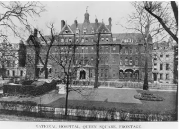
A century ago, there was an “epidemic” of neurologi-cal conversion symptoms, the condition neurologi-called shell shock. It is still fascinating for today’s neurologists and psychia-trists to watch the ilm documentaries and read the detailed case reports, such as those preserved in the archives of the National Hospital at Queen Square in London (Figure 4).
References
1. Linden SC, Hess V, Jones E. The neurological manifestations of trauma: lessons from World War I. Eur Arch Psychiatry Clin Neurosci. 2012;262(3):253-64. https://doi.org/10.1007/s00406-011-0272-9
2. Linden SC, Jones E. German battle casualties: the treatment of functional somatic disorders during World War One. J Hist Med Allied Sci. 2012;68(4):627-58. https://doi.org/10.1093/jhmas/jrs024
3. Linden S. They called it shell shock. Solhihull: Helion & Company; 2016.
4. Myers CS. A contribution to the study of shell shock: being an account of three cases of loss of memory, vision, smell, and taste, admitted into the Duchess of Westminster’s War Hospital, Le Touquet. Lancet. 1915;185(4772):316-20. https://doi.org/10.1016/S0140-6736(00)52916-X
5. Hurst A. Medical diseases of the war. London: E Arnold; 1917.
6. Linden SC, Jones E, Lees AJ. Shell shock at Queen Square: Lewis Yealland 100 years on. Brain. 2013;136(6):1976-88. https://doi.org/10.1093/brain/aws331
7. British Pathé. Wolderful shell shock recovery 1914-1918 [video]. 1917 [cited 2017 Jan 14]. Available from: http://www.britishpathe. com/video/wonderful-shell-shock-recovery
8. White RF, Steele L, O’Callaghan JP, Sullivan K, Binns JH, Golomb BA et al. Recent research on Gulf War illness and other health problems in veterans of the 1991 Gulf War: effects of toxicant exposures during deployment. Cortex. 2016;74:449-75. https://doi.org/10.1016/j.cortex.2015.08.022
9. Souques A, Rosanoff-Saloff M. La camptocormie: incurvation du tronc, consecutive aux traumatismes du dos et des lombes; considerations morphologiques. Rev Neurol (Paris). 1915;28:937-9.
10. Tatu L, Bogousslavsky J, Moulin T, Chopard J-L. The “torpillage” neurologists of World War I: electric therapy to send
hysterics back to the front. Neurology. 2010;75(3):279-83. https://doi.org/10.1212/WNL.0b013e3181e8e6fd
Figure 4. National Hospital for the Paralysed and Epileptic, around 1914, Queen Square Archives, QSA/15445.
Because medically-unexplained symptoms remain a major challenge and, considering the close relationship of symp-toms described in shell shock with clinical neurology, we should study their history in order to improve future care.