a
Diogo Filipe Amaro Santana Graça
Licenciado em Biologia
Biological Effects of Acrylic Engineered
Particulate
-
Systems
Dissertação para obtenção do Grau de Mestre em
Bioquímica
Orientadora: Ana Bettencourt, Professora Doutora,
Faculdade de Farmácia, Universidade de Lisboa
Co-orientadora: Maria João Silva, Professora Doutora,
Instituto Nacional de Saúde Doutor Ricardo Jorge
Júri:
Presidente: Prof. Doutor Pedro António de Brito Tavares Arguente: Prof. Doutora Lídia Maria Diogo Gonçalves Vogal: Prof. Doutora Ana Francisca de Campos Simão Bettencourt
II
Diogo Filipe Amaro Santana Graça
Licenciado em Biologia
Biological Effects of Acrylic Engineered
Particulate
-
Systems
Dissertação para obtenção do Grau de Mestre em
Bioquímica
Orientadora: Ana Bettencourt, Professora Doutora,
Faculdade de Farmácia, Universidade de Lisboa
Co-orientadora: Maria João Silva, Professora Doutora,
Instituto Nacional de Saúde Doutor Ricardo Jorge
Júri:
Presidente: Prof. Doutor Pedro António de Brito Tavares Arguente: Prof. Doutora Lídia Maria Diogo Gonçalves Vogal: Prof. Doutora Ana Francisca de Campos Simão Bettencourt
III
Copyright
“Biological Effects of Acrylic Engineered Particulate-Systems”
Copyright © Diogo Filipe Amaro Santana Graça, Faculdade de Ciências e Tecnologia, Universidade Nova de Lisboa
V
Acknowledgements
First of all, to my mentors, Professor Ana Bettencourt from Faculdade de Farmácia of Universidade de Lisboa and Doctor Maria João Silva from Instituto Nacional de Saúde Doutor Ricardo Jorge, for the tremendous effort and for the time spent on making this project a successful and very pleasant experience and without whom this project wouldn’t have been possible.
To my non-official mentors, Professor Lídia Gonçalves and Doctor Henriqueta Louro, as well as PhD Students Ana Matos and Inês Santos Ferreira, whose limitless patience, support and oversight helped to greatly improve the final result.
To Professor António Almeida from Faculdade de Farmácia of Universidade de Lisboa, Group leader of Nanostructured Systems for Overcoming Biological Barriers, who allowed me the opportunity to work in his “Laboratory 112”.
To INSA’s management board, Doctor Glória Isidro, Head of the Department of Genetics, and Doctor João Lavinha, Head of the Research & Development Unit, who allowed me the opportunity of working on such a praised institution with great facilities, as well as to all the researchers and colleagues, who have contributed to a fantastic working environment.
To Professor Ricardo Franco whose management and efforts allowed me to be a part of this project.
To Faculdade de Farmácia of Universidade de Lisboa and to Instituto Nacional de Saúde Doutor Ricardo Jorge for the accommodation and technical support over the duration of this project.
To Faculdade de Ciências e Tecnologia from Universidade Nova de Lisboa for being my second home during the first year of my master’s.
To all my teachers, who contributed to my improvement, both professional and personal
To my fellow lab colleagues, Diana Garcia, Paulo Roque Lino, Pedro Jogo, Inês de Mendonça, Mariana Pinhão, Ana Tavares, among many others who would make this a 10-pages section and who I see as valuable friends, for their support, both in and out of the lab work, and who helped me see the most troubled times as simple temporary setbacks. Also, for having the patience to deal with my craziness.
To my fellow, nearly masters, colleagues who, though I’ve met only last year, seemed like we’ve known each other for ages, for their friendship, support and comradery.
To all my friends and family, which are hard to distinguish between, for helping me to find the light whenever I fell onto the darkest pits of desperation.
To all those who, though might not be mentioned, have directly or indirectly contributed to the final result of this project.
I give you my most heartfelt thanks.
Thank You for helping to make a dream come true!
Financial support:
VII
Abstract
Polymeric particulate-systems are of great relevance due to their possible biomedical applications, among them as carriers for the nano- or microencapsulation of drugs. However, due to their unique specific properties, namely small size range, toxicity issues must be discarded before allowing its use on health-related applications.
Several polymers, as poly(methyl methacrylate) (PMMA), have proved to be suitable for the preparation of particulate-systems. However, a major drawback of its use refers to incomplete drug release from particles matrix. Recent strategies to improve PMMA release properties mention the inclusion of other acrylic polymers as Eudragit (EUD) on particles formulation. Though PMMA and EUD are accepted by the FDA as biocompatible, their safety on particle composition lacks sufficient toxicological data.
The main objective of this thesis was to evaluate the biological effects of engineered acrylic particulate-systems. Preparation, physicochemical characterization and in vitro toxicity evaluation were assessed on PMMA and PMMA-EUD (50:50) particles.
The emulsification-solvent evaporation methodology allowed the preparation of particles with spherical and smooth surfaces within the micrometer range (±500 nm), opposing surface charges and different levels of hydrophobicity. It was observed that particles physicochemical properties (size and charge) were influenced by biological media composition, such as serum concentration, ionic strength or pH. In what concerns to the in vitro toxicological studies, particle cellular uptake was observed on different cell lines (macrophages, osteoblasts and fibroblasts). Cytotoxicity effects were only found after 72 h of cells exposure to the particles, while no oxidative damage was observed neither on osteoblasts nor fibroblasts. Also, no genotoxicity was found in fibroblast using the comet assay to assess DNA damage. This observation should be further confirmed with other validated genotoxicity assays (e.g. Micronucleus Assay).
The present study suggests that the evaluated acrylic particles are biocompatible, showing promising biological properties for potential use as carriers in drug-delivery systems.
IX
Resumo
Os sistemas de partículas poliméricas têm adquirido uma grande relevância devido às suas possíveis aplicações biomédicas, entre elas para nano- ou microencapsulação de fármacos. No entanto, as propriedades específicas destas partículas, designadamente o reduzido tamanho, tornam necessário a avaliação da sua toxicidade de modo a garantir a sua utilização segura. Muitos polímeros têm demonstrado potencial para este tipo de aplicações, designadamente o poli(metil metacrilato) (PMMA). Contudo, a sua aplicação é muitas vezes limitada pela libertação incompleta dos fármacos através da matriz polimérica. De modo a aumentar a permeabilidade das partículas de PMMA, estratégias recentes têm vindo a incluir outros polímeros acrílicos na formulação, designadamente o Eudragit (EUD), visando melhorar a libertação dos fármacos. Apesar dos polímeros referidos serem aceites pela FDA como biocompatíveis, não existem estudos suficientes relacionados com a sua toxicidade na forma de partículas.
O principal objectivo desta tese foi avaliar os efeitos biológicos de partículas acrílicas. Em particular, realizou-se a preparação, caracterização físico-química e a avaliação de efeitos tóxicos em culturas celulares de partículas de PMMA e de PMMA-EUD (50:50).
As partículas, obtidas pelo método de emulsão-simples por evaporação de solvente, demonstraram uma distribuição de tamanhos semelhantes (±500 nm), cargas de sinal contrário e diferentes níveis de hidrofobicidade. Foi também observado que o tamanho e a carga das partículas eram influenciados pela composição do meio biológico (concentração do soro, força iónica e o pH). Relativamente aos ensaios de toxicidade, a internalização das partículas foi confirmada em várias linhas celulares (macrófagos, osteoblastos e fibroblastos). Só foram observados efeitos citotóxicos após 72 h de exposição às partículas. Não foi detetada a produção de espécies reativas de oxigénio em osteoblastos nem em fibroblastos. Também não foram observados quaisquer efeitos genotóxicos através da avaliação de lesões no ADN de fibroblastos através do ensaio do cometa. Estes resultados devem ser confirmados através de ensaios já validados para avaliação de genotoxicidade (ex: Ensaio dos Micronúcleos).
O presente estudo sugere que as partículas acrílicas avaliadas são biocompatíveis, tendo sido demonstrado que apresentam propriedades biológicas interessantes para uma possível aplicação em veiculação de fármacos.
XI
Index
Chapter 1. Introduction ... 3
1.1. Polymeric particulate-systems... 3
1.1.1. PMMA particulate-systems ... 3
1.1.2. Preparation techniques ... 5
1.1.3. Toxicological concerns ... 6
1.2. Toxicological evaluation of particulate-systems ... 7
1.2.1. The role of physicochemical properties ... 7
1.2.2. In vitro cellular assays ... 9
1.2.2.1. Cytotoxicity ... 10
1.2.2.2. Genotoxicity ... 11
1.2.2.3. Stress response ... 16
Chapter 2. Materials and Methods ... 17
2.1. Particles preparation ... 17
2.1.1. Materials ... 17
2.1.2. Methods ... 17
2.2. Particles characterization ... 19
2.2.1. Particles size distribution ... 19
2.2.2. Surface morphology ... 20
2.2.3. Surface charge evaluation ... 20
2.2.4. Chemical composition ... 20
2.2.5. Hydrophobicity ... 21
2.2.6. Study of the interactions between biological parameters and particles ... 22
2.2.6.1. Surface charge as a function of ionic strength, pH and serum concentration ... 22
2.2.6.2. Effect of serum concentration on size distribution ... 22
2.2.6.3. Protein adsorption assays ... 22
2.3. In vitro cellular assays ... 24
2.3.1. Cell lines, cell maintenance and particles dispersions ... 24
2.3.2. Cell Uptake Assays ... 25
2.3.3. Cytotoxicity Assays ... 26
2.3.4. Genotoxicity Assays ... 26
2.3.4.1. Materials ... 26
a) Comet Assay ... 26
b) Micronucleus assay ... 27
XII
a) Comet assay ... 27
b) Micronucleus assay ... 28
2.3.5. Stress Response Assays ... 28
2.3.6. Statistical Analysis ... 29
Chapter 3. Results and Discussion ... 31
3.1. Particles preparation ... 31
3.2. Particles characterization ... 33
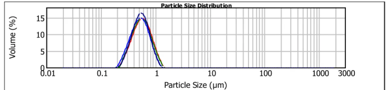
3.2.1. Particles size distribution ... 33
3.2.2. Surface morphology ... 34
3.2.3. Surface charge evaluation ... 35
3.2.4. Hydrophobicity ... 36
3.2.5. Chemical composition ... 37
3.2.6. Effect of biological conditions on particles properties ... 38
3.2.6.1. Effect of the ionic strength, serum concentration and pH on particles surface charge 39 3.2.6.2. Effect of serum concentration on size distribution ... 42
3.2.6.3. Protein adsorption assays ... 44
3.3. In vitro cellular assays ... 45
3.3.1. Cell uptake assays ... 46
3.3.2. Cytotoxicity assays ... 49
3.3.3. Genotoxicity ... 54
3.3.3.1. Comet Assay ... 54
3.3.3.2. Micronucleus Assay ... 57
3.3.4. Oxidative Stress ... 59
Chapter 4. Conclusions and Future Work ... 61
XIII
Index of Figures
Figure 1 - Chemical structure of PMMA repeating unit. ... 4
Figure 2 - Chemical structure of Eudragit RL 100. ... 4
Figure 3 - MTT conversion into formazan crystals inside the mitochondria. ... 10
Figure 4 - Conversion of resazurin into resorufin by viable cells. ... 10
Figure 5 - Critical steps for the comet assay on alkaline conditions. ... 14
Figure 6 - Potential fates of cells during CBMA after exposure to a potential genotoxic agent. ... 14
Figure 7 - Schematic representation of the experimental protocol for particles preparation. ... 18
Figure 8 - Experimental overlay for HIC assay. ... 22
Figure 9 - Representative Particle Size Distribution for PMMAp. ... 34
Figure 10 - Representative Particle Size Distribution for PMMA-EUDp... 34
Figure 11 - Panels showing particles shape from images taken with TEM spectroscopy for PMMAp and PMMA-EUDp, with and without NR. ... 35
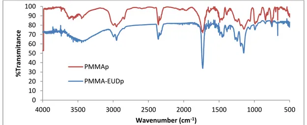
Figure 12 - FT-IR spectra with particles’ characteristic vibration peaks correspondent to the vibration frequencies of various identifiable bonds on their chemical composition. ... 38
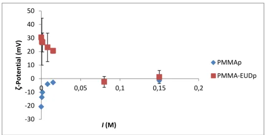
Figure 13 - Surface charge as a function of Ionic strength. ... 39
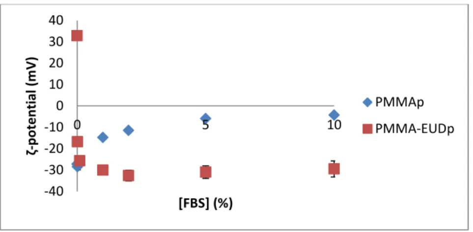
Figure 14 - Effect of FBS concentration (V/V) on the surface charge of both types of particles using water. ... 41
Figure 15 - Effect of FBS concentration (V/V) on the surface charge of both types of particles using culture medium. ... 41
Figure 16 - Surface charge as a function of pH for 0.12 mg of particles suspended in water solutions with different pH... 42
Figure 17 - BSA absorbance spectrum (concentration ranging between 0.125 – 1 mg/mL). ... 44
Figure 18 - Results from uptake assays after particle exposure for 1 and 24 h. ... 47
Figure 19 - Representative confocal microscopy images showing PMMAp and PMMA-EUDp (red signal) and L929 (above) and MG63 (below) cells (nuclei, blue signal) after 24 h of incubation. .... 49
Figure 20 - MTT results on L929 and MG63 cells viability variation to particle exposure. ... 52
Figure 21 - Examples of increasing DNA damage that can be found during Comet Assay analysis. ... 55
Figure 22 - Damage to the cells’ DNA is expressed as the mean DNA percentage on the comets’ tails. ... 56
XIV
Index of Tables
Table 1 - Different tested conditions for particles preparation. ... 18 Table 2 - Relationship between particles concentrations. ... 25 Table 3 - Yield of production per particle type. ... 33 Table 4 - Average results from size distributions of different batches (corresponding to different formulation conditions) evaluated through Laser Diffraction. ... 33 Table 5 - Surface charge of the particles diluted in dH2O. ... 36
XV
List of Abbreviations
ATA - Tocopherol acetate BCA – Bicinchoninic Acid BSA – Bovine Serum Albumin CB – Bi-nucleated cells
CBMA - Cytokinesis-block micronucleus assay CBMN – Number of micronuclei in bi-nucleated cells CBPI – Cytokinesis-block proliferation index
CPZ – Chlorpromazine DCM - Dichloromethane DMSO – Dimethyl sulfoxide DSB – Double-Strand Breaks
EMEA - European Medicines Agency EUD – Eudragit RL 100
FBS – Fetal Bovine Serum
FDA – Food and Drug Administration FF – Fast-Flow
FPG – Formamido-pyrimidine-DNA-glycosylase FRP – Free-radical polymerization
FT-IR – Fourier-Transform Infrared Spectroscopy HIC – Hydrophobic Interaction Chromatography
ICH - International Conference on Harmonisation of Technical Requirements for Registration of Pharmaceuticals for Human Use
IEP – Isoelectric Point IL-6 – Interleukin-6
LDH – Lactate Dehydrogenase
MCP-1 – Monocyte Chemoattractant Protein MEM – Minimum Essential Medium
MMA – Methyl methacrylate
XVI NR – Nile Red
OECD - Organization for Economic Co-operation and Development PBMAD - Poly(butadienemaleic anhydride-co-L-DOPA)
PBS – Phosphate buffered saline PCL - Poly-ε-caprolactone PEG – Polyethylene glycol PEI - Polyethileneimine PGE-2 – Prostaglandin E2 PLGA - Poly(lactide-co-glycolide) PLLA - Polylactide
PMA - Phorbol 12-myristate 13-acetate PMMA – Poly(methyl methacrylate) PMMAp – PMMA particles
PMMA-EUDp – PMMA/EUD particles (50:50) PS – Polystyrene
PVA – Polyvinyl acetate
ROS – Reactive Oxygen Species SCF – Supercritical Fluid
SESE – Single Emulsion by Solvent Evaporation SDS – Sodium Dodecyl Sulphate
SSB – Single-Strand Breaks
TEM – Transmission Electron Microscopy
XTT – 2,3-bis-(2-methoxy-4-nitro-5-sulfophenyl)-2H-tetrazolium-5-carboxanilide UV – Ultraviolet
1
Objectives and Thesis Structure
The thesis main objective was to evaluate the potential biological effects of engineered acrylic particulate-based systems.
Specific aims were:
- Optimization of the preparation method for obtaining two types of particles - PMMA and PMMA-Eudragit (50:50) - within the same micrometric size range and with opposite surface charges;
- Characterization of the particles physicochemical properties, as well as the effect of the biological conditions on those features;
- Evaluation of particles biological/toxicological effects by in vitro cellular assays.
The thesis is structured in four chapters including: Introduction, Materials and Methods, Results and Discussion and Conclusions and Future Work.
Chapter 1 – Introduction
Contains a brief description of the state of the art of various polymers that have already been described for particle formulation, followed by the specific case of the acrylic particulate-systems. Methods on particle formulation are then approached, as well as their influence on the particles potential toxicological effects. After, a description is made on several physicochemical properties with influence on particles’ toxicity. It ends up with a general description of several in vitro toxicological assays.
Chapter 2 – Materials and Methods
This chapter is organized in three main parts. The first and second parts detail the preparation and characterization of the particles, while the third part describes the in vitro cellular studies, as well as the conditions in which they were conducted. All of the reagents, materials, equipment and methods used in this work are presented in detail.
Chapter 3 –Results and Discussion
2 Chapter 4 –Conclusions and Future Work
3
Chapter 1. Introduction
1.1. Polymeric particulate-systems
Recent advances in polymer science have provided many innovations, underlining an increasing importance of polymeric particulate-systems in both therapeutic and diagnostic applications (Juneja and Joy, 2014; Papa et al., 2014).
In general, nanoencapsulation of drugs in particulate-systems involves forming drug-loaded particles with diameters ranging from 1 to 1000 nm (Reis et al., 2006, Naahidi et al., 2013). Particulate-systems offer relevant advantages in drug delivery by targeting molecules in specific cells and controlling drug release over time aiming to solve several problems related to the drugs themselves, such as low solubility, poor stability and unwanted side effects (Choi et al., 2012). The small size of these particles can have a huge impact over macro carrier systems, since it theoretically allow them to surpass barriers that other kind of carriers cannot. The use of polymeric materials for the synthesis of particles can constitute an improvement for the biomedical field, due to their flexibility in terms of size, mechanical stability, surface functionalization and hydrophilic/lipophilic properties (Papa et al., 2014).
Many biocompatible polymers are available to prepare particulate-systems that are being used in biomedical applications as carrier materials of molecules like DNA, proteins and drugs, as well as in cell tracking and labelling for fluorescence or magnetic resonance imaging (Höchler et al., 2012). There are multiple examples of such polymers including synthetic compounds as poly(lactide-co-glycolide) (PLGA) and poly-ε-caprolactone (PCL). For example, PLGA has been used in the preparation of microspheres (≈ 20 µm) for the sustained release of resperidone, an antipsychotic drug (D`Souza et al., 2014), and PCL has been used to formulate microspheres (28 – 43 µm) and nanospheres (750 nm), for insulin controlled-release studies (Mukerjee et al., 2007). Chitosan and its derivatives are another example of biocompatible polymers of natural origin used in drug delivery, as reported in the study of Gomathi et al. (2014), related to the preparation of particles (120-220 nm) loaded with lenalidomide, an anti-cancer drug.
In addition, the acrylic based polymers, such as poly(methyl methacrylate) (PMMA) and the Eudragit series, are a class of synthetic polymers with numerous biomedical applications.
1.1.1. PMMA particulate-systems
4 cement, in intra-ocular lenses (Pratt et al., 2006), as well as prosthetic (Reis et al., 2008) and mandibular dental material (Lye et al., 2011; Puricelli et al., 2011).
Figure 1 - Chemical structure of PMMA repeating unit.
The Eudragit polymer series is a trademark of Rohm GbmH & Co. KG. (Darmstadt, Germany) and comprise a number of (co-)polymers generally originated from polymerization reactions of acrylic and methacrylic acids or their esters. Each Eudragit compound has its own specific physicochemical properties derived from the added functional groups. Eudragit RL 100 (Figure 2) could be used in the preparation of polymeric particles because it can improve the matrix permeability, thus improving the drug release rates (Joshi, 2013).
Figure 2 - Chemical structure of Eudragit RL 100.
5 PMMA application as a vaccine coadjuvant has also been explored. Kreuter and Speiser (1976) showed the capacity for PMMAp, with the influenza virus inserted either before or after polymerization, as new vaccine adjuvants, though the first showed to have greater efficiency than the latter. In Voltan et al. (2007), PMMA core/EUD shell particles (< 300 nm) were prepared with reversibly-bound proteins (trypsin and lysozyme) which are able of aiding in the antigen incorporation, protecting it from oxidation and preserving its biological activity. Caputo et al. (2009) showed the safety of PMMA-core particles with a Eudragit L-100-55 outer-shell (with a size range between 0.22 - 2.00 µm) with hydrophilic surface properties, to be used as HIV-1 Tat protein-based vaccines, in mice by both mucosal and systemic administration.
The interest on such applications for PMMA is mainly due to a great number of advantages related to the kinetics of drug transport into the organism, but also to the possibility for pH- and thermo-dependent release, good mechanical stability and biocompatibility (Bettencourt and Almeida, 2012). In spite of the tremendous potential of PMMAp, a major drawback of its use refers to the incomplete drug release. To improve release profiles, recent strategies are focusing on formulating PMMA composites with permeable polymers, such as Eudragit RL 100 (Bettencourt and Almeida, 2012). The method chosen for particle formulation influences the particles properties and, therefore, will affect its potential toxicity. With this in mind, multiple techniques exist for the purpose of preparing PMMAp for a given target application, as described in the next section.
1.1.2. Preparation techniques
PMMAp, as spheres (monolithic devices) or capsules (reservoir devices), can be conveniently prepared by different methodologies. These techniques can be divided under two main types: 1) direct polymerization of the MMA monomer using polymerization reactions and 2) formulation from pre-formed PMMA polymer.
Polymerization techniques include conventional emulsion, surfactant-free emulsion and micro/mini-emulsion. These methods always require a physical or chemical initiation step (Bettencourt and Almeida, 2012).
6 SESE
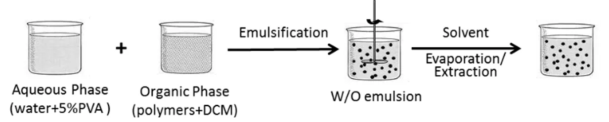
The single emulsion by solvent evaporation method is one of the most applied techniques for particle preparation, since it is one of the easiest methods (Bettencourt and Almeida, 2014). This technique is based on the emulsification of an organic phase, where the hydrophobic polymer is solubilized, into a water phase with a surfactant agent. Then, stirring conditions are applied, generally at room temperature, as the evaporation of the organic solvent occurs. This step results in the deposition of the polymer, forming particles. Most of the particles prepared by this method acquire a spherical conformation (Bettencourt and Almeida, 2012).
Some of the advantages of the use of this technique are their easy-to-apply steps and processes and the possibility to develop various types of particles, either empty or with encapsulated compounds. It is also one of the most affordable methods for obtaining emulsions. However, since it is also necessary to apply organic solvents (as dichloromethane) and surfactant agents (as polyvinyl acetate), there could be some toxicity events if these substances are not completely eliminated from the final formulations (Soomro et al., 2011; Bettencourt and Almeida, 2012).
1.1.3. Toxicological concerns
As previously mentioned, PMMAp can have a great positive impact in health-related fields and presents alternatives for numerous applications, such as drug delivery systems or the development of new vaccines. Having this in mind, an exhaustive characterization of the particles’ toxicological potential needs to be made in order to ensure its safe application in biological systems.
Biocompatibility of PMMA polymer
7 PMMA particulate-systems biocompatibility
In particular, when referring to PMMA in nano- and micro-size range, it is known that PMMA in the form of wear debris has been frequently present in patients’ bodies, but what happens to PMMA engineered particles for drug delivery lacks considerable scientific studies (Bettencourt and Almeida, 2014). The interaction of PMMApwith cells and the extracellular environment can trigger a sequence of biological effects considerably different from the material in the macro-size form. Particularly, some concern exists regarding the non-biodegradable behaviour of the nanoparticles, which can lead to their over-accumulation near organs and, eventually, to the development of harmful effects, which turns it into a toxicity-increasing factor (Rimessi et al., 2009). Besides the material toxicity, another important aspect to look for is the relevance of the preparation processes for particle safety. Preparation methods often require the use of surfactant agents and organic solvents, which may increase the risk of toxicity upon human exposure to the particles.
So far, the majority of studies that specifically address the evaluation of PMMApbiocompatibility did not find any significant toxicity. Ge et al. (2012) results are illustrative of this, as no toxicity from PMMAp conjugated with BSA was found with the MTT test, either in vitro with human colorectal cancer cells (HCT116) or in vivo assays with mice. The authors pinpoint the negative charge of the particles as responsible for the reduced toxicity because they interact less with the cells’ membranes since they are also anionic. Furthermore, PMMA–chitosan microspheres (50-500 µm) were shown to be hemocompatible and non-cytotoxic to mouse fibroblast cells (Changerath et al., 2009). Kundu et al. (2014) and Hazra et al. (2014) showed that PMMAp (<100 nm) coated with biosurfactants prepared through atomized microemulsion with and without ultrasounds, respectively, did not originate cytotoxicity on human peripheral blood mononuclear cells.Relatively to in vivo studies, Dhana lekshmi et al. (2010) concluded that no obvious toxicity was observed after oral administration of repaglinide-loaded PMMAp to albino rats.
Overall, there is insufficient data on PMMA particulate-based carriers’ toxicity, as happens in general with all engineered nanoparticles for drug delivery. Lack of standard biocompatibility evaluation criteria is a subject of intense discussion and in the future it is advisable that the toxicological evaluation should become an important part in the design of such particulate-systems, as well as the establishment of standardized protocols.
1.2. Toxicological evaluation of particulate-systems
1.2.1. The role of physicochemical properties
8 charge and hydrophobicity can greatly differ from the ones of the bulk material and, thereby, can also drive unpredictable biological interactions and effects (Louro et al., 2014).
Size plays an important role in biological interaction since the smaller the size of the particle, the higher will be its surface reactivity and the higher will be its capability to interact with multiple organelles. Another important property of smaller particles is their capability of surpassing barriers of subcellular structures, therefore, enabling them to reach structures like the nucleus. Though this potential biological interaction may be an advantage in terms of drug delivery innovative treatments for various conditions, it has also the reverse aspect of constituting a potential toxicity induction factor (Oberdörster et al., 2005).
Charge is also one of the determinant properties of biological interaction, since it relates with repulsion and attraction between different components. In fact, cationic particles have been shown to produce more effects on various cells than neutral or anionic particles. In Verma et al. (2008), uptake of anionic nanoparticles (≈ 6 nm) didn’t cause the formation of many pores in the cell membrane and cytotoxicity was minimal, while their cationic counter-parts showed to penetrate by generating pores on the cells’ membranes and produced cytotoxic effects on a mouse dendritic clone cell line. In Arvizo et al. (2010), it was demonstrated that the uptake of cationic gold nanoparticles caused decreased proliferation and cell viability of normal cells but not of cancer cells.
Another important property of the particles used as carriers is their hydrophobicity. This property concerns water-interactions and may affect not only the relation with the surrounding cells, but also with other compounds present in the biological media. Highly hydrophobic particles may be poorly stable and, therefore, are highly susceptible to aggregation when interacting with biological media (Ge et al., 2011). In consequence, particles may also have lower clearance rates, leading to their accumulation on various organelles/cells, which in turn may cause dose-dependent toxicity (Zhu et al., 2009; Murphy et al., 2011).
9
1.2.2.
In vitro
cellular assays
At an initial stage, toxicity of particulate-systems can be evaluated through in vitro cellular assays. In vitro assays allow an evaluation of cells exposure to diverse conditions, recurring to cell lines instead of a whole individual. In some cases, in vitro assays may be enough for toxicity assessment of a given substance, preventing the use of invasive methods on living organisms, e.g. rodents. However, their biggest downside is the difficulty of simulating a whole organism function and dynamics in aspects, such as cell-cell or surrounding tissues interactions, which may generate different effects based on parameters, as the location of the cells and their distribution over a generic area (Takhar and Mahant, 2011). Moreover, in vitro assays are restricted to observations at molecular and cellular levels, while in vivo studies account for the several layers of complexity inherent to the integrated response of a whole-organism and that may influence the outcome of the exposure to agents under study (Louro and Silva, 2010). Nevertheless, in vitro studies are valuable, rapid and cost-effective for the frontline toxicity assessment of new molecules or particle formulations and some limitations are being addressed with the development of 3D cultures, which is gaining increasing interest as researchers are realizing the limitations of 2D cultures (Caicedo-Carvajal et al., 2011).
As a first stage to assess particles toxicity it is important to evaluate their cellular uptake. For that evaluation, particles loaded with fluorescent compounds can be prepared. Some of the most commonly used fluorescent probes for this purpose are organic fluorescent dyes, such as Nile Red, which is widely used due to its absorption/emission wavelengths, high solubility on organic solvents and easy applicability on almost any particle formulation methods (Rose, 2010).
The use of fluorescent probes is an important tool for the identification of sites of interest and allows the visualization and evaluation of some effects over specific locations as far as the nuclear level (Forster et al., 2012).
10
1.2.2.1. Cytotoxicity
Cell viability is the most commonly investigated parameter in cytotoxicity testing. As cell viability is determined by various cellular processes, different endpoints are currently used to assess the actual state of the cells in vitro, such as the detection of mitochondrial activity and cellular membrane integrity (Kroll et al., 2009). One of the most common assays for the detection of mitochondrial activity is the MTT, a method first developed by Mosmann et al. (1983) to evaluate cell proliferation that enables a fast and quantitative measurement of living cells allowing the analysis of multiple samples at the same time. It is achieved through the reaction of of 3-(4,5-dimethyl-2-thiazolyl)-2,5-diphenyl-2H-tetrazolium bromide (MTT), a yellow, water-soluble tetrazolium dye, which is converted by the viable cells’ mitochondrial dehydrogenases into a water-insoluble, purple compound (formazan) by cleavage of the tetrazolium ring (Figure 3).
Figure 3 - MTT conversion into formazan crystals inside the mitochondria (adapted from Riss et al.,
2013).
Figure 4 - Conversion of resazurin into resorufin by viable cells (adapted from Riss et al., 2013).
11 Other method, which may be applied to evaluate cell viability, is by measuring membrane integrity using the Trypan Blue exclusion assay. The main principle applied in this methodology is that living cells possess intact cell membranes which exclude the internalization of certain dyes, such as trypan blue (Strober, 2001). Intact/viable cells will be presented as white/non-coloured, while damaged cells should be presented as blue cells. However, care should be taken in result interpretation as not always does the uptake of the dye indicate that a cell is unviable. In fact, as shown by Tran et al. (2011), some toxins may originate pores over the cellular membrane, therefore, increasing its permeability to the dye, even though the cell is still viable as its recovery mechanisms are capable of repairing most of the damage. This was shown by comparing cells coloured after 2 h of exposure to the toxin with cells to which the toxin was removed after that period and left to recover for 24 h. Furthermore, metabolic activity was found to be active since cells were still able to produce ATP.
Therefore, one must take into account the cell repair mechanisms since a cell membrane may be able to recover from damage, hence the cell may still viable. Also, the dye itself may induce damage to the cells after a long period of exposure, which ultimately leads to false positives. However, it has the advantage of being more accessible than other existing alternatives for cytotoxicity evaluation, such as MTT or Alamar Blue assays.
1.2.2.2. Genotoxicity
Genotoxicity can be derived from either primary or secondary effects, where the first can be sub-divided into direct and indirect effects.
Within the primary genotoxic effects, a direct effect derives from a direct interaction between the exogenous compounds and DNA, or any other molecule or process responsible for regulating its integrity. It might be caused, for instance, by the direct contact of particles with DNA, either through physical or chemical processes. An indirect effect will arise from oxidative stress generated from by-products of the reaction between particles and other organelles, either through generated reactive oxygen species (ROS) which can deplete the available anti-oxidants (Donaldson et al., 2010) or from ionic species resulting from soluble particles (Magdolenova et al., 2014).
12 interact with DNA, which can occur by various mechanisms. According to the literature, each particle may have its own mechanisms, though most particles show size-dependent entry (Chen and von Mikecz, 2005; Geiser et al., 2005; Nel et al., 2006).
Several studies (Park et al., 2008; Li et al., 2009; Wang et al., 2009) indicate that nanoparticles may decrease significantly the amount of anti-oxidants inside the cell, hence increasing the amount of free radicals and other reactive species which may affect DNA integrity. One of the by-products of mitochondrial processes, namely mitochondrial respiration, that can reproduce this mechanism is H2O2 originated by dismutation of superoxide anions, either spontaneously or through superoxide
dismutase activity. The inhibition of the DNA repair mechanism has also been reported for metal ions that can be released from metallic nanoparticles, which seem to inhibit the activity of some of the proteins involved in this biological process (Donaldson et al., 2010; Magdolenova et al., 2014). The same kind of effects may also underlie indirect genotoxicity when particles small enough to allocate near the nucleus are able to drag other small molecular weight compounds or reactive species generated on its surface, which can then diffuse into the nucleus and induce DNA damage. According to literature (Gedik et al., 2002; Schins et al., 2002), guanine is one of the most susceptible nucleotides to suffer damage from ROS forming 8-oxo-guanine adducts. The presence of these DNA adducts may be detected through the use of the enzyme formamido-pyrimidine-DNA-glycosylase (FPG) in a modified version of the comet assay (Donaldson et al., 2010).
ROS can also result from an inflammatory process, resulting in secondary genotoxicity as previously mentioned. This process can induce a high rate of oxygen consumption which can lead to the activation of NADPH-oxidase whose reaction can produce an elevated number of ROS and add to the already existing reactive species from other processes. Though these are produced as a self-defence mechanism they can produce damage in the surrounding cells and may transverse the nuclear membrane, causing oxidative damage in the DNA molecule (Donaldson et al., 2010). Most studies pinpoint surface area and its reactivity as the two main factors involved in dose-dependent genotoxic events. As previously stated, a protein corona may be formed on the particles surface, which could imply a variation on their genotoxic potential, while making it hard to identify the source of the effect. It is also difficult to study this process of corona formation in vitro since, in the native organism, the particles can transverse different combinations of proteins, impossible to simulate in the cell culture environment (Donaldson et al., 2010).
13 Genotoxicity evaluation
The assessment of genotoxic effects of a given substance generally follows the cytotoxicity analysis in order to evaluate only non-cytotoxic doses since cell death can increase the levels of DNA fragmentation, leading to false positive results. There are a few methods that can be employed for assessment of the genotoxicity of manufactured nanomaterials, e.g. the comet and the micronucleus assays. In addition, attention has to be given to the choice of the cell line since it should be biologically relevant and must not be affected by the method itself (Donaldson et al., 2010). Another issue in testing the genotoxic effects produced by nanoparticles is the choice of relevant positive and negative controls. Since most assays focus on the oxidative damage to DNA, some of the most employed positive controls are H2O2 and UV radiation. However, a number of
other positive control candidates are being tested (Donaldson et al., 2010).
14
Figure 5 - Critical steps for the comet assay on alkaline conditions (Adapted from Tice et al., 2000).
The micronucleus assay is based on the formation of very small nuclei during anaphase, when the nucleus divides forming two separate nuclei. It evaluates chromosomal abnormalities. Micronuclei contain either whole chromosomes or fragments which do not incorporate in none of the originated nuclei. It can be detected in the cytoplasm upon staining. This assay can be improved by adding a cytokinesis blocker with no effect over mitosis, such as Cytochalasin B, which allows obtaining multiple bi-nucleated cells and, therefore, by analysing only these, excluding interference from micronuclei generated before the assay. This method is also known as cytokinesis-block micronucleus assay (CBMA) and the possible occurrences on the cells are presented on Figure 6 (Fenech, 2000; Magdolenova et al., 2014).
Figure 6 - Potential fates of cells during CBMA after exposure to a potential genotoxic agent (Adapted from Fenech, 2000).
Other methods widely used in genotoxicity assessment include the Chromosomal Aberration assay, Ames test, HPRT gene mutation and the H2AX assay.
15 adapted to identify specific lesions on the chain, such as the ones produced through oxidative damage, but has the downside of being a somewhat subjective assay since it generally requires an operator to manually identify the comets. If not careful, an operator can be biased by knowing which sample was treated or not which can lead to ignoring some of the comets simply because they don’t fit with collected data resulting in a sort-of manipulation of the results. As for the micronucleus assay, it is a less quantitative assay than the comet assay (Magdolenova et al., 2014).
There are a number of variables which can influence the results obtained through genotoxicity-evaluating assays. From the method applied for the preparation of nanoparticles, to the choice of biologically significant concentrations, through the existence of impurities over the analysed samples, to the choice of the cell lines themselves, or even the way the particles are taken up at a certain cellular level (Magdolenova et al., 2014).
The vehicles used on particle dispersion may, in some cases, alter particles properties, such as their surface charge or hydrophobicity, which can have a direct or indirect impact over their potential for generating genotoxic responses. Hence, it must be a parameter to take into account when evaluating toxic effects. Care must also be taken while using very high concentrations of particles for they tend to agglomerate which may affect the amount of particles available for cell intake and, therefore, result in false positive/negative assessments (Magdolenova et al., 2014).
Another element which may affect these results is the presence of impurities or other chemical species mixed with the particles, such as free ionic species, ROS or proteins which promote agglomeration, since they can produce changes on the particles or simply have a direct effect over the cells during exposure, which could lead to false positive results (Magdolenova et al., 2014). One more variable is the cell line itself, since the same particles may interact differently with distinct cell lines, even if the cells derive from the same tissue origin. Factors as the receptors and transporter proteins present on the cells surface or even the amount of antioxidants and the metabolic pathways of each cell line may induce different responses when in contact with the materials/particles, since there are some lines more prone to damage than others (Magdolenova et al., 2014).
Apart from these issues, operator experience also plays a role on assay variability and it is often desirable that a set of assays are performed by the same operator, minimizing experimental variation, or following specific guidelines, such as the ones described by Fenech (2000) for the micronucleus assay.
16 There are some differences between what can be achieved, in terms of toxicity evaluation, through in vitro or in vivo assays. The main limitation of in vitro assays is that they can only evaluate primary genotoxicity due to the fact that it is very hard to reproduce all the in vivo conditions, in which secondary genotoxicity from the ROS originated through the inflammatory mechanism. However, in vitro studies can be useful to study concentrations, which may be excessive for many organisms. For biological relevance purposes the time of exposure for in vitro studies should be longer than the cell cycle period in order to maximize the accessibility of the cells to the particles. Also, the protein content must be taken into account since there are major differences between these systems in terms of both concentration and variability of proteins (Magdolenova et al., 2014).
Having all of the above in mind, PMMAp and PMMA-EUDp potential genotoxic effects were evaluated through the combination of Comet and Micronucleus in a fibroblast cell line.
1.2.2.3. Stress response
Both cyto- and genotoxicity are often mediated by oxidative stress from ROS or their excessive accumulation as reactional by-products. Several authors implicate the generation of ROS as early signs of nanoparticle-induced cellular damage. Some of the underlying mechanisms which originate oxidative stress are mitochondrial respiration and apoptosis, NADPH oxidase system activation, interference in calcium homeostasis or depletion of antioxidant enzymes. Also, ROS biological responses include the activation of signalling pathways, expression of inflammatory cytokines and chemokines, and increased transcription of inflammatory, genotoxicity, fibrosis and cancer associated factors (Manke et al., 2013).
Hence, oxidative stress is one of the most relevant outcomes of particles interaction with the biological medium. In fact, it is thought as one of the main reason for in vivo nanotoxicity (Zolnik et al., 2010; Chompoosor et al., 2010), especially if the particles can be sequestered by phagocytic cells in the reticuloendothelial system, which will target both liver and spleen (Schipper et al., 2009; Stark, 2011).
17
Chapter 2. Materials and Methods
PMMA and PMMA-EUD particles will be hereinafter represented by PMMAp and PMMA-EUDp, respectively.
2.1. Particles preparation
2.1.1. Materials
PMMA (Mw=120000) and polyvinyl alcohol (PVA, Mw=13000 – 23000; 87-89 % hydrolysed) were
provided by Sigma-Aldrich (UK). Dichloromethane (DCM), D(+)-Sucrose, Nile Red was purchased from Applichem (Germany). Poly(ethyl acrylate-co-methyl methacrylate-co-trimethylammonioethyl methacrylate chloride) (Eudragit RL 100, EUD; Mw=32000) was kindly provided by Evonik Degussa International AG (Zaragoza, Spain).
Sterile water used in the experiments was filtered through 0.22 µm pore size (Whatman filter).
2.1.2. Methods
Both particles were prepared at room temperature by single-emulsion (oil-in-water) through solvent evaporation (SESE). Preliminary assays were performed in order to optimize size distribution. After the preparation of each batch, size distribution was evaluated in order to verify changes in particles mean size and ensure that unimodal size populations were obtained.
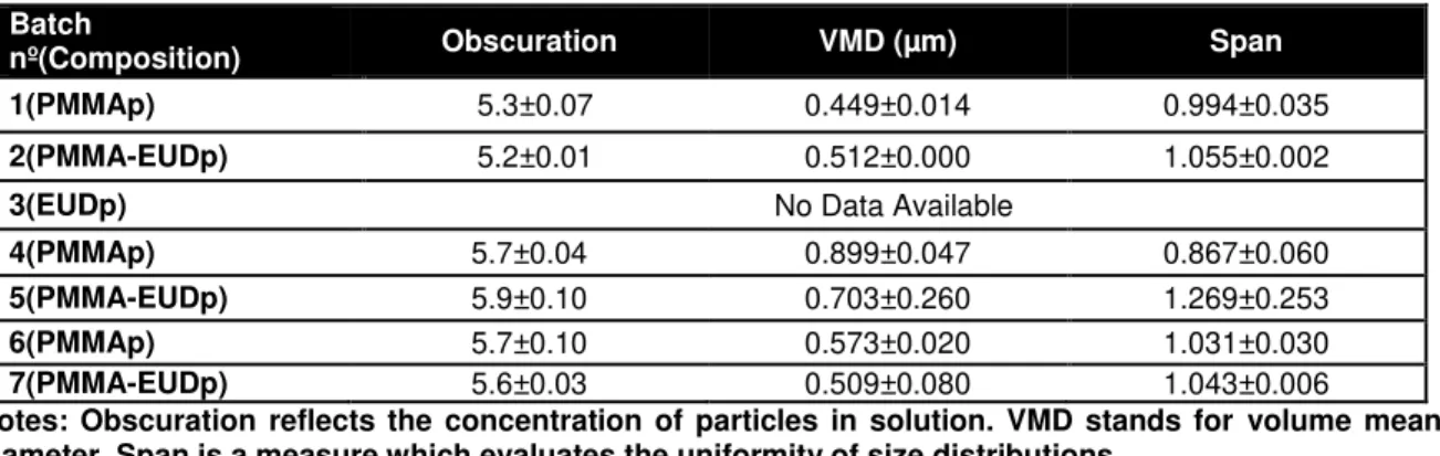
During this work we aimed to prepare particles with a size range around 500 nm in order to exclude different cellular effects due to this property. To achieve this goal, different formulation conditions were tested (Table 1).
Batches for PMMAp and PMMA-EUDp (50:50 polymer ratios) were prepared in pairs. Batches nº 1, 4 and 6 represent PMMAp, while batches nº 2, 5 and 7 correspond to PMMA-EUDp. Batch nº 3 corresponds to 100% EUD particles preparation.
Considering the best results in what refers to size distribution the selected protocol was as shown by Figure 7
18 successive centrifugations (64R, Beckman, USA) of 20000×g, during 20 min, for PMMAp and 5723×g, during 10 min, for PMMA-EUDp, both at 4 ºC. The particles were washed two times with 10 % D(+)-Sucrose.
Table 1 - Different tested conditions for particles preparation.
Batch nº(composition) PMMA (mg) Eudragit RL 100 (mg) Total Mass (mg) DCM (mL) PVA (5%)(mL) Time for emulsion at Silverson (min) Centrifugation
(×g, 4 ºC,Time)
1(PMMA) 25 - 25 3.5 10 10
2×20000,
20 min
2(PMMA-EUD) 62.5 62.5 125 5 30 10
5723, 10 min
20000, 20 min
3(EUD)* - 125 125 5 30 10
5723, 20 min
20000, 20 min
4(PMMA) 125 - 125 5 30 10
2×20000,
20 min
5(PMMA-EUD) 62.5 62.5 125 5 30 10 2×5723, 10 min
6(PMMA) 65 - 65 7.5 30 10
2×20000,
20 min
7(PMMA-EUD) 62.5 62.5 125 5 30 10 2×5723, 10 min
*Note: Eudragit (100%) particles were not possible to be prepared.
When the particles were prepared with intent to be lyophilized they were resuspended in 5 mL of filtered H2O and 1 mL of sucrose (0.5%).
19 In order to evaluate the Yield of Production (YP), aliquots were taken and lyophilized and weighted as a powder. The yield was calculated as follows: YP (%) = (Practical Yield/ Theoretical Yield) × 100.
Lyophilized particles were obtained after frozen samples submitted through a Freeze-Dryer (Alpha 1-4 (100-400), Christ, Germany) (n = 3).
Particles loaded with the fluorophore Nile Red (0.1 mM) were obtained using the same protocol as unloaded particles except that, after dissolving the polymers with DCM, 100 µL of the compound was added to the solution immediately before the emulsification step.
2.2. Particles characterization
2.2.1. Particles size distribution
Particle size distribution was evaluated by Laser Diffraction.
In Laser Diffraction the intensity of light, which is diffracted by a particle or set of particles is measured over a set of detectors distributed under a predetermined set of angles in accordance with Lorenz-Mie Theory, which states that light intensity is inversely proportional to the analysed particles size. Still there’s an associated residual error when using this technic, which must be minimized through an iterative process of measurements (Stojanović and Marković, 2012).
For Mie’s theory to be applied two parameters need to be known: the refraction index and the absorbance. The first can be obtained through a refractometer and the latter is obtained by observing the dispersed particles though microscopy. Before measurements the most suitable method must be chosen since results can be affected by a variety of factors, such as temperature, sample’s stability through time, agglomeration, among others. Two methods are available depending on sample properties: wet dispersion (used in the present study) and dry dispersion, the first easier to control and with higher signal to noise ratio and the latter faster and cheaper (Stojanović and Marković, 2012).
20
2.2.2. Surface morphology
Surface morphology was evaluated by Transmission Electron Microscopy (TEM). This technique is based on focusing an electron beam through a measurement chamber using electromagnetic lenses in a manner, which allows synthesizing an image with very high resolution. The system is run on vacuum and the scattering effect is produced by the electronic beam hitting any gas particles present in the solution (Höcherl, 2012).
The morphological characterization of PMMAp and PMMA-EUDp was performed by image analysis obtained by TEM in a Hitachi H8100 (Hitachi High-Technologies Europe GmbH, Germany) using an applied voltage of 200 kV, equipped with digital image acquisition with a CCD MegaView II bottom-mounted camera. To analyse the samples, a droplet of the suspension was deposited on the copper grid with a formvar film and dried at room temperature.
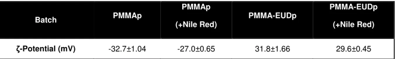
2.2.3. Surface charge evaluation
The surface charge of the particles was evaluated through ζ-potential measurement (Malvern Zetasizer Nano Z, Zen 2600, Malvern Instruments, UK). ζ-potential is based on dynamic light scattering methodologies while applying an electrical field that will polarize an electrophoretic cell and the surface charges will be evaluated due to electrophoretic mobility/velocity in the cell through the correlation equation of Smoluchowski which relates the velocity of the molecule through the electrical field with its charge (Domingues et al., 2009).
Standard electrophoretic cell was used with gold electrodes to apply the electric field. Termocaps were used to limit temperature variation. Samples were diluted in 3 mL filtered water and the cell was filled verifying for the existence of bubbles that could cause interference in the ζ-potential measurements.
2.2.4. Chemical composition
The chemical composition of the particles was assessed by FT-IR (Fourier transform infrared spectroscopy).
21
2.2.5. Hydrophobicity
Hydrophobicity was evaluated by hydrophobic interaction chromatography (HIC) for each type of particles.
HIC is generally used as a purification/separation method, usually for proteins, based on their different hydrophobicity. A compound which possesses a higher hydrophobic surface can be retained for a longer period of time in a column packed with a resin presenting many hydrophobic receptors than other compounds with less hydrophobic surfaces. This may happen when the first interacts by forming stronger bonds to the column’s surface where a selective resin (stationary phase) is immobilized. In HIC the substances are eluted several times with a neutral saline solution (mobile phase) until all different compounds have been separated. Strongly bonded compounds may only be released through an elution with a detergent, such as Triton X-100. However, it may occur that a compound presents such a strong bond with the chosen column package that it bounds to it irreversibly (Alpar and Almeida, 1994; Murphy et al., 2011).
This method may also be applied in order to relatively evaluate and compare particles with different hydrophobicity values (Alpar and Almeida, 1994). In order to do so, three types of column packages are usually used, each with increasing hydrophobicity in order to distinguish different bonding capabilities (Murphy et al., 2011).
In the present work, vertical disposable columns were used with three resins with different hydrophobicity as follows: Sepharose 4-FF < Butyl Sepharose 4-FF < Octyl Sepharose 4-FF, all provided by Ge Healthcare (UK). These resins were used as packaging for the columns after successive centrifugations
(
Megafuge 1.0R; Heraeus, UK), at 900 rpm for 10 min, at room temperature, in order to wash the preservation liquid (ethanol) in which they were suspended. For the third washing cycle the resins were re-suspended in mobile phase, a 0.6 M NaCl (Applichem, Germany) solution previously prepared. After the final centrifugation the resins were re-suspended in the mentioned mobile phase until 15 mL were obtained and sonicated (250, Branson, USA) for 10 min. Then 1 mL of resin suspension was applied to each empty column. The columns were washed with 10 mL mobile phase and then stored at 4-8 ºC overnight.The next day, 1 mL of the particles samples (20 mg/mL) was applied to each column (n = 3 per resin, per particle type) and the resulting elution was captured at vials for each column (Figure 8). This step was repeated for elutions with mobile phase, for partially retained particles, and 0.1% (w/V) Triton X-100 (Applichem, Germany), for strongly bound particles. 200 L from each vial were then transferred to a 96-well flat bottom microplate and OD600nm was measured (FLUOstar Omega,
22 Figure 8 - Experimental overlay for HIC assay.
2.2.6. Study of the interactions between biological parameters and particles
2.2.6.1. Surface charge as a function of ionic strength, pH and serum concentration
To evaluate the ionic strength effects, aqueous solutions of NaCl were set at 0.0008, 0.0015, 0.008, 0.015, 0.08 and 0.15 M though successive dilutions.To evaluate the serum percentage effect, solutions of Fetal Bovine Serum (FBS) (Life Technologies, UK) were prepared by successive dilutions in water and culture medium (RPMI 1640, Life Technologies, UK). The chosen concentrations were 0.01, 0.1, 1, 2, 5, and 10 % (V/V).
To evaluate the pH effect on the particles, sterile water was used and pH was adjusted with a pH measuring device (inoLab730, WTW, Germany) with 0.1 N HCl to pH=1.6, 2.3, 3.4, 5, 7.1 and with 0.1 N NaOH to pH=9.4, both provided by Merck (Germany).
All particle dispersions for the different evaluated conditions were prepared by adding 10 L of particles stock suspension (20 mg/mL) to 1 mL of testing condition (n = 3). The ζ-potential of the samples was then measured with Zetasizer Nano Z.
2.2.6.2. Effect of serum concentration on size distribution
Two particles suspensions of PMMAp and PMMA-EUDp (in culture medium containing 10% FBS) were incubated at 37 ºC. Samples were taken at 0, 1, 24, 48 and 72 h and introduced into Mastersizer 2000 - Hydro 2000S for particle size measuring. Mean particle size and standard deviation result from the mean of five consecutive measurements at constant stirring conditions. Measurements for PMMA-EUDp were made with and without 100 % sonication for aggregate stability evaluation.
2.2.6.3. Protein adsorption assays
23 (FLUOstar Omega, BMG Labtech, Germany) and the highest absorbance peaks were chosen for BSA adsorption analysis by measuring the OD at wavelengths 230 and 280 nm for BSA specific detection and 562 nm for total protein amount detection through the BCA method.
Particles dispersion (20 mg/mL) and BSA stock (1 mg/mL) solutions were prepared with 10 mM PBS (pH 7.4, Sigma-Aldrich, UK). Particles suspension with BSA were prepared in 1.5 mL microtubes at various concentrations depending on the assay and incubated at 37 ºC (Incubator INB 500, Memmert, Germany). Controls for BSA and both particles were also prepared in microtubes directly from the stock solutions. At pre-determined time points the samples were collected and then centrifuged (R11288 Sigma 112, Sigma-Aldrich, UK). The supernatants were transferred to 96-wells plates specially designed for UV measurements and non-adsorbed BSA was detected by UV-vis spectroscopy, as previously described.
In addition, a biochemical assay (bicinchoninic acid assay, BCA assay, Sigma-Aldrich, UK) for the detection of the BSA in the supernatants was also performed. The method was executed as follows: concentration of PMMAp and PMMA-EUDp was changed until OD600nm = 0.5, having been chosen
[PMMAp] = 500 µg/mL and [PMMA-EUDp] = 202 µg/mL. Then different studies were performed: i) BSA adsorption variation with time, ii) adsorption of particles at different concentration of BSA and iii) adsorption of BSA at different concentration of particles.
i) For BSA adsorption variation with time, dispersions of particles at the previously discussed concentrations were incubated with [BSA] = 1 mg/mL, at 37 ºC with steering conditions and analysed at 1, 2, 4, 6, 24 and 48 h.
ii) For [BSA] variation particles were incubated with 59.7; 67.6; 92.6; 113.6; 146.9; 208.3; 476.2 and 909.1 µg/mL of BSA for 24 h at 37 ºC.
iii) For [particles] variation, [BSA] was fixed at 1 mg/mL and was added to particles dispersions at 0.5, 1 and 2 mg/mL which were incubated for 24 h at 37 ºC.
After incubation, each sample was centrifuged (R11288 Sigma 112, Sigma-Aldrich, UK) for 15 min at 12000 rpm and 50 µL of the resulting supernatant were transferred into 96 wells plates specially designed for UV measurements. Then reaction was induced with 50 µL of BCA solution Kit (1:20 ratio between Copper (II) and Biocinchoninic Acid) which specifically identifies proteins through the reduction of Cu2+ to Cu+, which forms a purple chromogenic compound with absorbance maximum at 562 nm. The plates were incubated for 15 min at 37 ºC and then OD562nm was measured with the
24
2.3.
In vitro
cellular assays
2.3.1. Cell lines, cell maintenance and particles dispersions
Cell linesCell lines L929 (Mouse fibroblast cell line, ATCC® CCL-1™) and MG63 (human osteoblast cell line, ATCC CRL-1427™) were chosen for uptake evaluation, cytotoxicity and oxidative stress response. L929 alone was used for genotoxicity assays due to time concerns. THP1 cells (human monocytic cell line, ATCC TIB-202™) were also used for uptake evaluation because it can be differentiated into macrophages.
Cell maintenance
Each cell line was kept at optimal conditions for growth, i.e., incubated in RPMI 1640 culture medium supplemented with 10% FBS, 100 units of penicillin G (sodium salt), 100 µg of streptomycin sulfate and 2 mM L-glutamine, at 37 ºC with 5 % CO2, until confluence levels reached
at least 75 %. At this point cell lines were trypsinized with 1 mL of enzyme (TrypLE™Express) and subsequently transferred to new T-flasks at a tenth of its original volume. All the mentioned products were acquired from Life Technologies (UK).
The THP1 cell line was differentiated to macrophages for 3 days with 200nM of phorbol 12-myristate 13-acetate (PMA) (Sigma-Aldrich; UK) before exposition to the particles.
Particles dispersion
Stock particles dispersions were obtained by weighting an adequate amount of particles in purified sterile H2O to a final concentration of 20 mg/mL. Careful homogenization through sample inversion
was performed until no aggregation was visually detected. PMMA-EUDp stock dispersions were prepared immediately before its use due to their rapid aggregating properties. PMMAp stock solutions were prepared and used as seen fit since these didn’t show any physical signs of aggregation.
25 Table 2 - Relationship between particles concentrations.
Particle
concentration
(µg/mL)
96-well plates
(µg/cm2)
24-well plates
(µg/cm2)
10 3 2.8
100 30 28
500 147 139
1000 294 278
2000 588 556
5000 1471 1389
2.3.2. Cell Uptake Assays
Nile Red-loaded particles were used in these studies.
Cells were seeded in sterile flat bottom 96-well tissue culture plates (Greiner, Germany) at a cell density of 2×10⁵, 1×10⁵ and 2.5×10⁵ per well, respectively for L929, MG63 and THP1 cell lines. Cells were then exposed to 3, 30 and 147 µg/cm2 of each type of particles. Negative control refers to culture media-only.
After 24 h for L929 and MG63 or 72 h for THP1, culture medium was replaced with 100 µL of Nile Red loaded particles. The analysis for each concentration was made on 5 wells. The cells with the treatment medium were incubated for 1 and 24 h, after which they were washed three times with 250 µL of PBS containing 20 mM Glycine (Bio Rad, USA) at pH 7.4 at 37 ⁰C and then fluorescence was immediately measured at excitation wavelength 485 nm and emission 520 nm. PBS was then removed and the cells were disrupted with 100 µL of 1 % Triton X-100 after which fluorescence was again measured to determine the amount of internalized particles.
Confocal microscopy analysis was performed further for confirmation of cellular uptake.
26
2.3.3. Cytotoxicity Assays
The effect of particles on cell viability was first evaluated by two distinct methods which measure the cell mitochondrial activity, MTT and Alamar Blue assays.
Cells were seeded as in the uptake assay at a cell density of 2×10⁵ and 1×10⁵ per well, respectively for L929 and MG63 cell lines. Cells were then exposed to 3, 30, 147, 294 and 588 µg/cm2 of both types of particles. Negative control refers to culture media-only and Sodium Dodecyl Sulphate (SDS) (Merck, Germany) at 1 mg/mL was chosen as positive control. After 24 h, fresh media was applied with the different treatments and five wells per concentration were analysed. Cell viability was assessed after 24, 48 and 72 h of incubation at 37 ºC and 5 % CO2. After each time of
incubation, culture medium was replaced with medium containing 5 mM of resazurin (Applichem, Germany), or 0.25 mg/mL of MTT (Sigma-Aldrich, UK). This was followed by further 3 h of incubation in a fluorescence microplate reader after which fluorescence was measured at 530 nm for excitation wavelength and 590 nm for emission wavelength for the Alamar Blue assay.
For the plates containing MTT, the media was removed and intracellular formazan crystals were solubilized and extracted with 100 µL DMSO (Merck, Germany). After 15 min at room temperature absorbance was measured at 570 nm.
The relative cell viability (%) by comparison to the control cells was then calculated through the formulas [Fluorescence]sample/[Fluorescence]control x 100 for the Alamar Blue assay and [Absorvance 570nm]sample/[Absorvance 570nm]control x 100 for the MTT assay.
Alamar Blue assays had particle interference at the given fluorescence excitation and absorbance wavelengths and, therefore, results were not consistent and won’t be presented.
The Trypan Blue assay was further used to evaluate cell membrane integrity. Cell seeding was done as for the cytotoxicity assays previously described and the same for particle exposure but only for 24 h. For the Trypan Blue cell counting, a sample was taken from each well and a dilution factor of 5 was employed, by adding 200 µL of Trypan Blue (Sigma-Aldrich, UK) to 50 µL of the samples. Cells were then counted with a Neubauer chamber on an inverted microscope with phase contrast (TC5400, Meiji Techno). The number of unviable cells was compared to the number of total cells.