Original article
DOI: 10.2478/aiht-2018-69-3137
Occupational exposure to cytotoxic drugs: the importance
of surface cleaning to prevent or minimise exposure
Susana Viegas
1,2, Ana Cebola de Oliveira
3, Elisabete Carolino
1, and Mário Pádua
1H&TRC- Health & Technology Research Center, ESTeSL - Escola Superior de Tecnologia da Saúde, Instituto
Politécnico de Lisboa
1, Centro de Investigação em Saúde Pública, Escola Nacional de Saúde Pública, Universidade
NOVA de Lisboa
2, NOVA Medical School, Faculdade de Ciências Médicas, Universidade NOVA de Lisboa
3,
Lisbon, Portugal
[Received in April 2018; Similarity Check in April 2018; Accepted in August 2018]
Healthcare workers who prepare or administer cytotoxic agents run the risk of exposure, and the risks for health are real
even at doses lower than those applied in cancer patients, because, in theory, no dose is safe. The most common and
problematic route of exposure is through the skin, especially as work surfaces can remain contaminated even after cleaning.
This pilot study aimed to demonstrate the importance of having an effective surface decontamination protocol by
determining surface contamination with cyclophosphamide, 5-fluorouracil, and paclitaxel as the most common cytotoxic
drugs in an oncology day service. Samples were collected before and after drug handling and analysed with high performance
liquid chromatography with diode array detection (HPLC-DAD). Of the 29 samples collected before drug handling 23
were contaminated, five of which with more than one drug. Of the 30 samples collected after drug handling 25 were
contaminated, eight of which with more than one drug. The two time points did not significantly differ, which evidences
a widespread contamination and ineffective cleaning. This calls for revising the cleaning protocol and handling procedure
to place contamination under control as much as possible.
KEY WORDS:
5-fluoroacil; contamination control; cyclophosphamide; HPLC-DAD; oncology day service; paclitaxel
Cytotoxic drugs have been recognised as hazardous to
healthcare professionals such as medical doctors, oncology
nurses, pharmacists, and other staff, including maintenance,
since the late 1970s (1). The European Agency for Safety
and Health at Work (EU-OSHA) (2) and the European
Commission Directorate-General for Employment, Social
Affairs, and Inclusion (3) are well aware of the issue, but
the legislation of the member states still has not been
harmonised in terms of risk prevention in the healthcare
sector (4). The EU legislation, however, does require from
employers to provide a monitoring programme for
carcinogenic compounds, which includes most of the
cytotoxic drugs (4).
Essentially, their mechanism of action comes down to
inhibiting tumour growth and cell division by interfering
with the cell genetic material. However, the most common
cytotoxic drugs are not specific enough to target tumour
cells alone, but they also affect healthy cells of the exposed
individuals (5-11). This can lead to genotoxic effects that
can result in gene alterations and neoplasms in healthy
population or secondary tumours in treated cancer patients
(7, 9, 12).
Even exposure to doses lower than those received by
cancer patients can produce adverse health effects,
especially in chronically exposed healthcare workers
(13-16). There have been reports of reproductive toxicity that
can result in miscarriage, temporary or permanent infertility,
preterm births, congenital malformations, and learning
disabilities in the children of the exposed individuals (17,
18). In addition, long-term occupational exposure has been
associated with increased risk of hair loss, infections
(attributed to lower white blood cell count and
immunosuppression), organ toxicity (e.g. liver, kidney, lung,
and cardiac toxicity), myelotoxicity, mucosal ulcers, fatigue,
bleeding, and headaches (19). Furthermore, occupationally
exposed individuals have a higher incidence of DNA
damage, chromosomal abnormalities, and cancer consistent
with the inherent carcinogenic properties of these drugs
(15, 20, 21).
Exposure can occur at every stage of the cytotoxic drug
lifecycle: from production and distribution to its application
in hospital or home care settings and its disposal as waste.
The risk of exposure is not limited to healthcare workers
who prepare (pharmacists and pharmacy technicians) or
administer (nurses) cytotoxic agents. Anyone in touch with
contaminated air or objects or even patient excreta is at risk
(12, 22-26). One Dutch study (25) identified home care,
nursing homes, and laundry facilities as non-hospital
occupational settings with higher exposure, which were all
Correspondence to: Susana Patrícia Costa Viegas, ESTeSL - EscolaSuperior de Tecnologia da Saúde, Instituto Politécnico de Lisboa, Lisbon, Portugal; Av. D. João II, Lote 4.69.01, Lisboa, Portugal
related to the care of cancer patients. Clearly, a large number
of workers can be exposed to cytotoxic agents in three ways:
through inhalation of aerosolised drugs; through direct skin
contact, and rarely through ingestion from contaminated
hands and gloves (27). However, thanks to advanced
protection technology available at the workplaces, such as
biological safety cabinets, the most common and
problematic route of exposure is skin (4, 28).
Yet, even with improvements in safe handling practices
exposure to cytotoxic drugs is still a threat. Some studies
reported traces of several cytotoxic drugs on work surfaces
serving for receiving, storing, preparing, and validating
preparations in hospital pharmacies (29-33) and even on
surfaces in administration areas (33, 34). Some reported
cytotoxic drugs in biological samples (blood and urine)
from healthcare workers (19, 35-37) and some reported
contamination of work surfaces even after their cleaning
(4, 37-40).
The aim of this pilot study was therefore to establish
working surface contamination with cytotoxic drugs in an
outpatient oncology setting and see how effective the
adopted standard decontamination procedures are.
MATERIALS AND METHODS
Our study focused on the drug preparation unit and the
drug administration unit of an oncology day service (ODS),
which treated 375 patients (over 3145 visits) in 2015. It
also included the toilets used by patients and their
accompanying family members, who also run the risk of
exposure (Figure 1).
Before sampling began, we gathered information about
the number of chemotherapies prepared, most common
drugs used, application methods, and post-treatment
cleaning procedures. We also asked the staff to carry on
cleaning as usual to avoid bias. For sampling we chose a
day regarded as »normal« by the staff in terms of the number
of treatments. Samples from the working and other surfaces
were taken with wipes (24).
The preparation unit had four pharmacists, three
pharmacy technicians, and four assistants. It did not use a
closed-system drug transfer device and had only two
biosafety cabinets to prepare the cytotoxic drugs, but only
used one at a time. The cytotoxic drugs in this unit were
handled as follows: pharmacists would send the drugs to
pharmacy technicians to prepare them in one of the
biosafety cabinets and then check the preparations. Then
the assistants would carry the preparations in a specific
container similar to a biohazard box to the drug administration
unit (Figure 1).
The administration unit had thirteen infusion chairs and
three beds (plus two if needed) attended by 14 nurses and
seven assistants. There was also a reception desk for patient
registration on visits.
The drug preparation and administration surfaces are
cleaned at the end of the working day. The toilet is cleaned
several times a day and more thoroughly at the end of the
day. The cleaning products used were the same as in other
hospital facilities For the surfaces the staff used a product
containing sodium tripolyphosphate (2.5-10 %), sulphonic
acid (<2.5 %), sodium lauryl ether sulphate (2.5-10 %),
propan-2-ol (2.5-10 %), and a small percentage of alcohol.
For patient toilet they used a product with sodium
hypochlorite (<5 %).
Sampling
The choice of surfaces or objects to be wiped for
samples was based on the observed everyday activities at
the ODS and included surfaces and objects likely to be
contaminated with cytotoxic drugs either through work or
other routine behaviour (33).
Each spot was sampled twice: in the morning before
drug handling started and three hours after it started. In total
we collected 59 samples. Of the 29 morning samples, 12
were taken from the preparation unit, 12 from the
administration unit, and five from the patient toilet. Three
hours into the drug handling, we took 30 more samples: 12
from the preparation unit, 13 from the administration unit,
and five from the toilet.
Samples were taken with a 100 cm
2Kimtech Science
wipe (Kimberly-Clark Professional, Roswell, GA, USA)
soaked in ethyl-acetate and fastened to a stainless steel
frame (100 cm
2), as described elsewhere (33), except for
door handles, computer mice, and telephones, where we
could not use the stainless frame for obvious reasons. The
wipes were then placed in Petri dishes, sealed with Parafilm
M
®(Bemis, Neenah, WI, USA), stored at 2–8 °C for
transport to the laboratory in Lisbon (3 h), and then frozen
at -20 °C up to two months until analysis.
no contamination occurred during sampling and
transportation to the laboratory.
Sample analysis
The samples were analysed for cyclophosphamide (CP),
5-fluorouracil (5-FU), and paclitaxel (PTX) as the most
commonly and abundantly used drugs in our ODS and in
most of the oncology settings. Testing for all cytotoxic drugs
would be too cost-ineffective (31, 32, 40-44).
The wipes were placed in 15-mL capped tubes with
10 mL of mobile phase (see below) for extraction in a roll
homogeniser for 10 minutes.
The extracts were then forced through a 0.2 µm filter
and injected in a high-performance liquid chromatographer
with a diode array detector (HPLC-DAD) for separation
and quantification with a Thermo Unicam Surveyor
(Thermo Scientific
TM, San José, CA, USA). To do that we
used the C18 Hypersil-GOLD
®(Thermo Scientific
TM)
1 5 × 5 × 4 . 6 , a r e s p e c t i v e g u a r d c o l u m n , a n d
acetonitrile:methanol:water (19:13:68) as mobile phase.
The flow rate was 0.8 mL min
−1. All the HPLC-grade
solvents used were purchased from VWR International
(Radnor, PA, USA). CP, 5-FU, and PTX for calibration were
purchased from Merck KGaA (Darmstadt, Germany).
Calibration curves were obtained after their extraction from
spiked wipes. Each sample was injected in triplicate.
Chromatograms were integrated with the Xcalibur
TM2.0
software (Thermo Scientific
TM).
Method validation
Validation followed the US Food and Drug
Administration regulatory guidelines for analytical
procedures (57). Table 1 shows the results of method
validation, including the limit of detection (LOD) and limit
of quantification (LOQ), which were within the range for
this method (66-68). A sample was considered positive for
a particular drug if the value was above LOD.
All calibration curves with a correlation coefficient (R
2)
greater than 0.990 for the three drugs were linear over the
range. The LOD and LOQ correspond to the mean of five
calibration curves.
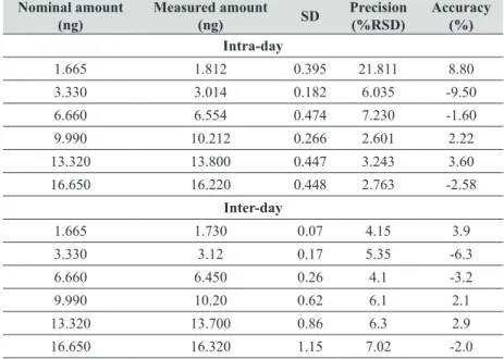
Tables 2, 3, and 4 show the intra- and inter-day accuracy
and precision parameters at six calibration levels. Table 5
summarises recoveries of 5-FU, CP, and PTX.
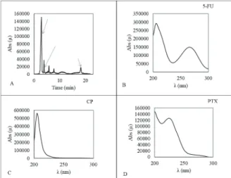
Their retention time was about 3.9, 5.8, and 18.4
minutes, respectively. Their typical chromatograms were
well resolved, which indicates that the assay was selective
(Figure 2).
Statistical analysis
Normality of distribution was tested with the
Shapiro-Wilk test. Prevalence of contamination before and after
drug handling was compared with the frequency analysis.
To compare drug concentrations at both time points (before
and after drug handling started) between the three locations
(drug preparation unit, drug administration unit, and patient
toilet) we used the Kruskal-Wallis test. Kruskal-Wallis
multiple comparison was also used for significantly
different results. To compare 5-FU, CP, and PTX
concentrations between the two time points we used the
Wilcoxon test. The level of significance was set at a 5 %.
RESULTS
Table 6 shows the distribution of positive samples with
quantifiable drugs (>LOQ) by sampling time. Of the 29
morning samples 21 were contaminated: seven in the
preparation unit, 10 in the drug administration unit, and
four in patient toilet (Table 7). Of these, three were
contaminated with more than one drug:, two in the drug
administration unit, and one in the patient toilet.
Of the 30 samples collected three hours into the drug
handling, 24 were positive, 19 of which from the drug
preparation and administration units and 5 from the patient
toilet (Table 8). Of these, five were contaminated with more
than one drug: four in the preparation unit, and one in the
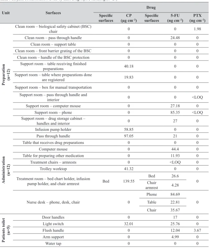
drug administration unit.
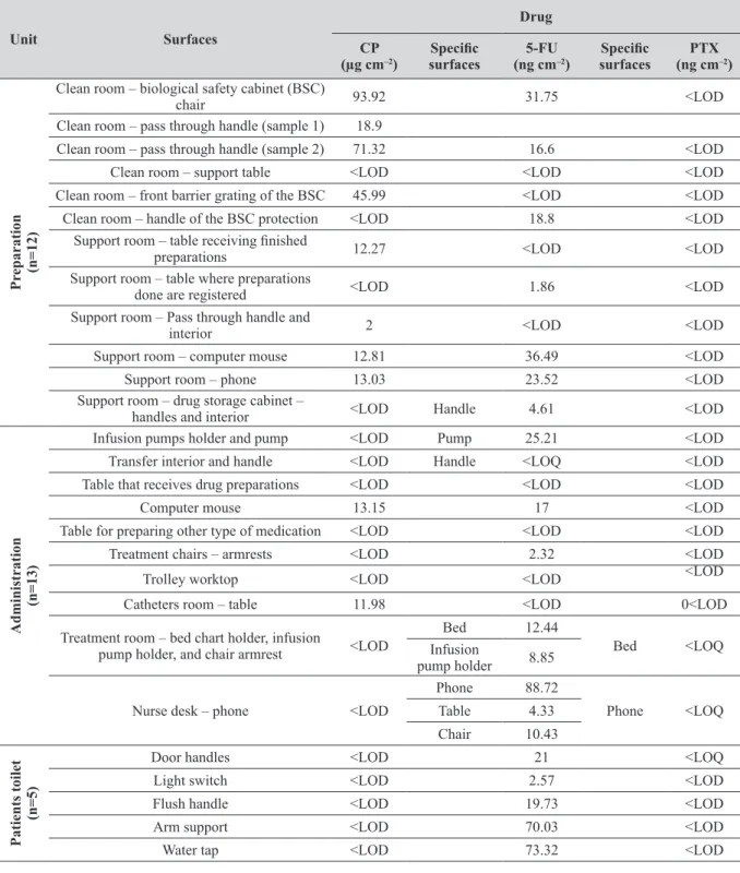
Table 9 shows total contamination of each unit by drug.
Figure 2
Typical chromatogram of a wipe spiked with the
drugs under study A: all three drugs; B, C, and D:
absorption spectra
Table 1 Method validation for each cytotoxic drug according to the FDA regulatory guidelines for analytical procedures (57)
Equation R2 LOD LLOQ Range
5-FU Peak area=35209+16473.8x5-FU 0.9983 0.900 ng 2.728 ng LOD-75 ng CP Peak area=35230.8+25147.7xCP 0.9936 1.78 µg 5.40 µg LOD-60 µg
Of the total fifty nine samples collected, 45 were
contaminated with one or more drugs. Considering only the
results higher than the LOQ, contamination before and
during drug handling did not differ significantly (p>0.05)
in any of the three units. However, the units did differ in
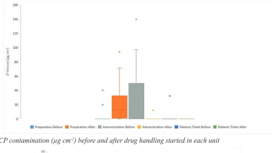
contamination with CP and 5-FU. CP was significantly
higher in the preparation unit than in other units three hours
into the drug handling, and significantly higher in the
administration unit than the others before drug handling
(Figure 3). In turn, 5-FU was significantly higher in the
preparation unit and the patient toilet three hours into the
drug handling, while in the administration unit the
concentration was higher before (Figure 4). Contamination
with PTX was not statistically analysed because there were
too few quantifiable samples to compare.
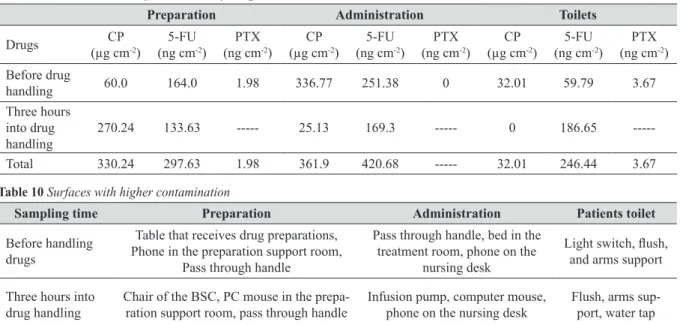
Table 10 shows the three sampling spots with
significantly higher contamination with any of the cytotoxic
drugs compared to the rest.
DISCUSSION
Our results clearly show that the current cleaning
procedure is ineffective in removing contamination with
the three representative cytotoxic drugs. They also suggest
that other variables contributed to contamination as well.
Our findings are similar to the contamination reported
in Portuguese hospitals (33), but we did not expect to find
higher contamination in the drug administration unit than
in the preparation unit, where drugs are handled and mixed
in high concentrations (33, 34).
Table 2 HPLC intra-day and inter-day (n=5) quantification accuracy and precision for 5-fluorouracil (injection volume 100 µL) Nominal amount
(ng)
Measured amount
(ng) SD
Precision (%RSD)
Accuracy (%) Intra-day
7.500 6.950 0.49 7.10 -7.33 15.000 15.300 0.09 0.60 2.00 30.000 30.840 0.08 0.27 2.80 45.000 44.360 0.89 2.00 -1.42 60.000 60.050 1.23 2.05 0.08 75.000 72.270 1.67 2.31 -3.64
Inter-day
7.500 7.050 0.18 2.56 -6.00 15.000 15.100 0.21 1.41 0.67 30.000 29.840 0.45 1.50 -0.53 45.000 45.380 0.53 1.16 0.84 60.000 58.550 0.24 0.41 -2.42 75.000 76.270 0.28 0.37 1.69
Table 3 HPLC intra-day and inter-day (n=5) quantification accuracy and precision for cyclophosphamide (injection volume 100 µL) Nominal amount
(µg)
Measured amount
(µg) SD
Precision (%RSD)
Accuracy (%) Intra-day
6.000 5.300 0.36 6.7 -11.7 12.000 12.300 0.43 3.5 2.5 24.000 25.160 0.55 2.2 4.8 36.000 35.240 0.74 2.1 -2.1 48.000 44.230 0.75 1.7 -7.9 60.000 51.000 1.79 3.5 -15.0
Inter-day
In the preparation unit the most contaminated surfaces
before drug handling were the ones in the support room and
three hours into the handling, the most contaminated
surfaces were the ones in the clean room (Table 7), which
is quite expected as handling goes. Clean rooms are the
central points for drug preparation but are also more
thoroughly cleaned than other rooms when handling is over.
This probably explains the distribution of contaminated
surfaces at both sampling times in both units (Figures 2 and
3 and Table 10). Similar findings were reported by
Fleury-Souverain et al. (30) for samples collected after the handling
started, as they did not collect samples after cleaning.
In our study, 5-FU was found in most samples at both
sampling time points, which confirms the findings reported
by Kiffmeyer et al. (24). They also reported that 5-FU was
the most used drug, which may explain our findings as well.
In both of our units and the patient toilet the most
contaminated surfaces were the ones handled without gloves
(phones, computer mouse, bed in the treatment room, toilet
light switch, flush handle, and handlebars) regardless of the
sampling time point. The same was reported in our previous
study (33), where we attributed contamination to
inappropriate cleaning and drug handling procedures. The
same pattern was observed in a study of Italian hospitals
(41), which reported that workers did not remove gloves
immediately after drug handling, and therefore spread the
contamination on phones, chairs, closets and other surfaces
not directly involved in drug preparation and administration.
The same conclusions were reported by Kiffmeyer et al.
(24), who found high concentrations of 5-FU on a closet
door knob that was not used to store 5-FU.
Contamination of infusion pumps after drug
administration started is similar to earlier reports (19, 33,
42). In fact, Hon et al. (34) claim that infusion pumps are
most often touched by nurses during drug administration.
This is of particular concern, because we found contamination
before and after drug administration and also observed that
the pumps were sometimes handled without gloves. Clearly,
this equipment is not being cleaned.
We would also like to draw attention to the contamination
of the beds in the treatment room, bed chart holders in
particular. We observed that this surface was handled/
touched by nurses and patients' families, implicating
exposure for all these persons. This is one more surface that
probably is not being cleaned well or often enough.
Furthermore, a study of health and safety practices by
nurses that administered cytotoxic drugs in 2011 reported
that 12 % of the 2069 respondents failed to use gloves, even
though 4 % reported skin contact with the drug during
handling and administration (44).
Figure 3
CP contamination (µg cm
-2) before and after drug handling started in each unit
We also have to draw attention to the widespread
contamination of the patient toilet before and after drug
handling started, as it puts family members at risk of
exposure, making this a public health issue as well. These
findings raise particular concern, because Moretti et al. (13)
showed in a recent study that handling cytotoxic drugs,
even under controlled conditions, involves a considerable
genotoxic risk (13).
What explains much of the contamination found in our
study is that our ODS had only a biological safety cabinet,
located in the clean room, but not a closed-system drug
transfer device. By default, biological safety cabinets are
heavily contaminated and are difficult to clean, and so are
the objects taken out of the cabinet (45). Closed-system
drug transfer devices serve to prevent transfer of
contamination from these objects to outside surfaces (4).
The benefits of closed drug transfer systems have already
been demonstrated by Sessink et al. (46) and Simon et al.
(47), but the latter also noted that the device could not
completely prevent chemical contamination.
Cleaning issues and recommendations
The recent EU recommendations (4) identify cleaning
as critical in reducing the spread of contamination. Cleaning
can reduce most of the contamination, but not all of it,
because there is no universal agent for different chemical
structures of cytotoxic drugs.
Several authors have tried to find a an agent capable of
inactivating, degrading, and reducing contamination with
cytotoxic drugs in oncology services (48-55), but the issue
remains. Our study only confirms that, as we found
contamination even on supposedly cleaned surfaces before
drug handling began.
Assessing the efficacy of the cleaning methods usually
employed in their healthcare facility to eliminate
environmental contamination with CP, Touzin et al. (55)
recommended combining sodium hypochlorite and sodium
thiosulfate to obtain optimal results. However, this and
many other studies address decontamination of a single
cytotoxic agent (48, 55-57), while in reality surfaces are
often contaminated with several agents. Lamerie et al. (52)
therefore assessed the efficiency of several chemical
solutions to decontaminate two types of work surfaces
intentionally contaminated with ten cytotoxic drugs.
Sodium hypochlorite showed the highest overall efficiency
of 98 %. Promising were also solutions containing anionic
surfactants with a high efficiency to safety ratio, as they
bind to impurities and particles that are suspended in the
liquid, which makes them effective cleaning agents in water.
In fact, the EU report (4) clearly points out that no single
chemical can completely clean, disinfect, and decontaminate
surfaces contaminated with cytotoxic drugs.
The cleaning product used in our ODS was the same as
the ones used in other hospital facilities not handling
cytotoxic agents. This suggests that there are no set criteria
to select a cleaning product that would be specific for an
ODS and effective enough to warrant chemical
Table 4 HPLC intra-day and inter-day (n=5) quantification accuracy and precision for paclitaxel (injection volume 100 µL)Nominal amount (ng)
Measured amount
(ng) SD
Precision (%RSD)
Accuracy (%) Intra-day
1.665 1.812 0.395 21.811 8.80 3.330 3.014 0.182 6.035 -9.50 6.660 6.554 0.474 7.230 -1.60 9.990 10.212 0.266 2.601 2.22 13.320 13.800 0.447 3.243 3.60 16.650 16.220 0.448 2.763 -2.58
Inter-day
1.665 1.730 0.07 4.15 3.9 3.330 3.12 0.17 5.35 -6.3 6.660 6.450 0.26 4.1 -3.2 9.990 10.20 0.62 6.1 2.1 13.320 13.700 0.86 6.3 2.9 16.650 16.320 1.15 7.02 -2.0
Table 5 Recovery of the cytotoxic drugs from spiked wipes (n=3)
Drug Nominal
amount
Recovery (%)
5-FU (ng)
15 95.80 45 86.30 75 91.03
CP (µg)
12 93.43 36 96.55 60 95.32
PTX (ng)
Table 6 Samples with values >LOQ
Sampling before (n=29)
Sampling after (n=30)
Drugs CP
(µg cm-2)
5-FU (ng cm-2)
PTX
(ng cm-2)
CP (µg cm-2)
5-FU (ng cm-2)
PTX
(ng cm-2)
Positive samples 7 16 2 10 20 0
Mean Range
16.5 LOD – 139.55
17 LOD - 85.35
0.24 LOD – 3.67
2.0 LOD – 93.92
19.0
LOD - 88.72 <LOQ
Table 7 Surface contamination levels by each drug before handling (n=29)
Unit Surfaces
Drug
Specific
surfaces
CP (µg cm–2)
Specific
surfaces
5-FU (ng cm–2)
PTX (ng cm–2)
Pr
eparation (n=12)
Clean room – biological safety cabinet (BSC)
chair 0 0 1.98
Clean room – pass through handle 0 24.48 0 Clean room – support table 0 0 0 Clean room – front barrier grating of the BSC 0 0 0 Clean room – handle of the BSC protection 0 0 0
Support room – table receiving finished
preparations 40.18 0 0
Support room – table where preparations done
are registered 19.83 0 0
Support room – box for manual transportation 0 0 0 Support room – pass through handle and
interior 0 0 <LOQ
Support room – computer mouse 0 27.18 0 Support room – phone 0 85.35 <LOQ Support room – drug storage cabinet –
handles and interior 0 27 0
Administration
(n=12)
Infusion pump holder 58.85 0 0 Pass through handle 97.05 21 0 Table that receives drug preparations 0 0 0
Computer mouse 0 44.4 0
Table for preparing other medication 0 11.93 0 Treatment chairs – armrests 0 <LOQ 0
Trolley worktop 41.32 0 0
Treatment room – bed chart holder, infusion
pump holder, and chair armrest Bed 139.55
Bed 26.6
0 Chair
armrest 4.28
Nurse desk – phone, desk, chair 0
Phone 84.69
0 Table 22.81
Chair 35.67
Patients toilet
(n=5)
Door handles 0 17 0
Light switch 32.01 25.76 0
Flush handle 0 12.04 3.67
Arm support 0 4.99 0
decontamination of the surfaces. The cleaning product used
in our ODS contained sodium tripolyphosphate, sulphonic
acid, sodium lauryl ether sulphate, propan-2-ol, and a small
percentage of alcohol. The toilet was cleaned with a product
containing sodium hypochlorite instead. However, despite
the use of sodium hypochlorite, almost all toilet samples
showed contamination. This points to other factors that may
render decontamination less efficient, such as dilution and
the agent contact time with the surface (55).
However, we also believe the criteria used to select
which surfaces to clean affected the findings reported by
this and previous studies. They are focused on rendering
the surfaces aseptic by removing microorganisms, because
oncology patients are most often immunocompromised.
Accordingly, the cleaning solutions are selected based on
their disinfection efficacy and not on chemical
decontamination.
Table 8 Surface contamination level by each drug after the beginning of tasks (n=30)
Unit Surfaces
Drug
CP (µg cm–2)
Specific
surfaces
5-FU (ng cm–2)
Specific
surfaces
PTX (ng cm–2)
Pr
eparation (n=12)
Clean room – biological safety cabinet (BSC)
chair 93.92 31.75 <LOD
Clean room – pass through handle (sample 1) 18.9
Clean room – pass through handle (sample 2) 71.32 16.6 <LOD Clean room – support table <LOD <LOD <LOD Clean room – front barrier grating of the BSC 45.99 <LOD <LOD Clean room – handle of the BSC protection <LOD 18.8 <LOD
Support room – table receiving finished
preparations 12.27 <LOD <LOD Support room – table where preparations
done are registered <LOD 1.86 <LOD Support room – Pass through handle and
interior 2 <LOD <LOD
Support room – computer mouse 12.81 36.49 <LOD Support room – phone 13.03 23.52 <LOD Support room – drug storage cabinet –
handles and interior <LOD Handle 4.61 <LOD
Administration
(n=13)
Infusion pumps holder and pump <LOD Pump 25.21 <LOD Transfer interior and handle <LOD Handle <LOQ <LOD Table that receives drug preparations <LOD <LOD <LOD Computer mouse 13.15 17 <LOD Table for preparing other type of medication <LOD <LOD <LOD Treatment chairs – armrests <LOD 2.32 <LOD Trolley worktop <LOD <LOD <LOD Catheters room – table 11.98 <LOD 0<LOD Treatment room – bed chart holder, infusion
pump holder, and chair armrest <LOD
Bed 12.44
Bed <LOQ Infusion
pump holder 8.85
Nurse desk – phone <LOD
Phone 88.72
Phone <LOQ Table 4.33
Chair 10.43
Patients toilet
(n=5)
Additionally, mainly the toilet floor was cleaned and
not the surfaces touched by hand. This explains why all the
toilet surfaces were contaminated before and after the drug
handling started at the ODS.
Surface monitoring recommendations
This study stresses the importance of each oncology
service having an effective monitoring programme in place.
Monitoring should be frequent and collect reliable and
detailed exposure information to identify the causes of
contamination and appropriate measures to avoid or
minimise exposure. Kiffmeyer et al. (24) reported a
sustained contamination control thanks to one such regular
monitoring programme. Furthermore, monitoring can show
where cleaning has failed. Sampling should precede the
implementation of preventive measures and be repeated
once they are in place to establish their efficiency (60).
Viegas et al. (64) demonstrated the usefulness of surface
monitoring, as it not only gives an indirect measure of
dermal exposure but also relevant guidelines to prevent
further exposure.
Although this study findings call for changes in the
workflow and cleaning that would reduce contamination
and exposure to cytotoxic drugs, it has several limitations
to bear in mind. One is the limited number of positive
samples for PTX, even though it is the most used cytotoxic
drug in this ODS. This points to the need to revise the
sampling method and frequency. The other limitation is that
we did not sample exactly the same surfaces before and
three hours into the drug handling because some of the
surfaces were covered by objects used in everyday
operations and therefor inaccessible. For this reason we
cannot reliably follow how contamination developed
between the two sampling times.
CONCLUSIONS
Our results have shown a widespread contamination in
the ODS even after it has been cleaned and call for
improvements to define an optimise cleaning. This includes
the selection of proper cleaning agents or combinations
thereof, optimal dilution, and contact time with the surfaces.
This can only be accomplished in collaboration with trained
cleaners, who have been made aware of the risks of poor
cleaning practices.
Operating procedures, including technical resources
should also be improved to avoid or reduce the spread of
contamination. A surface monitoring programme could
greatly contribute to achieve that goal, as it can provide
guidelines for improvement.
Acknowledgements
This study was developed with the support of Escola
Superior de Tecnologia da Saúde de Lisboa, Instituto
Politécnico de Lisboa. The authors would like to thank to
Dr Xavier Guardino Sola for the important comments that
greatly improved the manuscript.
Conflicts of interest
None to declare.
REFERENCES
1. Falck K, Gröhn P, Sorsa M, Vainio H, Heinonen E, Holsti LR. Mutagenicity in urine in nurses handling cytotoxic drugs. Lancet 1979;8128:1250-1. PMID: 87722
2. European Agency for Health and Safety at Work (EU-OSHA). Current and emerging occupational safety and health (OSH) issues in the healthcare sector, including home and community care, 2014 [displayed 16 August 2018]. Available
Table 9 Total contamination for each unit by drug
Preparation Administration Toilets
Drugs CP (µg cm-2)
5-FU (ng cm-2)
PTX
(ng cm-2)
CP (µg cm-2)
5-FU (ng cm-2)
PTX
(ng cm-2)
CP (µg cm-2)
5-FU (ng cm-2)
PTX
(ng cm-2)
Before drug
handling 60.0 164.0 1.98 336.77 251.38 0 32.01 59.79 3.67 Three hours
into drug handling
270.24 133.63 --- 25.13 169.3 --- 0 186.65
---Total 330.24 297.63 1.98 361.9 420.68 --- 32.01 246.44 3.67
Table 10 Surfaces with higher contamination
Sampling time Preparation Administration Patients toilet
Before handling drugs
Table that receives drug preparations, Phone in the preparation support room,
Pass through handle
Pass through handle, bed in the treatment room, phone on the
nursing desk
Light switch, flush,
and arms support
Three hours into drug handling
Chair of the BSC, PC mouse in the prepa-ration support room, pass through handle
Infusion pump, computer mouse, phone on the nursing desk
at https://osha.europa.eu/en/publications/reports/current-and- emerging-occupational-safety-and-health-osh-issues-in-the-healthcare-sector-including-home-and-community-care/view 3. European Commission Directorate-General for Employment,
Social Affairs (EC). Occupational health and safety risks in the healthcare sector - Guide to prevention and good practice, 2011 [displayed 16 August 2018]. Available at http://ec. europa.eu/social/BlobServlet?docId=7167&langId=en 4. European Parliament (EP). Preventing occupational
expo-sure to cytotoxic and other hazardous drugs: European policy recommendations, 2016 [displayed 16 August 2018]. Available at http://www.europeanbiosafetynetwork.eu/wp-content/uploads/2016/05/Exposure-to-Cytotoxic-Drugs_ Recommendation_DINA4_10-03-16.pdf
5. Besse JP, Latour JF, Garric J. Anticancer drugs in surface waters. What can we say about the occurrence and environmental significance of cytotoxic, cytostatic and endocrine therapy drugs? Environ Int 2012;39:73-86. doi: 10.1016/j.envint.2011.10.002
6. Deblonde T, Hartemann P. Environmental impact of medical prescriptions: assessing the risks and hazards of persistence, bioaccumulation and toxicity of pharmaceuticals. Public Health 2013;127:312-7. doi: 10.1016/j.puhe.2013.01.026 7. Gajski G, Gerić M, Domijan A-M, Garaj-Vrhovac V.
Combined cyto/genotoxic activity of a selected antineoplastic drug mixture in human circulating blood cells. Chemosphere 2016;165:529-38. doi: 10.1016/j.chemosphere.2016.09.058 8. Kosjek T, Heath E. Occurrence, fate and determination of
cytostatic pharmaceuticals in the environment. TrAC Trends Anal Chem 2011;30:1065-87. doi: 10.1016/j.trac.2011.04.007 9. Toolaram AP, Kümmerer K, Schneider M. Environmental
risk assessment of anti-cancer drugs and their transformation products: a focus on their genotoxicity characterization-state of knowledge and short comings. Mutat Res-Rev Mutat 2014;760:18-35. doi: 10.1016/j.mrrev.2014.02.001 10. Zhang J, Chang VWC, Giannis A, Wang JY. Removal of
cytostatic drugs from aquatic environment: a review. Sci Total Environ 2013;445-446:281-98. doi: 10.1016/j. scitotenv.2012.12.061
11. Zounkova R, Kovalova L, Blaha L, Dott W. Ecotoxicity and genotoxicity assessment of cytotoxic antineoplastic drugs and their metabolites. Chemosphere 2010;81:253-60. doi: 10.1016/j.chemosphere.2010.06.029
12. Kopjar N, Milas I, Garaj-Vrhovac V, Gamulin M. Alkaline comet assay study with breast cancer patients: evaluation of baseline and chemotherapy-induced DNA damage in non-target cells. Clin Exp Med 2006;6:177-90. doi: 10.1007/ s10238-006-0113-8
13. Moretti M, Grollino MG, Pavanello S, Bonfiglioli R, Villarini M, Appolloni M, Carrieri M, Sabatini L, Dominici L, Stronati L, Mastrangelo G, Barbieri A, Fatigoni C, Bartolucci GB, Ceretti E, Mussi F, Monarca S. Micronuclei and chromosome aberrations in subjects occupationally exposed to antineoplastic drugs: a multicentric approach. Int Arch Occ Env Hea 2015;88:683-95. doi: 10.1007/s00420-014-0993-y 14. Turci R, Minoia C, Sottani C, Coghi R, Severi P, Castriotta
C, Del Bianco M, Imbriani M. Occupational exposure to antineoplastic drugs in seven Italian hospitals: The effect of quality assurance and adherence to guidelines. J Oncol Pharm Pract 2011;17:320-32. doi: 10.1177/1078155210381931 15. Yoshida J, Koda S, Nishida S, Yoshida T, Miyajima K,
Kumagai S. Association between occupational exposure
levels of antineoplastic drugs and work environment in five
hospitals in Japan. J Oncol Pharm Pract 2011;17:29-38. doi: 10.1177/1078155210380485
16. Zhang X, Zheng Q, Lv L, An M, Zhang Y, Wei Y, Feng W. Evaluation of adverse health risks associated with antineoplastic drug exposure in nurses at two Chinese hospitals: the effects of implementing a pharmacy intravenous admixture service. Am J Ind Med 2016;59:264-73. doi: 10.1002/ajim.22553
17. Connor TH, Lawson CC, Polovich M, McDiarmid MA. Reproductive health risks associated with occupational exposures to antineoplastic drugs in health care settings: A review of the evidence. J Occup Environ Med 2014:56:901-10. doi: 2014:56:901-10.1097/JOM.0000000000000249
18. Fransman W, Roeleveld N, Peelen S, de Kort W, Kromhout H, Heederik D. Nurses with dermal exposure to antineoplastic drugs: Reproductive outcomes. Epidemiology 2007;18:112-9. doi: 10.1097/01.ede.0000246827.44093.c1
19. Fransman W, Peelen S, Hilhorst S, Roeleveld N, Heederik D, Kromhout H. A pooled analysis to study trends in exposure to antineoplastic drugs among nurses. Ann Occup Hyg 2007;51:231-9. doi: 10.1093/annhyg/mel081
20. McDiarmid MA, Oliver MS, Rogers B, Escalante C. Chromosome 5 and 7 abnormalities in oncology personnel handling anticancer drugs. J Occup Environ Med 2010;52:1028-34. doi: 10.1097/JOM.0b013e3181f73ae6 21. Rombaldi F, Cassini C, Salvador M, Saffi J, Erdtmann B.
Occupational risk assessment of genotoxicity and oxidative stress in workers handling anti-neoplasic drugs during a working week. Mutagenesis 2009;24:143-8. doi: 10.1093/ mutage/gen060
22. Mahboob M, Rahman MF, Rekhadevi PV, Sailaja N, Balasubramanyam A, Prabhakar PV, Singh SP, Reddy UA, Rao GS, Grover P. Monitoring of oxidative stress in nurses occupationally exposed to antineoplastic drugs. Toxicol Int 2012;19:20-4. doi: 10.4103/0971-6580.94510
23. Connor TH. Hazardous anticancer drugs in health care: environmental exposure assessment. Ann NY Acad Sci 2006;1076:615-23. doi: 10.1196/annals.1371.021
24. Kiffmeyer TK, Tuerk J, Hahn M, Stuetzer H, Hadtstein C, Heinemann A, Eickmann U. Application and assessment of a regular environmental monitoring of the antineoplastic drug contamination level in pharmacies - the MEWIP project. Ann Occup Hyg 2013;57:444-55. doi: 10.1093/annhyg/mes081 25. Meijster T, Fransman W, Veldhof R, Kromhout H. Exposure
to antineoplastic drugs outside the hospital environment. Ann Occup Hyg 2006;50:657-64. doi: 10.1093/annhyg/mel023 26. Sessink PJ, Bos RP. Drugs hazardous to healthcare workers.
Evaluation of methods for monitoring occupational exposure to cytostatic drugs. Drug Safety 1999;20:347-59. PMID: 10230582
27. Turci R, Sottani C, Spagnoli G, Minoia C. Biological and environmental monitoring of hospital personnel exposed to antineoplastic agents: a review of analytical methods. J Chromatogr B 2003;789:169-209. doi: 10.1016/S1570-0232(03)00100-4
Spectrom 1998;12:1485-93. doi:
10.1002/(SICI)1097-0231(19981030)12:20<1485::AID-RCM333>3.0.CO;2-N
29. Connor TH, Anderson RW, Sessink PJ, Broadfield L, Power LA. Surface contamination with antineoplastic agents in six cancer treatment centers in Canada and the United States. Am J Health-Syst Ph 1999;56:1427-32. PMID: 10428450 30. Fleury-Souverain S, Mattiuzzo M, Mehl F, Nussbaumer S,
Bouchoud L, Falaschi L, Gex-Fabry M, Rudaz S, Sadeghipour F, Bonnabry P. Evaluation of chemical contamination of surfaces during the preparation of chemotherapies in 24 hospital pharmacies. Eur J Hosp Pharm 2015;22:333-41. doi: 10.1136/ejhpharm-2014-000549
31. Hedmer M, Georgiadi A, Bremberg ER, Jönsson BA, Eksborg S. Surface contamination of cyclophosphamide packaging and surface contamination with antineoplastic drugs in a hospital pharmacy in Sweden. Ann Occup Hyg 2005;49:629-37. doi: 10.1093/annhyg/mei042
32. Schmaus G, Schierl R, Funck S. Monitoring surface contamination by antineoplastic drugs using gas chromatography-mass spectrometry and voltammetry. Am J Health Syst Pharm 2002;59:956-61. PMID: 12040735 33. Viegas S, Pádua M, Veiga A, Carolino E, Gomes M.
Antineoplastic drugs contamination of workplace surfaces in two Portuguese hospitals. Environ Monit Assess 2014;186:7807-18. doi: 10.1007/s10661-014-3969-1 34. Hon CY, Teschke K, Chua P, Venners S, Nakashima L.
Occupational exposure to antineoplastic drugs: identification
of job categories potentially exposed throughout the hospital medication system. Saf Health Work 2011;2:273-81. doi: 10.5491/SHAW.2011.2.3.273
35. Sessink PJ, Cerná M, Rössner P, Pastorková A, Bavarová H, Franková K, Anzion RB, Bos RP. Urinary cyclophosphamide excretion and chromosomal aberrations in peripheral blood lymphocytes after occupational exposure to antineoplastic agents. Mutat Res 1994;309:193-9. doi: 10.1016/0027-5107(94)90092-2
36. Sottani C, Rinaldi P, Leoni E, Poggi G, Teragni C, Delmonte A , M i n o i a C . S i m u l t a n e o u s d e t e r m i n a t i o n o f cyclophosphamide, ifosfamide, doxorubicin, epirubicin and daunorubicin in human urine using high-performance liquid chromatography/electrospray ionization tandem mass spectrometry: bioanalytical method validation. Rapid Commun Mass Spectrom 2008;22:2645-59. doi: 10.1002/ rcm.3657
37. Sottani C, Porro B, Imbriani M, Minoia C. Occupational exposure to antineoplastic drugs in four Italian health care settings. Toxicol Lett 2011;213:107-15. doi: 10.1016/j. toxlet.2011.03.028
38. Schierl R, Böhlandt A, Nowak D. Guidance values for surface monitoring of antineoplastic drugs in German pharmacies. Ann Occup Hyg 2009;53:703-11. doi: 10.1093/annhyg/ mep050
39. Sessink PJM, Connor TH, Jorgenson JA, Tyler TG. Reduction in surface contamination with antineoplastic drugs in 22 hospital pharmacies in the US following implementation of a closed-system drug transfer device. J Oncol Pharm Pract 2011;17:39-48. doi: 10.1177/1078155210361431
40. Touzin K, Bussières JF, Langlois E, Lefebvre M. Evaluation of surface contamination in a hospital hematology–oncology pharmacy. J Oncol Pharm Pract 2009;15:53-61. doi: 10.1177/1078155208096904
41. Castiglia L, Miraglia N, Pieri M, Simonelli A, Basilicata P, Genovese G, Guadagni R, Acampora A, Sannolo N, Scafarto MV. Evaluation of occupational exposure to antiblastic drugs in an Italian hospital oncological department. J Occup Health 2008;50:48-56. PMID: 18285644
42. Kopp B, Schierl R, Nowak D. Evaluation of working practices and surface contamination with antineoplastic drugs in outpatient oncology health care settings. Int Arch Occup Environ Health 2013;86:47-55. doi: 10.1007/s00420-012-0742-z
43. Larson RR, Khazaeli MB, Dillon HK. Monitoring method for surface contamination caused by selected antineoplastic agents. Am J Health Syst Pharm 2002;59:270-7. PMID: 11862639
44. Boiano JM, Steege AL, Sweeney MH. Adherence to safe handling guidelines by health care workers who administer antineoplastic drugs. J Occup Environ Hyg 2014;11:728-40. doi: 10.1080/15459624.2014.916809
45. Vyas N, Turner A, Clark JM, Sewell GJ. Evaluation of a closed-system cytotoxic transfer device in a pharmaceutical isolator. J Oncol Pharm Pract 2014;22:10-9. doi: 10.1177/1078155214544993
46. Sessink PJ, Trahan J, Coyne JW. Reduction in surface contamination with cyclophosphamide in 30 US hospital pharmacies following implementation of a closed-system drug transfer device. Hosp Pharm 2013;48:204-12. doi: 10.1310/hpj4803-204
47. Simon N, Vasseur M, Pinturaud M, Soichot M, Richeval C, Humbert L, Lebecque M, Sidikou O, Barthelemy C, Bonnabry P, Allorge D, Décaudin B, Odou P. Effectiveness of a closed-system transfer device in reducing surface contamination in a new antineoplastic drug - compounding unit: a prospective, controlled, parallel study. PLoS One 2016;11:e0159052. doi: 10.1371/journal.pone.0159052 48. Barek J, Cvacka J, de Méo M, Laget M, Michelon J,
Castegnaro M. Chemical degradation of wastes of antineoplastic agents amsacrine, azathioprine, asparaginase and thiotepa. Ann Occup Hyg 1998;42:259-66. doi: 10.1016/ S0003-4878(98)00023-4
49. Benvenuto JA, Connor TH, Monteith DK, Laidlaw JL, Adams SC, Matney TS, Theiss JC. Degradation and inactivation of antitumor drugs. J Pharm Sci 1993;82:988-91. doi: 10.1002/ jps.2600821003
50. Castegnaro M, De Méo M, Laget M, Michelon J, Garren L, Sportouch MH, Hansel S. Chemical degradation of wastes of antineoplastic agents. 2: six anthra-cyclines: darubicin, doxorubicin, epirubicin, pirarubicin, aclarubicin, and daunorubicin. Int Arch Occup Environ Health 1997;70:378-84. doi: 10.1007/s004200050232
51. Hansel S, Castegnaro M, Sportouch MH, De Méo M, Milhavet JC, Laget M, Duménil G. Chemical degradation of wastes of antineoplastic agents: cyclophosphamide, ifosfamide and melphalan. Int Arch Occup Env He 1997;69:109-14. doi: 10.1007/s004200050124
52. Lamerie TQ, Nussbaumer S, Décaudin B, Fleury-Souverain S, Goossens JF, Bonnabry P, Odou P. Evaluation of
decontamination efficacy of cleaning solutions on stainless
steel and glass surfaces contaminated by 10 antineoplasic agents. Ann Occup Hyg 2013;57:456-69. doi: 10.1093/ annhyg/mes087
Profesionalna izloženost citotoksičnim lijekovima: koliko je važno čišćenje površina za njezino sprječavanje
odnosno smanjenje
U zdravstvenih radnika koji pripremaju ili primjenjuju citotoksične lijekove zdravstveni rizici zbog izloženosti su realni,
čak i pri dozama nižima od onih koje se primjenjuju u bolesnika jer, načelno, nijedna doza nije neškodljiva za zdravlje.
Najčešći i najproblematičniji put izlaganja jest koža, napose zato što radne površine gdjekad ostanu kontaminirane i nakon
njihova čišćenja. Cilj ovoga preliminarnog istraživanja bio je pokazati koliko je važno osmisliti djelotvoran protokol za
dekontaminaciju na temelju pokazatelja kontaminacije radnih površina jedinice za pripremu lijekova, jedinice za njihovu
primjenu te bolesničkoga zahoda onkološke ambulante trima najčešćim citotoksičnim onkološkim lijekovima:
ciklofosfamidom, 5-fluoroacilom i paklitakselom. Uzorke smo prikupljali prije rada s lijekovima te tri sata od početka
rada s njima te ih analizirali tekućinskom kromatografijom s detektorom s nizom dioda (engl.
high performance liquid
chromatography with diode array detection
, krat. HPLC-DAD). Od 29 uzoraka prikupljenih prije rada s lijekovima, 23
su bila kontaminirana, od kojih pet s više lijekova. Od 30 uzoraka prikupljenih tri sata nakon početka rada s lijekovima,
njih 25 bilo je kontaminirano, od kojih osam s više lijekova. Kontaminacija površina prije i nakon početka rada s lijekovima
nije bila značajno različita, što upozorava na raširenu kontaminaciju i nedjelotvorno čišćenje. Stoga bi trebalo revidirati
postojeći protokol čišćenja i rukovanja lijekovima te svesti kontaminaciju na najmanju moguću mjeru.
KLJUČNE RIJEČI:
5-fluoroacil; ciklofosfamid; HPLC-DAD; onkološka ambulanta; paklitaksel; suzbijanje kontaminacije
their metabolites in the rine of patients administered antineoplastic therapy. Environ Mol Mutagen 1987;10:341-56. PMID: 3315656
54. Shea JA, Shamrock WF, Abboud CA, Woodeshick RW,
Nguyen LQ, Rubino JT, Segretario J. Validation of cleaning
procedures for highly potent drugs. I. Losoxantrone. Pharm Dev Technol 1966;1:69-75. doi: 10.3109/10837459609031420 55. Touzin K, Bussières JF, Langlois E, Lefebvre M, Métra A.
Pilot study comparing the efficacy of two cleaning techniques
i n r e d u c i n g e n v i r o n m e n t a l c o n t a m i n a t i o n w i t h cyclophosphamide. Ann Occup Hyg 2010;54:351-9. doi: 10.1093/annhyg/meq004
56. Lunn G, Sansone EB, Andrew AW, Hellwig LC. Degradation and disposal of some antineoplastic drugs. J Pharm Sci 1989;78:652-9. doi: 10.1002/jps.2600780811
57. Raghavan R, Burchett M, Loffredo D, Mulligan JA. Low-level (PPB) determination of cisplatin in cleaning validation (rinse water) samples. II. A high-performance liquid chromatographic method. Drug Dev Ind Pharm 2000;26:429-40. doi: 10.1081/DDC-100101250
58. Ladeira C, Viegas S, Costa-Veiga A. How to deal with uncertainties regarding the occupational exposure to antineoplastic mixtures: additive effect should always be
considered? In: Topical Scientific Workshop - New Approach
Methodologies in Regulatory Science, Proceedings of a
Scientific Workshop Helsinki, 19-20 April 2016 [displayed
17 August 2018]. Available at https://echa.europa.eu/ documents/10162/22816069/scientific_ws_proceedings_ en.pdf
59. Cavallo D, Ursini CL, Perniconi B, Francesco AD, Giglio M, Rubino FM, Marinaccio A, Iavicoli S. Evaluation of genotoxic effects induced by exposure to antineoplasic drugs in lynphocytes and exfoliated buccal cells of oncologu nurses and pharmacy employees. Mutat Res 2005;587:45-51. doi: 10.1016/j.mrgentox.2005.07.008
60. Fučić A, Jazbec A, Mijić A, Šešo-Šimić D, Tomek R. Cytogenetic consequences after occupational expo-sure to
antineoplastic drugs. Mutat Res 1998;416:59-66. doi: 10.1016/S1383-5718(98)00084-9
61. Roland C, Ouellette-Frève JF, Plante C, Bussières J-F. Surface contamination in a teaching hospital: a 6 year perspective. Pharm Technol Hosp Pharm 2016;1:187-93. doi: 10.1515/pthp-2016-0016
62. Viegas S, Ladeira C, Costa-Veiga A, Perelman J, Gajski G. Forgotten public health impacts of cancer - an overview. Arh Hig Rada Toksikol 2017;68:287-97. doi: 10.1515/aiht-2017-68-3005
63. Cavallo D, Ursini CL, Omodeo-Salè E, Iavicoli S. Micronucleus induction and FISH analysis in buccal cells and lymphocytes of nurses administering antineoplastic drugs. Mutat Res 2007;628:11-8. doi: 10.1016/j. mrgentox.2006.10.014
64. Gajski G, Ladeira C, Gerić M, Garaj-Vrhovac V, Viegas S. Genotoxicity assessment of a selected cytostatic drug mixture in human lymphocytes: a study based on concentrations relevant for occupational exposure. Environ Res 2018;161:26-34. doi: 10.1016/j.envres.2017.10.044
65. Sabatini L, Barbieri A, Tosi M, Violante FS. A new high-performance liquid chromatographic/electrospray ionization tandem mass spectrometric method for the simultaneous determination of cyclophosphamide, methotrexate and
5-fluorouracil as markers of surface contamination for
occupational exposure monitoring. J Mass Spectrom 2005;40:669-74. doi: 10.1002/jms.840
66. Castagne V, Habert H, Abbara C, Rudant E, Bonhomme-Faivre L. Cytotoxics compounded sterile preparation control by HPLC during a 16-month assessment in a French university hospital: importance of the mixing bags step. J O n c o l P h a r m P r a c t 2 0 1 1 ; 1 7 : 1 9 1 - 6 . d o i : 10.1177/1078155210376846