Contents lists available atScienceDirect
Physiological and Molecular Plant Pathology
journal homepage:www.elsevier.com/locate/pmpp
REVIEW: Novel sources and functions of microbial lipases and their role in
the infection mechanisms
Ananias Pascoal
a,∗, Letícia M. Estevinho
a, Ivone M. Martins
a,b,1, Altino B. Choupina
aaCIMO-Mountain Research Center, Department of Biology and Biotechnology, Agricultural College of Bragança, Polytechnic Institute of Bragança, Campus Santa Apolónia, E 5301-855, Bragança, Portugal
bCEB - Centre of Biological Engineering, University of Minho, 4710-057, Braga, Portugal
A R T I C L E I N F O
Keywords:
Lipases
Phytophthora cinnamomi
Biocatalysts
Biotechnological applications
A B S T R A C T
Lipases belong to the family of serine hydrolases, which in turn include various esterase enzymes (E.C.3.1.1.1). They are involved in the cleavage of triacylglycerols to free fatty acids and glycerol in many important biological processes, as for instance routine metabolism of dietary triglycerides to cell signalling and inflammation. Lipases constitute a ubiquitous group of enzymes able to catalyse a number of different reactions, many of them of industrial interest. Particularly, microbial lipases exhibit a wide range of industrial applications, namely in pharmaceutical, food and detergents industry. The aim of this review is to summarize the recent achievements illustrating the importance and the versatility of microbial lipases, including their involvement in infection mechanisms.
1. Introduction
Lipases (triacylglycerol acyl hydrolases, E.C. 3.1.1.3) belong to the family of serine hydrolases, which include several esterase enzymes. These enzymes are able to catalyse the hydrolysis of fats and oils in the oil-water interface with the release of free fatty acids, diglycerides, monoglycerides, and glycerol [1,2]. Furthermore, they also have the ability to catalyse esterification reactions, transesterification and in-teresterification in organic solvents [3,4]. Lipases as catalysts have been reported to play a special and important role in the detergents industry, food production and processing, pharmaceutical, paper, cosmetics and chemical synthesis industries [2,5]. Several studies have been published reporting the different applications of lipases (Table 1). In fact, the use of lipases in thefield of biotechnology has been known as a rentable business involving billions of dollars [6]. In the production of cosmetic products, they can function as active ingredients or as biocatalysts in the synthesis of specific cosmetic chemicals, mainly esters, aromatic compounds and active agents [7].
Lipases can split emulsified esters of glycerine and long-chain fatty acids such as triolein (trioleilglicerol, TC18) [8]. For esterases, the substrate is considered the standard tributyrin (tributirilglicerol, TC4), which in turn can be hydrolysed by lipases [9]. It is generally accepted that esterases can hydrolyse ester and triglyceride esters of short carbon chain (< 10 carbons) bonds, releasing fatty acids of low molecular
weight, and therefore are also called non-lipolytic esterases [6] or carboxylesterases [10], since lipases preferentially hydrolyse triglycer-ides of long chain (> 10 carbons) bonds.
Lipases are widely distributed in nature [3] being found in animals, such as pancreatic lipase, lipases of the tongue, milk, adipose tissue, etc.; in plants (mainly in oily seeds); in bacteria, mainly in species of the genera Chromobacterium, Pseudomonas andStaphylococcus; in yeasts, mainly in species of the generaCandidaandYarrowia lipolyticaand in fungi especially in species of the generaAspergillus, Geotrichum, Rhi-zopus,Mucor, Penicillium[3,11].
Lipases are quite diverse regarding their enzymatic properties and substrate specificity, which together with their easy availability makes them very attractive for multiple biological functions and biotechno-logical applications. Microbial lipases isolated from bacteria and fungi, are the most appealing in biotechnology and organic chemistry, mainly due to their properties, such as the shortest generation time, high yield of conversion of substrate into product, versatility and ease of mass production [3,12].
2. Kinetic and physicochemical characteristics
Lipases are usually acidic glycoproteins [29], with a molecular mass between 20 and 60 kDa and the isoelectric point may vary between 4 and 5 [30]. Most lipases have an optimal range of activity and stability
https://doi.org/10.1016/j.pmpp.2018.08.003
Received 23 October 2017; Received in revised form 25 July 2018; Accepted 8 August 2018 ∗Corresponding author.
E-mail address:[email protected](A. Pascoal). 1Current a
ffiliation.
Available online 21 August 2018
0885-5765/ © 2018 Elsevier Ltd. All rights reserved.
for pH values between 6.0 and 8.0 and temperature between 30 and 40 °C. However, these properties may vary significantly depending on the source, or even between isoforms produced by one microorganism. Lipases are water soluble enzymes that act on the metabolism and digestion of triacylglycerols [31,32], thus, the reactions catalysed by lipases occur using a lipidic substrate in the form of emulsion. However, they have a better performance when the substrate concentration is sufficiently high to form micellar aggregates or emulsions, as they can interact with the user interface of the aggregate substrate [31]. This increased activity in the presence of micelles or emulsions is due to the phenomenon known as interfacial activation [33], which in lipases has been early observed in studies undertaken with pancreatic lipases of pig [34,35]. The purified lipases allowed the study of the phenomenon in detail, where the lipase activity of triacetin increased remarkably, since it exceeded its limit of solubility.
The model of the pancreatic lipase enzyme was proposed with the assumption that the interfacial activation could answer a conforma-tional change suffered by lipases [36]. These reactions cannot be de-scribed by a Michaelis-Menten reaction, since the hydrolysis process has several steps [36]. With respect to the model substrate, the acti-vation is explained with changes in the lipid substrate interface: in-creasing the local concentration of the substrate instead of the active site, will reduce the orientation and conformation of lipids in the in-terface states related to the geometry of the active site of the enzyme, or lessen the degree of hydration of the substrate [37,38].
3. Structural characteristics
Pleiss and co-workers, have compared the structure of the active site of various esterases and lipases and they observed that those from li-pases are normally wider and deeper than the ones from esterases,
which could explain the ability of lipases to recognize a more varied and longer chains substrates (e.g. triolein) [38,39].
When a lipase in the aqueous phase is under its solubility limit or in the absence of/or at low concentration emulsion of a lipid substrate, exists typically a helix covering the active site, in particular, an oligo-peptide helix motif termed “lid” which protects the active site. This amphiphilic cover consists of polar amino acids on the outside and nonpolar amino acids on the inner side, in contact with the active site [39]. It has been reported that lipases with the lid covering the active site are in a closed conformation and, in the presence of an emulsion (upon contact with the interface formed by the lipid lipases), undergo a conformational change in the lid region exposing their active site for the hydrolysis of triacylglycerol molecules [40]. The active site is only exposed when the cover opens,i.e.the enzyme only catalyses a reaction in hydrophobic-hydrophilic interfaces or in the presence of a hydro-phobic solvent. The mechanism of conformational change form“closed to open”is called interfacial activation [26].
The active site of lipases is generally characterized by a triad of serine, histidine and an acid residue (aspartic or glutamic acid)
classi-fied as serine hydrolases, which are essential for all the reactions cat-alysed by these enzymes [6,41]. The mechanisms involved in the cat-alysis of serine hydrolases has been previously proposed [41,42]. The
first step consists of the removal of a proton from serine, mechanism by which aspartate and histidine residues are required. The hydroxyl group of serine attacks the carbonyl carbon forming an intermediate substrate. The presence of an oxoanionic spacer contributes to the stabilization of the charge distribution and also to reduce the minimum energy for the formation of a tetrahedral intermediate. The last step is the deacylation: the acyl group is transferred to the enzyme and re-leased by the attack of a nucleophile (e.g. H2O), thus the catalytic centre of the enzyme is regenerated.
Table 1
Somefields of industrial applications of microbial lipases.
Applicationfield Remarks References
Biotechnological
•
Catalyse a several reactions (transesterification and etherification), production of antidepressants, anti-hypertensive and vasodilators;•
Generate enantiomerically enriched primary and secondary alcohols;•
Bioremediation;•
It is used for removing the triglyceride waxes industry, removing hydrophobic components that cause problems in paper manufacture.[13,14] [15] [16,17] [3,18,19]
Food industry
•
Flavour enhancement in dairy products such as cheese butter and margarine;•
Brewing industry;•
Improveflavours of rice and alcoholic beverages;•
It is used in tea processing;•
Hydrolysis of milk fat; production of substitutes for butter and other additives used in cereals, snacks and chewing.[3,18–20] [21] [18] [3,18] [22]
Detergent industry
•
Enhance the ability to remove tough stains;•
Industrial or household cleaners; dishwashing;•
Remove fat (lipids) stains.[5] [3,18,23] [3,18]
Pharmaceutical and cosmetics
•
It is an important compound used in the synthesis of anti-inflammatory drugs (ibuprofen and Naxopreno); hair-waving preparation; skin inflammations; slimming down out by fat removal;•
Stereo-selectivity•
2-phenyl-propanoic acid, important compound used in the synthesis of anti-inflammatory drugs, it can be obtained by transesterification reactions.[20,24,25] [26] [20,24,25]
Medical applications
•
It is used as digestive aids; treatment of malignant tumours; treatment of gastrointestinal disturbances, dyspepsia, cutaneous manifestations of digestive allergies.[3]
Other applications
•
Leather manufacture (Degreasing of leather);•
It is used to improve the recovery of humidity and facilitates to the removal or dissolving fatty stains in textile industry;•
Has an enormous advantage in oleo-chemical industry because it saves energy and minimizes thermal degradation during the reactions of hydrolysis, glycerolysis, and alcoholysis•
In the biodiesel production catalysed by lipases through transesterification of triglycerides with short-chain alcohols. In this process, different types of oils, such as soybean, sunflower, corn, jatropha, karanja, waste cocking oil,Chlorella protothecoides, Chlorella protothecoides, animal fat, groundnut, canola, coconut and palm oil has been used;•
Fine and bulk chemical industries (acrylamide, wastewater treatment, papermaking, pesticide formulation, soil erosion prevention and gel electrophoresis);•
For the resolution of Racemic Acids and Alcohols;•
In the Enantioselective catalysis;•
Biosensors;•
Also is used in waste treatment (degradation of organic debris and sewage treatment, cleaning of holding tanks, septic tanks, grease traps, etc.)The serine residue involved in the catalytic triad typically is a pentapeptide with a conserved consensus sequence (Ser-Gly-X-Gly-X), where X represents any of 20 amino acids. Currently, some databases of conserved domains, for example the PROSITE [43,44] and the Pfam [44] use the sequence of pentapeptide and the region around it to classify a given amino acid sequence encoding a lipase or esterase.
It was observed that all tertiary structures solved with lipases pos-sess the α/β hydrolase configuration [45] (Fig. 1), which has been identified in 1992 by comparingfive enzymes with completely different catalytic functions: dienolactona hydrolase, haloalkane dehalogenase, serine carboxypeptidase II, wheat acetylcholinesterase andGeotrichum candidumlipase. These enzymes have no similarity between sequences, do not act on similar substrates, and do not have the same nucleophile donor. However, they have structural similarities, combined with the preservation of the arrangement of the catalytic residues [46]. Ac-cording to these authors, the number of groups of enzymes withα/β
hydrolase configuration has increased, making this one of the most versatile and widely distributed configuration type of proteins.
Besides having the same reaction mechanism, the carboxylesterases and lipases have some structural similarities. The main one is the pre-sence of theα/βhydrolase configuration, consisting of severalα-helices andβ-sheets interspersed. This is a fairly common configuration drolases, found also in other families of enzymes such as epoxide hy-drolases (EC 3.3.2.3) [47] and haloperoxidases (EC 1.11.1.) [48].
4. Lipases role in pathogenicity mechanisms
Pathogenicity has been defined as the ability of a pathogen to infect [49], being their pathogenicity expressed by means of their virulence [50].
The lipase 8 (LIP8) gene plays an importance role in the virulence of Candida albicans. These researchers observed that ReducingLIP8 ex-pression found in reduced growth in lipid-containing media and the deficit of mutants inLIP8 gene were significantly less virulent in a murine intravenous infection model [51]. Also, Feng [52] and Silva [53], reported that Lip5 and Lip8 fromC. albicanspromote host pa-thogen interaction. Moreover, extracellular phospholipase and pro-teases enzymes have been pointed as a virulence factor in the patho-genesis of haematogenous infections caused by C. albicans, altering their surface characteristics as adherence and penetration [54], Other studies revealed that some pathogenic microorganisms can grow in the host by using haemoglobin as their source of iron.Candidaspecies can produce haemolysins that degrade haemoglobin and extract elemental iron from host cells. Haemolysins have been appointed to be the key of virulence factors because they might promote the survival and persis-tence of the pathogen in the host [55].
Candida albicansis known as an opportunistic pathogen involved in oral, vaginal, and systemic infections and responsible for a high mor-tality rate in the United States due to their infection in the bloodstream
[56]. This pathogen has been pointed as the most prevalent human fungal pathogen, with an ability to inhabit diverse host niches and cause disease in both immunocompetent and immunocompromised patients [57,58].
Candida albicans, in order to exercise its function of infection in the host niches, needs a wide range of virulence factors andfitness attri-butes [59]. Some of those factors like morphological transition between yeast and hyphal forms, adhesins and invasins, thigmotropism, the formation of biofilms, phenotypic switching, surface hydrophobicity, have been pointed as affecting theC. albicansvirulence [51,53,60]. Also
fitness attributes as rapid adaptation tofluctuations in environmental pH, metabolic flexibility, powerful nutrient acquisition systems and robust stress response machineries have been pointed as virulence factors [59]. Given that, the secretion of aspartic proteinases byC. al-bicans has been reported as the key of their virulence [61–63]. Re-garding biofilms production, lipases have a role in helping the pathogen to evade host immune response and the mechanisms of their virulence has been clearly described [64]. According to these researchers, thefirst step was characterized by the adhesion and colonization of the yeast to the host cell surfaces by the expression of adhesins. Followed by thig-motropism, invasion of host cell through induced endocytosis, biofilm formation, evasion from the host immune response, inflammatory re-sponse andfinally the host infection; among other factors that influence fungal pathogenicity as i) robust stress response mediated by heat shock proteins, ii) auto-induction of hypha formation iii) excretion of am-monia and concomitant extracellular alkalinisation, etc. Other studies revealed thatC. albicanscan use two mechanisms to invade host cells as induced endocytosis and active penetration [13,65,66]. During the in-duced endocytosis, the fungus expresses specialized proteins on the cell surface that mediate binding to host ligands thereby triggering the engulfment of the fungal cell into the host cell [14]. The lipases Lip5, Lip6, Lip8 and Lip9 have been reported [67] as some of the secreted by C. albicanswhich enable the organism to adhere and colonize during infection in humans.
Also in mechanisms of infection in plants by fungi, in particular of the genusPhytophthorassp. several authors report the participation of lipases in several steps of these mechanisms [68–73].
One such plant pathogen,Phytophthora cinnamomibelongs to the class of Oomycetes, that includes several of the most devastating om-nivore pathogens for the natural ecosystems worldwide [52,73,74]. Phytophthora cinnamomiis a destructive and widespread soil-borne pa-thogen which infects woody plant hosts and, in the northeast of Por-tugal and in the Spanish region, has been reported to affect mainly chestnut-tree(Castanea sativa), cherry-tree(Prunus avium)and cork oak (Quercus suber), [73,75–77]. In fact,P. cinnamomi, due to its capacity to destroy natural plant communities and causing diseases with and eco-nomically important impact in forestry, horticulture, and in the nursery industry, it's called by some authors as“biological bulldozer”[78].
our group in previous studies [75]. Some proteins as endo-1,3-beta-glucanase, endo-endo-1,3-beta-glucanase, glucanase inhibitor protein (GIP), necrosis-inducing Phytophthora protein 1 (NPP1) and transglutaminase have been pointed to be involved in the mechanisms of infection of this pathogen [79–81]. Pathogenicity of many biotrophic and hemi-bio-trophic pathogens has been clearly described [82]. According to these researchers the biotrophic fungi and Oomycetes produce virulence proteins that are recognized and are pathogenic for plants. Their in-fection process is related to the formation of haustoria within living plant cells. The pathogen penetrates the cell wall and invaginates the plasma membrane during infection, where it forms this specialized feeding structure. Therefore, the haustorium appears to play an essen-tial role in nutrient acquisition and may be involved in the redirection of host metabolism and the suppression of host defences. Among the biotrophic fungi,Melampsora linihas yielded the most information re-garding the virulence proteins [83] and four proteins (AvrL567, AvrM, AvrP123 and AvrP4) involved in virulence have been reported [84].
In the available nucleotide databases we canfind two genomes of P. cinnamomi [85,86]. The study of these sequences, their structure functions and interactions will allow elucidating the genetic and mo-lecular mechanisms, defence and attack strategies of the fungusP. cin-namomiin order to establish more efficient and resilient control stra-tegies.
In the nucleotide databaseshttp://www.ebi.ac.uk/enaandhttps:// www.ncbi.nlm.nih.gov/, there are two genome assembly contig set [85] and one transcriptome assembly contig set, from P. cinnamomi [86].
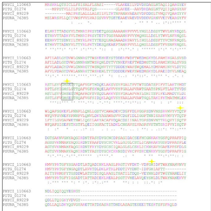
The analysis of these sequences has allowed us to identify several ORF's that encode proteins with relevant role in the virulence of P. cinnamomi. We have also identified two ORF's that codify proteins with esterase/lipase's domains with similarity to lipases described in other Phytophthora species involved in pathogenicity mechanisms. These sequences are in the database “fungidb”, with the references PHYCI_76143 and PHYCI_89229 and encode proteins with 309 and 425 amino acids respectively. The primary sequence of these lipases and their alignment by the Clustal algorithm with other lipases of the genus Phytophthorais shown inFig. 2. In addition to the similarity between Phytophthora lipases, we can also observe in the figure the common serine residue involved in the catalytic triad with a conserved consensus sequence (Ser-Gly-X-Gly-X), where X represents any of 20 amino acids. The structural prediction of these P. cinnamomi lipases was per-formed on the Phyre2 server [88], based on the homology of these li-pases with the crystallized dog's (Canis lupusfamiliaris) gastric lipase, with access code 1K8Q in Protein Data Bank [89] (Fig. 3).
The extracellular lipases of some pathogens as Staphylococcus aureus, Staphylococcus epidermidis, Propionibacterium acnes and Pseudomonas aeruginosa, as well as pathogenic fungi, such asMalassezia furfurandHortaea werneckii, have been proposed as potential virulence factors [51].
5. Biotechnological applications of lipases
Lipases are produced by plants, animals and microbes but only microbial lipases are found to be industrially important with an en-ormous biotechnological potential, due to the diverse enzymatic prop-erties, substrate specificity, chemoselectivity, regioselectivity and ste-reoselectivity [9,90]. Even though plant lipases are also available, they are not commercially exploited because of the low yield and the com-plexity of the processes involved. Regarding the lipases from animal origin, some disadvantages have been reported, among which: the presence of bitter tasting amino acids, the presence of residual animal hormones or viruses and the undesirable effects in the processing of vegetarian diets [11].
Current discoveries about fungal lipases associated with patho-genicity have given these enzymes great versatility with multiple functions and applications.
Esterases and lipases are capable of catalysing a series of reactions, possessing good stability in organic solvents. For that reason, they can be considered excellent biocatalysts in intermediate stages of conven-tional chemical processes, and in catalysis of chemical reactions in-volving substrates insoluble in aqueous media. Moreover, they are used in the resolution of racemic mixtures and in the selective removal of certain compounds.
Concerning the microbial lipases, these exhibit a wide range of in-dustrial applications, because of the higher stability, high yield of conversion of substrate into product, the great versatility to adapt to environmental conditions and the simplicity in genetic manipulation and growth conditions [11,90]. A high synthesis rate is obtained with simple processes and low investment and, hence these enzymes are mostly extracellular, their extraction, isolation and purification is simple [91].
The biotechnological potential of lipases is related with their ability to catalyse various reactions. Typically, they preserve the structure and are stable in organic solvents, not requiring the presence of cofactors but requiring stable conditions of temperature and pH. In addition, these enzymes exhibit a broad substrate specificity and high enantios-electivity [24,91–94].
Nowadays, lipases are used for the generation of enantiomerically enriched primary and secondary alcohols and, to a lesser extent, to obtaining chiral carboxylic acids and secondary amines [15]. The ste-reoselectivity of lipases is useful in the synthesis of biopolymers such as, for example, polyesters and polyphenols in the kinetic resolution of racemic mixtures in the secondary hydrolysis reactions, the transes-terification and alcohol etherification [16,25].
The panorama of lipases use encompasses many industries: enzymes of microbial origin are used in food industry, in the manufacture of detergents (hydrolysis of fats), cosmetics (removal of lipids) and was-tewater treatment (decomposition and removal of oily substances). As these enzymes have enormous catalytic potential, they are also used as ideal biocatalysts in organic chemistry,fine chemicals (synthetic esters) in the pharmaceutical industry, and in the production of food additives (flavour enhancement) [11,91,93].
5.1. Pharmaceutical industry
The synthesis of bioactive substances has been practiced over the years by means of conventional organic chemistry. This synthesis route, in some cases, can result in problems such as instability of the molecule on the reaction conditions and the formation of racemic mixture [26]. The ability of lipases to resolve racemic mixtures by the synthesis of a single enantiomer is currently exploited for drug production by the pharmaceutical industry.
Microbial lipases are used to concentrate polyunsaturated fatty acids from animal and botanical lipids, and their mono and diacylgly-cerides are used to produce a variety of pharmaceuticals. The use of lipases in pharmaceutical industry is due, in particular, to the antioselectivity exhibited by many of these enzymes. The en-antioselective catalysis allows the obtention of optically pure products, since these enzymes are chiral molecules capable of recognizing and acting preferably in the isomers of a racemic mixture. This feature is extremely advantageous because, in many cases, the isomers (R and S) have a biological activity, while others are less active or even toxic [20].
One important compound used in the synthesis of non-steroids anti-inflammatory drugs (Ibuprofen and Naxopreno) is 2-phenyl-propanoic acid, which can be obtained by transesterification reactions or hydro-lysis of the corresponding ester catalysed by lipases [19]. Another ex-ample is the use of a Candida antarcticalipase resolution of racemic thiotetronic acid derivatives to obtain the compound (R)-thiolacto-mycin which has a chiral quaternary carbon atom C5 [15,19,95].
5.2. Food industry
These enzymes have been extensively used in the food industry, especially in the dairy industry for the hydrolysis of milk fat, the modification of the fatty acid chain lengths to enhance the cheese characteristics and, more recently, to accelerate cheese ripening and the lipolysis of butter fat and cream. In addition, lipases have been used in selective hydrolysis of fat, allowing its use in the formation offlavoured products, in the production of substitutes for butter and other additives used in cereals, snacks and chewing. The addition of these hydrolysates provides a variety of food sensory characteristics [96]. Lipases are also used to modify the taste of food, the synthesis of esters of fatty acids, and short chain alcohols, these being the basic flavour and aroma compounds [20].
Another example can be given in the baking industry, in the man-ufacture of bread lipase degrade lipids from wheat changing its
interaction with the gluten, obtaining a dough conditioner result in increasing bread volume and improved texture. Hydrolysis performed by specific lipases is applied to obtain monoacylglycerol which are used as emulsifying agents [97].
5.3. Detergent industry
The use of enzymes in detergent formulations is common in devel-oped countries, with over half of all detergents presently available containing enzymes. Lipases are used in the detergent industry to fa-cilitate the breaking of bonds present in the triglycerides and hence solubilise grease adhered to the fabric, therefore enhancing the ability to remove tough stains. These enzymes make the detergent en-vironmentally safe, by reducing the environmental load, as they save energy by enabling a lower wash temperature. Also, the products are mostly biodegradable and leave no harmful residues [5].
Examples of enzymes used in detergents are Lipolase®(Novozymes)
obtained from the fungus Thermomyces lanuginosa and expressed in Aspergillus oryzae; the Lumafast®(Genencor, USA) and Lipomax®
(Gist-Brocades, The Netherlands), bacterial lipases from Pseudomonas men-docina, andPseudomonas alcaligenes[27].
The most important commercial application area for hydrolytic li-pases is industrial or household cleaners [23], which is generally used in combination with one or more enzymes such as proteases, amylases and cellulases, responsible for the removal of various fats [18].
Final remarks
The recent DNA sequencing techniques with platforms that allow massive sequencing, combined with advanced bioinformatics cap-abilities allow us to deduce the role of many molecular factors in tabolic pathways. It is possible that many molecules have roles in me-tabolic pathways that until our days are unknown. The recent associations of lipases in the pathogenicity mechanisms of some mi-croorganisms confirms the high versatility of these enzymes. However, much is unknown about the concrete action of these enzymes in the metabolic pathways involved in pathogenicity mechanisms. We think that the silencing of lipase genes in phytopathogenic fungi and sub-sequent plant infection with these fungi may help to better understand the role of these enzymes in the infection.
The discovery of molecule factors and their interactions in the metabolic pathways of infection is an important step in the develop-ment of prevention strategies, therapy processes and control of fungal infections in plants.
Conflicts of interest
The authors declare that there are no conflicts of interest. Also, are indebted to the careful and constructive criticisms of the reviewers.
Acknowledgments
A. Pascoal would like to thank Foundation for Science and Technology (FCT, Portugal), Programa Operacional Pontencial Humano (POPH) and European Union (EU) for his Postdoctoral grant SFRH/ BPD/91380/2012. The authors also are grateful to the Centre of Molecular and Environmental Biology, funded by FCT, UID/BIA/ 04050/2013 (POCI-01-0145-FEDER-007569) and by the European Regional Development Fund (ERDF) through the COMPETE2020 –
Programa Operacional Competitividade e Internacionalização (POCI).
References
[1] S.C. Gopinath, P. Anbu, T. Lakshmipriya, A. Hilda, Strategies to characterize fungal lipases for applications in medicine and dairy industry, BioMed Res. Int. 2013 (2013) 154549.
[2] A. Guldhe, B. Singh, T. Mutanda, K. Perrnaul, F. Bux, Advances in synthesis of biodiesel via enzyme catalysis: novel and sustainable approaches, Renew. Sustain. Energy Rev. 41 (2015) 1447–1464.
[3] A.K. Singh, M. Mukhopadhyay, Overview of fungal lipase: a review, Appl. Biochem. Biotechnol. 166 (2) (2012) 486–520.
[4] P. Villeneuve, J.M. Muderhwa, J. Graille, M. Haas, Customizing lipases for bioca-talysis: a survey of chemical, physical and molecular biological approaches, J. Mol. Catal. B Enzym. 9 (2000) 113–148.
[5] K.E. Jaeger, M.T. Reetz, Microbial lipases form versatile tools for biotechnology, Trends Biotechnol. 16 (1998) 396–403.
[6] K.E. Jaeger, B.W. Dijkstra, M.T. Reetz, Bacterial biocatalysts: molecular biology, three-dimensional structures, and biotechnological applications of lipases, Annu. Rev. Microbiol. 53 (1999) 315–351.
[7] M.B. Ansorge-Schumacher, O. Thum, Immobilised lipases in the cosmetics industry, Chem. Soc. Rev. 42 (2013) 6475–6490.
[8] A.R. Macrae, R.C. Hammond, Present and future applications of lipase, Biotech Engin Rev 3 (1985) 193–217.
[9] R. Sharma, Y. Chisti, U.C. Banerjee, Production, purification, characterization, and applications of lipases, Biotechnol. Adv. 19 (2001) 627–662.
[10] E.S. Lin, S.C. Sung, Cultivating conditions influence exopolysaccharide production by the edible BasidiomyceteAntrodia cinnamomeain submerged culture, Int. J. Food Microbiol. 108 (2006) 182–187.
[11] P. Ellaiah, T. Parabhakar, B. Ramakrishna, A.T. Taleb, K. Adinarayana, Production of lipase by immobilized cells ofAspergillus niger, Process Biochem. 39 (2004) 525–528.
[12] H. Chahinian, Y.B. Ali, A. Abousalham, S. Petry, L. Mandrich, G. Manco, S. Canaan, L. Sarda, Substrate specificity and kinetic properties of enzymes belonging to the hormone-sensitive lipase family: comparison with non-lipolytic and lipolytic car-boxylesterases, Biochim. Biophys. Acta 1738 (2005) 29–36.
[13] W. Zhu, S.G. Filler, Interactions ofCandida albicanswith epithelial cells, Cell Microbiol. 12 (3) (2010) 273–282.
[14] Q.T. Phan, C.L. Myers, Y. Fu, D.C. Sheppard, M.R. Yeaman, W.H. Welch, A.S. Ibrahim, J.E. Edwards Jr., S.G. Filler, Als3 is aCandida albicansinvasin that binds to cadherins and induces endocytosis by host cells, PLoS Biol. 5 (3) (2007) e64.
[15] D. Isaksson, K. Sjodin, H.E. Hogberg, Enantiomerically enriched cryptone by lipase catalysed kinetic resolution, Tetrahedron: Asymmetry 17 (2006) 275–280. [16] K.E. Jaeger, T. Eggert, Lipases for biotechnology, Curr. Opin. Biotechnol. 13 (2002)
390–397.
[17] A. Kumar, S.S. Kanwar, Catalytic potential of a nitrocellulose membrane-im-mobilized lipase in aqueous and organic media, J. Appl. Polym. Sci. 1 (2012) E37–E44.
[18] F. Hasan, A.A. Shah, A. Hameed, Industrial application of microbial lipase, Enzym. Microb. Technol. 39 (2006) 235–251.
[19] A. Pandey, S. Benjamin, C.R. Soccol, P. Nigam, N. Krieger, V.T. Soccol, The realm of microbial lipases in biotechnology, Biotechnol. Appl. Biochem. 29 (Pt 2) (1999) 119–131.
function, mechanism, and assay, Methods Enzymol. 400 (2005) 569–588. [21] I. Eş, J.D.G. Vieira, A.C. Amaral, Principles, techniques, and applications of
bio-catalyst immobilization for industrial application, Appl. Microbiol. Biotechnol. 99 (2015) 2065–2082.
[22] M. Virto, C. F, M.A. Bustamante, L.J. Barron, M. Aramburu, M.S. Vicente, F.J. Pérez-Elortondo, M. Albisiu, M. De Renobales, Lamb rennet paste in ovine cheese man-ufacture, Int. Dairy J. 13 (2003) 391–399.
[23] K.T. Lee, T.A. Foglia, K.S. Chang, Production of alkyl ester as biodiesel from frac-tionated lard and restaurant grease, J. Am. Oil Chem. Soc. 79 (2002) 191–195. [24] E. Rigo, J.L. Ninow, M. Di Luccio, V. Oliveira, E. Polloni, D. Remonatto, F. Arbter,
R. Vardanega, D. Oliveira, H. Treichel, Lipase production by solid fermentation of soybean meal with different supplements, LWT - Food Sci. Technol. (Lebensmittel-Wissenschaft -Technol.) 43 (2010) 1132–1137.
[25] [2T. Schulz, J. Pleiss, R.D. Schmid, Stereoselectivity ofPseudomonas cepacialipase toward secondary alcohols: a quantitative model, Protein Sci. 9 (2000) 1053–1062. [26] A.M. Brzozowski, H. Savage, C.S. Verma, J.P. Turkenburg, D.M. Lawson,
A. Svendsen, S. Patkar, Structural origins of the interfacial activation in
Thermomyces (Humicola) lanuginosalipase, Biochemistry 39 (2000) 15071–15082. [27] H. Horchani, H. Mosbah, N.B. Salem, Y. Gargouri, A. Sayari, Biochemical and
molecular characterisation of a thermoactive, alkaline and detergent-stable lipase from a newly isolatedStaphylococcus aureusstrain, J. Mol. Catal. B Enzym. 56 (2009) 237–245.
[28] S. Abraham, N.R. Kamini, M.K. Gowthaman, Process strategies for alkaline lipase production usingAspergillus NigerMTCC, 2594, Int. J. Appl. Pharm. 1 (2011) 126–133.
[29] U.T. Bornscheuer, Microbial carboxyl esterases: classification, properties and ap-plication in biocatalysis, FEMS Microbiol. Rev. 26 (1) (2002) 73–81.
[30] A. Hiol, M.D. Jonzo, D. Druet, L. Comeau, Production, purification and character-ization of an extracellular lipase fromMucor hiemalisf.hiemalis, Enzym. Microb. Technol. 25 (1–2) (1999) 80–87.
[31] A. Aloulou, J.A. Rodriguez, S. Fernandez, D. van Oosterhout, D. Puccinelli, F. Carriere, Exploring the specific features of interfacial enzymology based on lipase studies, Biochim. Biophys. Acta 1761 (2006) 995–1013.
[32] A. Svendsen, Lipase protein engineering, Biochim. Biophys. Acta 1543 (2000) 223–238.
[33] L. Sarda, P. Desnuelle, Actions of pancreatic lipase on esters in emulsions, Biochim. Biophys. Acta 30 (1958) 513–521.
[34] K. Holwerda, P.E. Verkade, A.H.A. de Willigen, Vergleichende Untersuchungen über die Verseifungsgeschwindigkeit einiger Einsäuriger Triglyceride unter Einfluss von Pankreasextrakt. I. Der Einfluss des Verteilungszustandes der Triglyceride auf die Verseifungsgeschwindigkeit, Recl Trav Chim Pay B 55 (1936) 43–57. [35] F. Schonheyder, K. Volqvartz, On affinity of pig pancreas lipase for tricaproin in
heterogeneous solution, Acta Physiol. Scand. 9 (1945) 57–67.
[36] P. Desnuelle, L. Sarda, G. Ailhaud, Inhibition of pancreatic lipase by diethyl-p-ni-trophenyl phosphate in emulation, Biochim. Biophys. Acta 37 (1960) 570–571. [37] J.M. Muderhwa, H.L. Brockman, Lateral lipid distribution is a major regulator of
lipase activity. Implications for lipid-mediated signal transduction, J. Biol. Chem. 267 (1992) 24184–24192.
[38] J.M. Smaby, J.M. Muderhwa, H.L. Brockman, Is lateral phase separation required for fatty acid to stimulate lipases in a phosphatidylcholine interface? Biochemistry 33 (1994) 1915–1922.
[39] J. Pleiss, M. Fischer, R.D. Schmid, Anatomy of lipase binding sites: the scissile fatty acid binding site, Chem. Phys. Lipids 93 (1998) 67–80.
[40] N. Kamiya, H. Kasagi, M. Inoue, K. Kusunoki, M. Goto, Enantioselective recognition mechanism of secondary alcohol by surfactant-coated lipases in nonaqueous media, Biotechnol. Bioeng. 65 (1999) 227–232.
[41] M.T. Reetz, Lipases as practical biocatalysts, Curr. Opin. Chem. Biol. 6 (2002) 145–150.
[42] L. Brady, A.M. Brzozowski, Z.S. Derewenda, E. Dodson, G. Dodson, S. Tolley, J.P. Turkenburg, L. Christiansen, B. Huge-Jensen, L. Norskov, et al., A serine pro-tease triad forms the catalytic centre of a triacylglycerol lipase, Nature 343 (1990) 767–770.
[43] N. Hulo, A. Bairoch, V. Bulliard, L. Cerutti, B.A. Cuche, E. de Castro, C. Lachaize, P.S. Langendijk-Genevaux, C.J. Sigrist, The 20 years of PROSITE, Nucleic Acids Res. 36 (2008) D245–D249.
[44] C.J. Sigrist, L. Cerutti, N. Hulo, A. Gattiker, L. Falquet, M. Pagni, A. Bairoch, P. Bucher, PROSITE: a documented database using patterns and profiles as motif descriptors, Briefings Bioinf. 3 (2002) 265–274.
[45] R.D. Finn, J. Tate, J. Mistry, P.C. Coggill, S.J. Sammut, H.R. Hotz, G. Ceric, K. Forslund, S.R. Eddy, E.L. Sonnhammer, A. Bateman, The Pfam protein families database, Nucleic Acids Res. 36 (2008) D281–D288.
[46] P. Fojan, P.H. Jonson, M.T. Petersen, S.B. Petersen, What distinguishes an esterase from a lipase: a novel structural approach, Biochimie 82 (2000) 1033–1041. [47] C. Schmidt-Dannert, Recombinant microbial lipases for biotechnological
applica-tions, Bioorg. Med. Chem. 7 (1999) 2123–2130.
[48] M. Nardini, B.W. Dijkstra, Alpha/beta hydrolase fold enzymes: the family keeps growing, Curr. Opin. Struct. Biol. 9 (1999) 732–737.
[49] P. van Baarlen, A. van Belkum, R.C. Summerbell, P.W. Crous, B.P.H.J. Thomma, Molecular mechanisms of pathogenicity: how do pathogenic microorganisms de-velop cross-kingdom host jumps? FEMS Microbiol. Rev. 31 (3) (2007) 239–277. [50] A. Casadevall, L.A. Pirofski, Host-pathogen interactions: redefining the basic
con-cepts of virulence and pathogenicity, Infect. Immun. 67 (8) (1999) 3703–3713. [51] A. Gacser, F. Stehr, C. Kroger, L. Kredics, W. Schafer, J.D. Nosanchuk, Lipase 8
affects the pathogenesis of Candida albicans, Infect. Immun. 75 (10) (2007) 4710–4718.
[52] J. Feng, F. Wang, G. Liu, D. Greenshields, W. Shen, S. Kaminskyj, G.R. Hughes,
Y. Peng, G. Selvaraj, J. Zou, Y. Wei, Analysis of a Blumeria graminis-secreted lipase reveals the importance of host epicuticular wax components for fungal adhesion and development, Mol. Plant Microbe Interact. 22 (12) (2009) 1601–1610. [53] S. Silva, M. Negri, M. Henriques, R. Oliveira, D.W. Williams, J. Azeredo, Adherence
and biofilm formation of non-Candida albicansCandida species, Trends Microbiol. 19 (5) (2011) 241–247.
[54] A.S. Ibrahim, F. Mirbod, S.G. Filler, Y. Banno, G.T. Cole, Y. Kitajima, J.E. Edwards Jr., Y. Nozawa, M.A. Ghannoum, Evidence implicating phospholipase as a virulence factor ofCandida albicans, Infect. Immun. 63 (5) (1995) 1993–1998.
[55] J.M. Manns, D.M. Mosser, H.R. Buckley, Production of a hemolytic factor by Candida-albicans, Infect. Immun. 62 (11) (1994) 5154–5156.
[56] M.A. Pfaller, R.N. Jones, S.A. Messer, M.B. Edmond, R.P. Wenzel, National sur-veillance of nosocomial blood stream infection due to species of Candida other than Candida albicans: frequency of occurrence and antifungal susceptibility in the SCOPE Program. SCOPE Participant Group. Surveillance and Control of Pathogens of Epidemiologic, Diagn. Microbiol. Infect. Dis. 30 (2) (1998) 121–129. [57] M.M. Harriott, M.C. Noverr, Importance of Candida-bacterial polymicrobial
bio-films in disease, Trends Microbiol. 19 (11) (2011) 557–563.
[58] M. Park, E. Do, W.H. Jung, Lipolytic enzymes involved in the virulence of human pathogenic fungi, MYCOBIOLOGY 41 (2) (2013) 67–72.
[59] S. Nicholls, D.M. MacCallum, F.A. Kaffarnik, L. Selway, S.C. Peck, A.J. Brown, Activation of the heat shock transcription factor Hsf1 is essential for the full viru-lence of the fungal pathogenCandida albicans, Fungal Genet. Biol. 48 (3) (2011) 297–305.
[60] J.E. Cutler, Putative virulence factors ofCandida albicans, Annu. Rev. Microbiol. 45 (1991) 187–218.
[61] M. Monod, Z.M. Borg-von, Secreted aspartic proteases as virulence factors of Candida species, Biol. Chem. 383 (7–8) (2002) 1087–1093.
[62] M. Schaller, H.C. Korting, W. Schafer, J. Bastert, W.C. Chen, B. Hube, Secreted aspartic proteinase (Sap) activity contributes to tissue damage in a model of human oral candidosis, Mol. Microbiol. 34 (1) (1999) 169–180.
[63] B.N. Taylor, P. Staib, A. Binder, A. Biesemeier, M. Sehnal, M. Rollinghoff, J. Morschhauser, M. Schroppel, Profile of Candida albicans-secreted aspartic pro-teinase elicited during vaginal infection, Infect. Immun. 73 (3) (2005) 1828–1835. [64] R. Gupta, A. Kumari, P. Syal, Y. Singh, Molecular and functional diversity of yeast and fungal lipases: their role in biotechnology and cellular physiology, Prog. Lipid Res. 57 (2015) 40–54.
[65] F. Dalle, B. Wachtler, C. L'Ollivier, G. Holland, N. Bannert, D. Wilson, C. Labruere, A. Bonnin, B. Hube, Cellular interactions ofCandida albicanswith human oral epithelial cells and enterocytes, Cell Microbiol. 12 (2) (2010) 248–271. [66] J.R. Naglik, D.L. Moyes, B. Wachtler, B. Hube,Candida albicansinteractions with
epithelial cells and mucosal immunity, Microb. Infect. 13 (12–13) (2011) 963–976. [67] F. Stehr, A. Felk, A. Gacser, M. Kretschmar, B. Mahnss, K. Neuber, B. Hube,
W. Schafer, Expression analysis of theCandida albicanslipase gene family during experimental infections and in patient samples, FEMS Yeast Res. 4 (4–5) (2004) 401–408.
[68] T.A. Richards, D.M. Soanes, M.D. Jones, O. Vasieva, G. Leonard, K. Paszkiewicz, P.G. Foster, N. Hall, N.J. Talbot, Horizontal gene transfer facilitated the evolution of plant parasitic mechanisms in the oomycetes, Proc. Natl. Acad. Sci. U. S. A. 108 (37) (2011) 15258–15263.
[69] J.M. Mancheno, M.A. Pernas, M.J. Martinez, B. Ochoa, M.L. Rua, J.A. Hermoso, Structural insights into the lipase/esterase behavior in the Candida rugosa lipases family: crystal structure of the lipase 2 isoenzyme at 1.97 angstrom resolution, J. Mol. Biol. 332 (5) (2003) 1059–1069.
[70] H. Zorn, H. Bouws, M. Takenberg, M. Nimtz, R. Getzlaff, D.E. Breithaupt, R.G. Berger, An extracellular carboxylesterase from the basidiomycete Pleurotus sapidus hydrolyses xanthophyll esters, J. Biol. Chem. 386 (2005) 435–440. [71] P. Langcake, Uptake of sterols byphytophthora-infestans, their
intracellular-dis-tribution and metabolism, Trans. Br. Mycol. Soc. 64 (Feb) (1975) 55–65. [72] W.D. Nes, A.E. Stafford, Evidence for metabolic and functional discrimination of
sterols byPhytophthoracactorum, Proc. Natl. Acad. Sci. U. S. A. 80 (11) (1983) 3227–3231.
[73] C.A. Voigt, W. Schafer, S. Salomon, A secreted lipase of Fusarium graminearum is a virulence factor required for infection of cereals, Plant J. 42 (3) (2005) 364–375. [74] G. Devescovi, J. Bigirimana, G. Degrassi, L. Cabrio, J.J. LiPuma, J. Kim, I. Hwang,
V. Venturi, Involvement of a quorum-sensing-regulated lipase secreted by a clinical isolate of Burkholderia glumae in severe disease symptoms in rice, Appl. Environ. Microbiol. 73 (15) (2007) 4950–4958.
[75] A.B. Choupina, L. Estevinho, I.M. Martins, Scientifically advanced solutions for chestnut ink disease, Appl. Microbiol. Biotechnol. 98 (9) (2014) 3905–3909. [76] S. Kamoun, Molecular genetics of pathogenic oomycetes, Eukaryot. Cell 2 (2)
(2003) 191–199.
[77] J.E. Rookes, M.L. Wright, D.M. Cahill, Elucidation of defence responses and sig-nalling pathways induced in Arabidopsis thaliana following challenge with
Phytophthoracinnamomi, Physiol. Mol. Plant Pathol. 72 (4–6) (2008) 151–161. [78] S. Kamoun, O. Furzer, J.D. Jones, H.S. Judelson, G.S. Ali, R.J. Dalio, S.G. Roy,
L. Schena, A. Zambounis, F. Panabieres, D. Cahill, M. Ruocco, A. Figueiredo, X.R. Chen, J. Hulvey, R. Stam, K. Lamour, M. Gijzen, B.M. Tyler, N.J. Grunwald, M.S. Mukhtar, D.F. Tome, M. Tor, G. Van Den Ackerveken, J. McDowell, F. Daayf, W.E. Fry, H. Lindqvist-Kreuze, H.J. Meijer, B. Petre, J. Ristaino, K. Yoshida, P.R. Birch, F. Govers, The Top 10 oomycete pathogens in molecular plant pa-thology, Mol. Plant Pathol. 16 (4) (2015) 413–434.
[80] S. Meirinho, M. Carvalho, A. Domínguez, A. Choupina, Isolation and character-ization by asymmetric PCR of the ENDO1 gene for glucan endo-1,3-B-D-glucosidase inPhytophthora cinnamomiassociated with the ink disease of Castanea sativa Mill, Braz Arch Biol 53 (3) (2010) 513–518.
[81] Y.J. Yu, S.C. Wu, H.H. Chan, Y.C. Chen, Z.Y. Chen, M.T. Yang, Overproduction of soluble recombinant transglutaminase fromStreptomyces netropsisinEscherichia coli, Appl. Microbiol. Biotechnol. 81 (3) (2008) 523–532.
[82] P.N. Dodds, M. Rafiqi, P.H. Gan, A.R. Hardham, D.A. Jones, J.G. Ellis, Effectors of biotrophic fungi and oomycetes: pathogenicity factors and triggers of host re-sistance, New Phytol. 183 (4) (2009) 993–1000.
[83] G.J. Lawrence, P.N. Dodds, J.G. Ellis, Rust offlax and linseed caused by Melampsora lini, Mol. Plant Pathol. 8 (4) (2007) 349–364.
[84] A.M. Catanzariti, P.N. Dodds, G.J. Lawrence, M.A. Ayliffe, J.G. Ellis, Haustorially expressed secreted proteins fromflax rust are highly enriched for avirulence elici-tors, Plant Cell 18 (1) (2006) 243–256.
[85] D.J. Studholme, R.L. McDougal, C. Sambles, E. Hansen, G. Hardy, M. Grant, R.J. Ganley, N.M. Williams, Genome sequences of sixPhytophthoraspecies asso-ciated with forests in New Zealand, Genom Data 7 (2016) 54–56.
[86] A. Reitmann, D.K. Berger, N. van den Berg, Putative pathogenicity genes of
Phytophthora cinnamomiidentified via RNA-Seq analysis of pre-infection structures, Eur. J. Plant Pathol. 147 (1) (2017) 211–228.
[87] M.A. Larkin, G. Blackshields, N.P. Brown, R. Chenna, P.A. McGettigan, H. McWilliam, et al., Clustal W and clustal X version 2.0, Bioinformatics 23 (2007) 2947–2948.
[88] L.A. Kelley, S. Mezulis, C.M. Yates, M.N. Wass, M.J. Sternberg, The Phyre2 web portal for protein modeling, prediction and analysis, Nat. Protoc. 10 (6) (2015)
845–858.
[89] A. Roussel, N. Miled, L. Berti-Dupuis, M. Riviere, S. Spinelli, P. Berna, V. Gruber, R. Verger, C. Cambillau, Crystal structure of the open form of dog gastric lipase in complex with a phosphonate inhibitor, J. Biol. Chem. 277 (3) (2002) 2266–2274. [90] N. Cihangir, E. Sarikaya, Investigation of lipase production by a new isolate of
Aspergillussp, World J. Microbiol. Biotechnol. 20 (2004) 193–197.
[91] J.F. Burkert, F. Maugeri, M.I. Rodrigues, Optimization of extracellular lipase pro-duction byGeotrichumsp. using factorial design, Bioresour. Technol. 91 (2004) 77–84.
[92] P.O. Carvalho, P.R.B. Campos, M.D. Noffs, J.G. Oliveira, M.T. Schimizu, D.M. Silva, Application of microbial lipases to concentrate polyunsaturated fatty acids, Quim. Nova 26 (2003) 75–80.
[93] M. Elibol, D. Ozer, Response surface analysis of lipase production by freely sus-pendedRhizopus arrhizusProcess, Biochemistry 38 (2002) 367–372.
[94] H.F. Castro, A.A. Mendes, J.C. Santos, C.L. Aguiar, Modification of oils and fats by biotransformation, Quim. Nova 27 (2004) 146–156.
[95] K. Toyama, T. Tauchi, N. Mase, H. Yoda, K. Takabe, Lipase-catalyzed kinetic re-solution of thiotetronic acid derivatives bearing a chiral quaternary carbon: total synthesis of (R)-thiolactomycin and its O-analogue, Tetrahedron Lett. 47 (2006) 7163–7166.
[96] M. Virto, C. F, M.A. Bustamante, L.J. Barron, M. Aramburu, M.S. Vicente, F.J. Pérez-Elortondo, M. Albisiu, M. De Renobales, Lamb rennet paste in ovine cheese man-ufacture, Int. Dairy J. 13 (2003) 391–399.