http://dx.doi.org/10.1590/0037-8682-0210-2015
Major Article
INTRODUCTION
Corresponding author: Dra. Juliana Figueirêdo da Costa Lima. Lab. de Imunoepidemiologia/Depto. de Imunologia/CPqAM/FIOCRUZ. Av. Professor Moraes Rêgo s/n, 4º andar, Cidade Universitária, 50670-420 Recife, Pernambuco, Brasil.
Phone: 55 81 2101-2569; Mobile: 55 81 98822-7743
e-mail:jfcl@cpqam.fi ocruz.br / jujufi [email protected]
Received 17 August 2015
Accepted 10 November 2015
Single-tube nested PCR assay with in-house DNA
extraction for Mycobacterium tuberculosis
detection in blood and urine
Juliana Figueirêdo da Costa Lima
[1],[2], Gabriela de Moraes Rêgo Guedes
[1],[3],
Juliana Falcão de Araújo Lima
[1], Laís Ariane de Siqueira Lira
[1],
Fabiana Cristina Fulco Santos
[1], Mercia Eliane de Arruda
[1],
Lílian Maria Lapa Montenegro
[1]and Haiana Charifker Schindler
[1][1]. Laboratório de Imunoepidemiologia, Centro de Pesquisas Aggeu Magalhães, Fundação Oswaldo Cruz, Recife, Pernambuco, Brasil. [2]. Programa de Pós-Graduação Stricto Sensu em Clínica Médica, Universidade Federal do Rio de Janeiro, Rio de Janeiro, Rio de Janeiro, Brasil. [3]. Laboratório Central, Universidade Federal de Pernambuco, Recife, Pernambuco, Brasil.
ABSTRACT
Introduction: Molecular analyses are auxiliary tools for detecting Koch’s bacilli in clinical specimens from patients with
suspected tuberculosis (TB). However, there are still no effi cient diagnostic tests that combine high sensitivity and specifi city and yield rapid results in the detection of TB. This study evaluated single-tube nested polymerase chain reaction (STNPCR) as a molecular diagnostic test with low risk of cross contamination for detecting Mycobacterium tuberculosis in clinical samples.
Methods:Mycobacterium tuberculosis deoxyribonucleic acid (DNA) was detected in blood and urine samples by STNPCR
followed by agarose gel electrophoresis. In this system, reaction tubes were not opened between the two stages of PCR (simple and nested). Results: STNPCR demonstrated good accuracy in clinical samples with no cross contamination between microtubes.
Sensitivity in blood and urine, analyzed in parallel, was 35%–62% for pulmonary and 41%–72% for extrapulmonary TB. The specifi city of STNPCR was 100% in most analyses, depending on the type of clinical sample (blood or urine) and clinical form of disease (pulmonary or extrapulmonary). Conclusions: STNPCR was effective in detecting TB, especially the extrapulmonary
form for which sensitivity was higher, and had the advantage of less invasive sample collection from patients for whom a spontaneous sputum sample was unavailable. With low risk of cross contamination, the STNPCR can be used as an adjunct to conventional methods for diagnosing TB.
Keywords:Mycobacterium tuberculosis. Molecular diagnostic test. Nested polymerase chain reaction. Blood. Urine.
Tuberculosis (TB) is a major cause of morbidity and mortality affecting individuals of various ages and social classes (1). In
2013, 83,310 cases were recorded in Brazil, including 34.7 new cases per 100,000 habitants in the Northeastern region(2) (3).
Conventional bacteriological tests for detecting TB do not combine high sensitivity and specifi city ( 4). For example,
acid-fast bacillus (AFB) smear microscopy is rapid and low cost, but has low sensitivity and it is not specifi c for Mycobacterium tuberculosis (5).
Molecular approaches have been used as alternative tools for diagnosing infectious diseases(5) (6) ( 7) (8 ) (9). One of these is
polymerase chain reaction (PCR), which has been applied to the detection of M. tuberculosis (4) (5) (7) (8) ( 10) (11) ( 12) (13) at
concentrations of < 10 cells/mL in clinical samples(7) ( 14) (15).
Nested PCR is a variation of this technique that involves two amplifi cation steps (7) (8) (13 ) (16) (17), yielding greater sensitivity and
specifi city(8) (16) (17). Single-tube nested polymerase chain reaction
(STNPCR) is more rapid than conventional nested PCR(8) (14)
with less probability of cross-contamination and requiring a smaller amount of reagent. In STNPCR, both amplifi cation reactions occur consecutively and the microtubes do not need to be opened or changed for the addition of new reagents(4) (8).
There are still no effi cient diagnostic tests that combine high sensitivity and specifi city and yield rapid results in the detection of TB(18); this is especially challenging when few bacilli are
present, for example in cases of extrapulmonary TB, pulmonary TB with a negative AFB test, childhood TB, and co-infection with TB-human immunodefi ciency virus (HIV)(7) (10) (12).
METHODS
especially the paucibacillary and extrapulmonary forms(10) –
remains a challenge. STNPCR can achieve a more rapid diagnosis of TB than culture methods and has higher sensitivity than the AFB test using urine and blood(7) (13) (19 ) (20). However, AFB, culture
methods, and the GeneXpert Mycobacterium tuberculosis (MTB)/ rifampicin (RIF) test are still considered gold standards for diagnosing pulmonary TB in many regions(20) (21) (22) (23 ).
Various studies have reported the molecular diagnosis of TB using extrapulmonary samples, including in cases of negative AFB results(12) (13) (19). The present study evaluated
the performance of STNPCR in detecting the presence of M. tuberculosis complex in blood and urine using an in-house DNA extraction method from patients with suspected TB. The results demonstrate that the STNPCR method can improve clinical practice by providing early diagnosis of TB so as to avoid unnecessary invasive procedures and inappropriate treatment.
Patients were non-randomly selected from public hospitals located in Recife between July 2006 and August 2009. Patients sought health services spontaneously with diverse simptons and suspected of various diseases, for example, tuberculosis or pneumonia. Sample size was determined by convenience according to the number of patients with suspected TB who sought medical attention at participating hospitals. Control patients showing pulmonary symptoms but without TB were recruited from the same hospitals.
Blood [3-5mL collected in a vacuum tube with anti-coagulant ethylenediaminetetraacetic acid (EDTA)] and urine (10ml/day for 3 consecutive days) samples were obtained from each patient suspected of TB before specifi c treatment was initiated. The samples were sent to Aggeu Magalhães Research Center, Oswaldo Cruz Foundation [Centro de Pesquisas Aggeu Magalhães/Fundação Oswaldo Cruz (CPqAM/FIOCRUZ)] within 4h of collection and stored at 4°C-8°C for up to 24h. Urine samples were decontaminated using Petroff’s method(24) (25).
Blood cells were separated from whole blood at room temperature by density gradient centrifugation using Ficoll-Paque Plus (GE Healthcare, Uppsala, Sweden).
Ethical considerations
The study protocol was approved by the Research Ethics Committee of CPqAM/FIOCRUZ (no. 0133.0.095.000-08) with the consent of participating hospitals. All participants provided written, informed consent for collection of biological samples; in cases of minors or those unable to sign or decide for themselves, this was provided by a guardian or legal representative.
Study population
Patients of both genders between the ages of 15 and 69 years and suspected of TB infection after clinical examination were recruited from public hospitals in Recife, the capital of the State
the physician who attended to them at the hospital, without data from molecular tests. Clinical diagnoses were made and STNPCR performed in a double-blinded fashion. Active TB was defi ned as a positive AFB test or TB culture according to the criteria of the Ministry of Health of Brazil(22) (26) and
American Thoracic Society modifi ed criteria(23) (27). In cases
where conventional tests were negative, inconclusive, or were not conducted, individuals were considered TB-positive if they had a clinical or radiological profi le consistent with active TB and if they showed clinical improvement after treatment for 1 month with antibiotics. Latent TB was defi ned as a positive skin test for tuberculin and negative bacteriological exams (when it was available on patient) and with no clinical, radiological, or bacteriological evidence of active TB(23). The
negative control group consisted of individuals who used health services for reasons other than TB, who had a scar corresponding to the Bacillus Calmette-Guérin (BCG) TB vaccination, negative tuberculin test, had not had contact with a bacillipherous adult, and showed no clinical, epidemiological, or laboratory evidence compatible with TB. Patients who were human immunodefi ciency virus/acquired immunodefi ciency syndrome (HIV/AIDS)-positive, had chronic diseases such as kidney or heart disease, cystic fi brosis, or other chronic lung diseases, or who were using immunosuppressive drugs for more than 15 days were excluded from the study.
DNA extraction
Deoxyribonucleic acid (DNA) was extracted using a modifi ed version of a previously published in-house p rotocol(28).
After DNA was bound to the Sephaglas BandPrep kit resin (Amersham Pharmacia Biotech, Little Chalfont, UK), 200μl of sodium iodide (0.9g/mL) was added to each microtube to increase bond strength. A tube without DNA served as a negative control.
Single-tube nested polymerase chain reaction
The assay was based on a previously described STNPCR method(9) and was optimized for human blood and urine. The
IS6110 insert sequence (GenBank accession no. X52471) was the target for amplifi cation. The outer primers TJ5 (5'-CCG CAA AGT GTG GCT AAC-3') and TJ3 (5'-ATC CCC TAT CCG TAT GGT G-3') amplifi ed a 409-bp fragment, and the inner primers OLI5 (5'-AAC GGC TGA TGA CCA AAC-3') and STAN3 (5'-GTC GAG TAC GCC TTC TTG TT-3') amplifi ed a 316-bp fragment( 29). The primers have been previously tested in blood
and urine samples using a conventional nested PCR system(13).
Amplifi cation was carried out on an automatic Eppendorf Gradient thermal cycler (Hamburg, Germany). The first stage of PCR consisted of 15 cycles (94°C for 1min, 57°C for 1 min, and 72°C for 1 min) and the second of 45 cycles (94°C for 1 min, 60°C for 1 min, and 72°C for 1 min). For the fi rst 15 cycles, each tube contained 0.5pmoles of outer primers in a fi nal volume of 50μl containing 200mM Tris-HCl (pH 8.4),
RESULTS
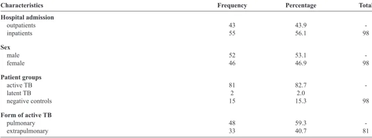
TABLE 1 -Clinical and epidemiological characteristics of study participants.
Characteristics Frequency Percentage Total
Hospital admission
outpatients 43 43.9
-inpatients 55 56.1 98
Sex
male 52 53.1
-female 46 46.9 98
Patient groups
active TB 81 82.7
-latent TB 2 2.0
negative controls 15 15.3 98
Form of active TB
pulmonary 48 59.3
-extrapulmonary 33 40.7 81
TB: tuberculosis.
of 50 pmoles of each primer. This mixture was placed on the inner surface of the microtube cap followed by incubation at 37°C for 30 min, and then eluted from the cap surface into the reaction mixture by briefl y interrupting the PCR after the 15th
cycle and repeatedly inverting the tubes(9) (30). A 10-μl volume
of Tris-EDTA served as a negative control and a 2-μl volume of M. tuberculosis strain H37Rv DNA with 8μl Milli-Q-purifi ed water served as a positive control for each set of reactions. PCR was repeated for all samples yielding negative results as confi rmation.
Electrophoresis
Amplicons generated by STNPCR were resolved by 2% agarose gel electrophoresis. The gel was stained with ethidium bromide (20μl per liter of 1× Tris-EDTA buffer) and visualized under ultraviolet light. The Low Mass Ladder (Invitrogen) was used as a marker.
Statistical analysis
A database was created using Statistical Package for the Social Sciences (SPSS) v.10.0 software (SPSS Inc., Chicago, IL, USA) to store clinical, epidemiological, and laboratory data for all patients. PCR sensitivity and specifi city and positive and negative predictive values were analyzed in parallel in leukocytes, plasma and urine( 31) for each clinical sample along
with their 95% confi dence intervals. Note that denominated blood sample means leukocytes and plasma analyzed in parallel. The χ2 test was used to evaluate the results, along with Fisher’s
Exact test when necessary. OpenEpi v.2.3.1 (http://www. openepi.com/oe2.3/DiagnosticTest/DiagnosticTest.htm) and
We collected 167 blood and urine samples from 98 individuals (mostly male) with a mean age of 33.4 ± 18.3. A total of 83 patients were diagnosed with active or latent TB (Table 1) that were of two clinical forms, pulmonary and
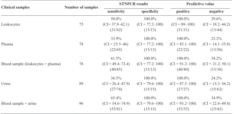
extrapulmonary. Clinical forms of extrapulmonary TB are described on Table 2. The performance and accuracy of STPCR were analyzed using blood and urine samples according to the clinical form of TB (Table 3). When previously tested using M. tuberculosis strain H37Rv genomic DNA, the STNPCR system showed a detection limit of 1ag (0.001fg) (results not shown)(14).
The sensitivity of STNPCR for detecting DNA in leukocytes, plasma and urine samples from patients with or without TB was 50%, 34% and 36.5%, respectively, while the specifi city was 100% relative to negative samples (Table 4). The
p value for STNPCR on leukocytes was <0.001 and on plasma p = 0.007. The sensitivity and specifi city in blood samples (leukocytes and plasma analyzed as one sample) were also determined(31), where a result was positive when
at least one sample was positive by PCR. The sensitivity and specifi city in blood were 61.5% and 100%, respectively
(Table 4), as compared to 65.4% and 100%, respectively, for
TABLE 2 -Frequency of clinical forms of extrapulmonary TB.
Clinical form of TB Frequency Percentage
Pulmonary 47 59.5
Pleural 17 21.5
Peripheral lymph node 3 3.8
Meningoencephalitis 1 1.3
Bone 2 2.5
Cutaneous 1 1.3
Renal 1 1.3
Other* 7 8.8
Total 79 100.0
TB: tuberculosis; *Data were not actualized in our database (missed values) for various reasons.
TABLE 3 -Determination of clinical forms of TB (pulmonary or extrapulmonary) by STNPCR analysis of blood and urine samples from individuals with or without TB.
Pulmonary TB (95% CI)
Clinical samples Number of samples
sensitivity positive predictive value specifi city negative predictive value
58.1% 100.0% 100.0% 41.9%
Blood samples* 56 (43.3–71.6) (77.2–100.0) (86.7–100.0) (26.4–59.2)
25/43 13/13 25/25 13/31
35.0% 100.0% 100.0% 36.6%
Urine 55 (22.1–50.5) (79.6–100) (78.5–100) (23.6–51.9) 14/40 15/15 14/14 15/41
61.7% 100.0% 100.0% 45.5%
Blood and urine* 62 (47.4–74.2) (79.6–100.0) (88.3–100.0) (29.8–62.0)
29/47 15/15 29/29 15/33
Extrapulmonary TB (95% CI) Clinical samples Number of samples
sensitivity positive predictive value specifi city negative predictive value
Blood samples* 65.0% 100.0% 100.0% 65.0%
33 (43.3–81.9) (77.2–100.0) (77.2–100.0) (43.3–81.9)
13/20 13/13 13/13 13/20
40.6% 100.0% 100.0% 44.1%
Urine 47 (25.5–57.7) (79.6–100) (77.2–100) (28.9–60.6) 13/32 15/15 13/13 15/34
Blood and urine* 71.9% 100.0% 100.0% 62.5%
47 (54.6–84.4) (79.6–100) (85.7–100) (42.7–78.8)
23/32 15/15 23/23 15/24
TB: tuberculosis; STNPCR: single-tube nested polymerase chain reaction; CI: confi dence interval. *The patient was considered positive when at least one
TABLE 4 -Accuracy of STNPCR for clinical samples from TB patients and healthy individuals.
STNPCR results Predictive value
Clinical samples Number of samples
sensitivity specifi city positive negative
50.0% 100.0% 100.0% 29.6%
Leukocytes 75 (CI= 37.9–62.1) (CI = 77.2–100) (CI = 89–100) (CI = 18.2–44.2)
(31/62) (13/13) (31/31) (13/44)
33.9% 100.0% 100.0% 23.2%
Plasma 78 (CI = 23.5–46) (CI = 77.2–100) (CI = 85.1–100) (CI = 14.1–35.8)
(22/65) (13/13) (22/22) (13/56)
61.5% 100.0% 100.0% 34.2%
Blood sample (leukocytes + plasma) 78 (CI = 49.4–72.4) (CI = 77.2–100) (CI = 91.2–100) (CI = 21.2–50.1)
(40/65) (13/13) (40/40) (13/38)
36.5% 100.0% 100.0% 24.2%
Urine 89 (CI = 26.4–47.9) (CI = 79.6–100) (CI = 87.5–100) (CI = 15.3–36.2)
(27/74) (15/15) (27/27) (15/62)
65.4% 100.0% 100.0% 34.9%
Blood sample + urine 96 (CI = 54.6–74.9) (CI = 79.6–100) (CI = 93.2–100) (CI = 22.4–49.8)
(53/81) (15/15) (53/53) (15/43)
STNPCR: single-tube nested polymerase chain reaction; TB: tuberculosis; CI: confi dence interval. Note: Sensitivity and specifi city were
calculated for non-TB (negative control + discarded TB).
DISCUSSION
A posteriori hematogenous lymphatic dissemination of Koch bacilli follows the establishment of Ghon’s complex in infected patients(32), and previous studies have detected circulating
bacilli in blood and urine by PCR(7) (12) (13). In the present study, we
evaluated the effi ciency of detecting the M. tuberculosis complex in clinical samples by STNPCR. This method has the advantage of using samples that are easily collected with minimal invasiveness from patients with suspected pulmonary or extrapulmonary TB. This is especially useful for children. The gold standard of gastric lavage cultures requires hospitalization of the patient and is extremely invasive, and should be reserved for patients with negative AFB and extrapulmonary TB(10) (12) (13) (14) (33).
Although the STNPCR showed high sensitivity for detecting M. tuberculosis in blood and urine samples, is unclear why circulating bacilli were found in these samples in patients that do not have a bacteremia nor genitourinary TB(34); the presence
of M. tuberculosis in the blood and urine of active TB cases is typically very low, since these are paucibacillary samples. It is possible that patients with active infection have mycobacterial DNA in their macrophages and in other immune cells(35).
Recently, two automated tests for diagnosing TB directly from clinical samples have been evaluated(3 6), i.e., GeneXpert
MTB/RIF for pulmonary TB and GenoType MTBDRplus (Hain Lifescience GmbH, Nehren, Germany). Both methods can detect TB in clinical samples as well as resistance to RIF and/or isoniazid(36) (37) (38). Despite these advantages, the tests
are only indicated for use with sputum. Therefore, a subset of TB patients with no sputum or with the extrapulmonary form of the disease cannot be diagnosed by these methods according to World Health Organization recommendations(1).
The nested PCR approach increases accuracy of detection(8) (14).
In STNPCR, reactions take place in one tube for each sample, and the tubes do not need to be opened between the fi rst and nested cycles. This decreases the risk of cross contamination between the two steps(8) and thereby increases sensitivity and
specifi city relative to the conventional nested PCR(13) method.
STNPCR can amplify a smaller quantity of genomic DNA than is present in a single bacillus (5fg DNA/bacillus)( 14)(3 9) (40),
implying that STNPCR can detect M. tuberculosis in samples containing only a single or even fragments of a cell(14). It may
therefore be useful in cases with no bacteriological confi rmation, which would eliminate the possibility of patients receiving nonspecifi c treatments. Around 30% of suspected TB cases are treated in an empirical manner without bacteriological confi rmation based on a set of clinical, epidemiological, and laboratory criteria or on radiological evaluation(41).
The main limitation of this study was the reference test that was used; it was neither the gold standard culture method, which has high sensitivity and specifi city (10-100 bacilli/ml is considered as positive, but the test requires 3 to 8 weeks to obtain a fi nal result(22) (23)), nor the AFB test, which is rapid and low-cost but
less sensitive (> 5,000 bacilli/mL is considered as positive(5) (42)).
Given that patients were paucibacillary, the reference test was based on an set of criteria and not one test(43); using this approach,
greater than that of gold standard methods(12) (39), suggesting that
it is reliable for confi rming the presence of TB.
Tuberculosis is endemic in the Recife region(44), and without
bacteriological confi rmation the status of excluded TB cases is unclear. Patients classifi ed as non-TB were diagnosed by a physician based solely on clinical suspicion(1); TB was
excluded if the patient did not respond to the treatment that was administered(1). It is therefore possible that some excluded TB
cases were falsely diagnosed as negative. On the other hand, in some cases the STNPCR was positive but these were classifi ed as falsely positive, although some studies indicate that PCR can be considered the gold standard for infectious diseases owing to the high specifi city(45) (46).
The heme group of hemoglobin in whole blood acts as an inhibitor of PCR. When leukocytes and plasma were analyzed separately, this factor was eliminated( 14) (47) (48). In these samples,
STNPCR had higher sensitivity than conventional nested PCR using whole blood(7) (13) (39). The separation of blood into
leukocytes and plasma as well as different primer sequences(29)
may improve the results of STNPCR(7) (8) (13).
We analyzed the sensitivity and specifi city of STNPCR for each sample regardless of the clinical form of TB
(Table 4). We concluded that the sensitivity of STNPCR was
higher for detection of M. tuberculosis in plasma and leukocytes as compared to plasma alone. The sensitivity in blood samples in the present study was higher than the previously reported value(13), indicating that it is important to analyze patient plasma
instead of leukocytes only.
Despite the low sensitivity of STNPCR in urine samples, when these were analyzed in parallel with blood samples, sensitivity in the latter was increased by 8%. This is consistent with another study that found that analysis of urine samples increased detection sensitivity in blood samples by 10%(12). For
this reason, the use of urine samples in PCR is recommended for TB diagnosis, especially for extrapulmonary forms or when using extrapulmonary samples(39) (49). The use of more than one
clinical sample from the same patient enhances sensitivity(12)
since it increases the probability of detecting DNA circulating in body fl uids(10). Another advantage of the STNPCR system
is that it can be applied to blood and urine regardless of the infection site and clinical form of TB.
As for AFB and culture methods, the main diffi culty in analyzing extrapulmonary TB samples by STNPCR is that they are paucibacillary(39). PCR has been proposed as the method
of choice for diagnosing TB when AFB results are negative and infection or disease is strongly suspected(40). However,
GeneXpert MTB/RIF has been suggested as being more accurate in detecting TB in smear-negative cases(50). For positive
AFB cases, PCR may still be useful in determining whether or not bacilli of the M. tuberculosis complex are present(38).
Nonetheless, STNPCR should only be used as an auxiliary tool for diagnosing TB since it cannot distinguish between active and latent forms of the disease(39).
M. tuberculosis. As such, STNPCR can be useful for confi rming the disease in cases that would not otherwise be diagnosed or that would be submitted to therapeutic trial. This system also has the advantages of rapidity and high specifi city and sensitivity as compared to AFB and culture methods(14) (51).
ACKNOWLEDGMENTS
The authors declare that there is no confl ict of interest. CONFLICT OF INTEREST
FINANCIAL SUPPORT
REFERENCES
The authors thank to Bruno César Silva and Marta Maciel Lyra for clinical assistance; Carlos Luna for statistical support; and Regina Bressan for input in the writing of the manuscript.
This work was supported by the International Clinical, Operational, and Health Services Research and Training Award Program (NIH #U2RTW006885 and #5U2RTW006883-02 AI066994); Centro de Pesquisas Aggeu Magalhães/Fundação Oswaldo Cruz; Programa de Desenvolvimento Tecnológico em Insumos para Saúde/FIOCRUZ (RID-08); and Ministério da Ciência e Tecnologia/CNPq 15/2007 – Projetco Universal and Fundação de Amparo à Ciência e Tecnologia do Estado de Pernambuco/PP-SUS.
1. World Health Organization (WHO). Global Tuberculosis Report
2013. Genève: WHO; 2013. 289p. (Acessed 2014 May 15th)
Available at: http://www.who.int/tb/publications/global_report/en/
2. Ministério da Saúde (MS). Sistema de Informação de Agravos
de Notifi cação. DATASUS. Brasília: Ministério da Saúde; 2014.
(Acessed 2015 Jul 12th). Available at: http://dtr2004.saude.gov.br/
sinanweb/tabnet/tabnet?sinannet/tuberculose/bases/tubercbrnet.
def.
3. World Health Organization (WHO). Global Tuberculosis Report 2014.
Genève: WHO; 2014. 171p. (Accessed 2015 April 16). Available at: http://apps.who.int/iris/bitstream/10665/137094/1/9789241564809_ eng.pdf?ua=1
4. Tortora GJ, Funke BR, Case CL. Microbiologia. 8th ed. Porto
Alegre (RS): Artmed; 2005.
5. Moure R, Muñoz L, Torres M, Santin M, Martín R, Alcaide F. Rapid Detection of Mycobacterium tuberculosis Complex and
Rifampin Resistance in Smear-Negative Clinical Samples by Use of an Integrated Real-Time PCR Method. J Clin Microbiol 2011;
49:1137-1139.
6. Kocagöz T, Yilmaz E, Ozkara S, Kocagöz S, Hayran M,
Sachedeva M, et al. Detection of Mycobacterium tuberculosis in
sputum samples by polymerase chain reaction using a simplifi ed
8. Silva MAL, Soares CRP, Medeiros RA, Medeiros Z, Melo FL.
Optimization of single-tube nested PCR for the diagnosis of visceral leishmaniasis. Exp Parasitol 2013; 134:206-210.
9. Abath FGC, Werkhauser R, Melo FL. Single-tube nested PCR
using immobilized internal primers. Biotechniques 2001; 33:
1210-1214.
10. Torrea G, Perre PV, Ouedraogo M, Zougba A, Sawadogo A,
Dingtoumda B, et al. PCR-based detection of the Mycobacterium tuberculosis complex in urine of HIV-infected and uninfected
pulmonary and extrapulmonary tuberculosis patients in Burkina
Faso. J Med Microbiol 2005; 54:39-44.
11. Broccolo F, Scarpellini P, Locatelli G, Zingale A, Brambilla AM,
Cichero P, et al. Rapid diagnosis of Mycobacterial infections and quantifi cation of Mycobacterium tuberculosis load by two Real-time calibrated PCR assays. J Clin Microbiol 2003; 41:4565-4572. 12. Rebollo MJ, Garrido RSJ, Folgueira D, Palenque E, Díaz-Pedroche
C, Lumbreras C, et al. Blood and urine samples as useful sources
for direct detection of tuberculosis by polymerase chain reaction.
Diag Microbiol Infect Dis 2006; 56:141-146.
13. Cruz HLA, Montenegro RA, Lima JFA, Poroca DR, Lima
JFC, Montenegro LML, et al. Evaluation of a Nested-PCR for
Mycobacterium tuberculosis detection in blood and urine samples.
Braz J Microbiol 2011; 42:321-329.
14. Lima JFC. Detecção do Mycobacterium tuberculosis em amostras
de sangue e urina através da Nested-PCR em único tubo. 2009. 111p. (Master’s Dissertation). Centro de Pesquisas Aggeu Magalhães. Fundação Oswaldo Cruz; 2009 Recife.
15. Butt T, Ahmad RN, Kazmi SY, Afzal RK, Mahmood A. An update
on the diagnosis of tuberculosis. J Coll Physicians Surg Pak 2003;
13:728-734.
16. Assis NCS, Lopes ML, Cardoso NC, Costa MM, Souza CO, Lima
KVB. Diagnóstico molecular da tuberculose pulmonar. J Bras
Patol Med Lab 2007; 43:1-7.
17. Sankar S, Balakrishnan B, Nandagopal B, Thangaraju K, Natarajan
S. Comparative Evaluation of Nested PCR and conventional smear methods for the detection of Mycobacterium tuberculosis in
sputum samples. Mol Diagn Ther 2010; 14:223-227.
18. Ruffi no-Netto A. Programa de Controle da Tuberculose no
Brasil: Situação Atual e Novas Perspectivas. Infor Epidemio SUS
(periódico na Internet) 2001; 10:129-138.
19. Mehta PK, Raj A, Singh N, Khuller GK. Diagnosis of
extrapulmonary tuberculosis by PCR. FEMS Immunol Med
Microbiol 2012; 66:20-36.
20. Piatek AS, Cleef MV, Alexander H, Coggin WL, Rehr M, Kampen
SV, et al. GeneXpert for TB diagnosis: planned and purposeful implementation. Glob Health Sci Pract 2013; 1:18-23.
21. Ministério da Saúde (MS), Departamento de Gestão e Incorporação
de Tecnologias em Saúde da Secretaria de Ciência, Tecnologia e Insumos Estratégicos (CONITEC). Proposta de incorporação do Xpert MTB-RIF como teste para diagnóstico de tuberculose e para indicação de resistência à rifampicina. Technical Report Number 49. Brasília (DF): MS, CONITEC; 2013.
22. Silva Jr JB. Tuberculose: Guia de Vigilância Epidemiológica.
J Bras Pneumol (online). 2004; 30:S57-S86.
23. American Thoracic Society. Diagnostic Standards and
Classifi cation of Tuberculosis in adults and children. Am J Respir
Crit Care Med 2000; 161:1376-1395.
24. Barreto AMW, Campos CED, Martins FM. Manual de Bacteriologia da Tuberculose. 2nd ed. Brasília, DF: Ministério da
Saúde; 1994.
25. Ministério da Saúde (MS). Manual de Bacteriologia da Tuberculose. 3rd ed. Brasília, DF: MS; 2005.
26. Ministério da Saúde (MS), Secretaria de Políticas de Saúde, Departamento de Assistência Primária. Manual técnico para
o controle da tuberculose: cadernos de atenção básica. 6th. ed.
Rev Ampl, Brasília, DF: MS; 2002.
27. American Thoracic Society. Diagnostic standards and classifi cation of tuberculosis. Am Rev Respir Dis 1990; 142:725-735.
28. Rossetti MLR, Jardim SB, Rodrigues VFS, Moura AR, Oliveira H,
Zaha A. Improvement of Mycobacterium tuberculosis detection in
clinical samples using DNA purifi ed by glass matrix. J Microbiol Methods 1997; 28:139-146.
29. Ritis K, Tzoanopoulos D, Speletas M, Papadopoulos E, Arvanitidis
K, Kartali S, et al. Amplifi cation of IS6110 sequence for detection
of Mycobacterium tuberculosis complex in HIV-negative
patients with fever of unknown origin (FUO) and evidence of
extrapulmonary disease. J Intern Med 2000; 248:415-424. 30. Abath FGC, Werkhauser RP, Melo FL. Método, kit e iniciadores para
a identifi cação de sequências específi cas de nucleotídeos através da reação em cadeia da polimerase tipo Nested em um único tubo de reação. Número de Registro: BR n. PI 015740-5, 29 nov 2001.
31. Medronho RA. Epidemiologia. 2nd ed. São Paulo: Ed. Atheneu; 2002.
32. Campos HS. Tuberculosis: etiopathogenesis and clinical presentations.
Pulmão RJ 2006; 15:29-35.
33. Petera J, Greenc CD, Hoelschere MF, Mwaba P, Zumlac AD, Dheda
K. Urine for the diagnosis of tuberculosis: current approaches, clinical applicability, and new developments. Curr Opin Pulm Med
2010; 16:262-270.
34. Banada PP, Koshy R, Alland D. Detection of Mycobacterium tuberculosis in Blood by Use of the Xpert MTB/RIF Assay. J Clin Microbiol 2013; 51:2317-2322.
35. Mirza S, Restrepo BI, McCormick JB, Fisher-Hoch SP. Diagnosis
of tuberculosis lymphadenitis using polymerase reaction on peripheral blood mononuclear cells. Am J Trop Med Hyg 2003;
69:461-465.
36. Friedrich SO, Venter A, Kayigire XA, Dawson R, Donald
PR, Diacon AH. Suitability of Xpert MTB/RIF and Genotype
MTBDRplus for patient selection for a Tuberculosis Clinical Trial. J Clin Microbiol 2011; 49:2827-2831.
37. Lacoma A, Garcia-Sierra N, Prat C, Ruiz-Manzano J, Haba
L, Roses S, et al. GenoType MTBDRplus Assay for Molecular Detection of Rifampin and Isoniazid Resistance in Mycobacterium tuberculosis Strains and Clinical Samples. J Clin Microbiol 2008; 46:3660-3667.
38. Piatek AS, Cleef MV, Alexander H, Coggin WL, Rehr M, Kampen
SV, et al. GeneXpert for TB diagnosis: planned and purposeful implementation. Glob Health Sci Pract 2013; 1:18-23.
39. Kox LFF, Rhienthong D, Miranda AM, Udomsantisuk N, Ellis
K, van Leeuwen J, et al. A more reliable PCR for detection of
Mycobacterium tuberculosis in clinical samples. J Clin Microbiol 1999; 32:672-678.
40. Portillo-Gómez L, Morris SL, Panduro A. Rapid and effi cient detection of extrapulmonary Mycobacterium tuberculosis by PCR analysis. Int J Tuberc Lung Dis 2000; 4:361-370.
41. Mello FCQ, Bastos LGV, Soares SLM, Rezende VM, Conde
MB, Chaisson RE, et al. Predicting smear negative pulmonary tuberculosis with classifi cation trees and logistic regression: a cross-sectional study. BMC Public Health 2006; 6:1-8.
42. Khan MA, Mirza SH, Abbasi SA, Butt T, Anwar M. Peripheral
blood-based Polymerase Chain Reaction in diagnosis of pulmonary
tuberculosis. J Ayub Med Coll Abbottabad 2001; 18:25-28. 43. Lin LI. Assay Validation Using the Concordance Correlation
44. Ministério da Saúde (MS). Controle do Brasil Bol Epidemiol
Ministério da Saúde 2014; 44:1-13.
45. Centers for Disease Control and Prevention (CDC). Guidance
for Clinicians on the Use of Rapid Infl uenza Diagnostic Tests. v. unique. 13p. CDC; 2011. (Acessed 2014 March 13). Available at: http://www.cdc.gov/fl u/pdf/professionals/diagnosis/clinician_ guidance_ridt.pdf.
46. Centers for disease control and prevention (CDC). CDC
Researchers Verify 'Nested' PCR Assay as Gold Standard for Malaria Dx. Abril 2010. (Acessed 2014 March 13). Available at:
http://www.genomeweb.com/pcrsample-prep/cdc-researchers-verify-nested-pcr-assay-gold-standard-malaria-dx
47. An SF, Fleming KA. Removal of inhibitors of the polymerase chain
reaction from formalin fi xed, paraffi n-wax embedded tissues. J Clin Pathol 1991; 44:924-927.
48. Barea JA, Pardini MIMC, Gushiken T. Extração de DNA de
materiais de arquivo e fontes escassas para utilização em reação de polimerização em cadeia (PCR). Rev Bras Hematol Hemoter
2004; 26:274-281.
49. Myeong-Hee K, Yang HY, Suh JT, Lee HJ. Comparison of
In-house PCR with conventional techniques and Cobas Amplicor
M. tuberculosisTM kit for detection of Mycobacterium tuberculosis.
Yonsei Med J 2008; 49:537-544.
50. Theron G, Peter J, Meldau R, Khalfey H, Gina P, Matinyena
B, et al. Accuracy and impact of Xpert MTB/RIF for the
diagnosis of smear-negative or sputum-scarce tuberculosis using
bronchoalveolar lavage fl uid. Thorax 2013; 68:1043-1051.