Cop
yright
© ABE&M t
odos os dir
eit
os r
eser
vados
.
Diagnosis and treatment of Paget’s
disease of bone: a mini-review
Diagnóstico e tratamento da doença de Paget dos ossos: uma minirrevisão
Bruno Ferraz-de-Souza1, Pedro Henrique Silveira Correa1
ABSTRACT
Paget’s disease of bone (PDB) is a chronic progressive disorder of bone metabolism that may go undetected for many years, and endocrinologists should be alert to its clinical signs and promptly diagnose and treat PDB before it results in irreversible complications, such as deformity, fracture or neurological sequelae. Most commonly, PDB is suspected upon the incidental inding of elevated serum alkaline phosphatase levels or a radiographic abnormality in an otherwise healthy individual above 55 years of age. Some of these individuals may have symptoms such as bone pain or enlar-gement with increased warmth. In general, a basic laboratory evaluation of bone metabolism, plain radiographies of affected bones and bone scintigraphy are suficient to corroborate the diagnosis. Antiresorptive therapy with bisphosphonates is the mainstay of treatment of symptomatic PDB, and intravenous zoledronic acid has emerged as an effective and safe treatment option, leading to sustai-ned remission and improved quality of life. It is extremely important, though, to ensure calcium and vitamin D suficiency before and during treatment in order to prevent hypocalcemia. The beneit of treating all asymptomatic patients is not clear, but treatment is warranted if the pagetic lesion is loca-ted in a site where progression to fracture, deformity, or compression would signiicantly impair the patient quality of life. This mini-review focuses on important aspects of the diagnosis and treatment of PDB. Arq Bras Endocrinol Metab. 2013;57(8):577-82
Keywords
Paget’s disease of bone; bisphosphonates; zoledronic acid; osteoclast; antiresorptive treatment
RESUMO
A doença de Paget dos ossos (PDB) é uma doença progressiva e crônica do metabolismo ósseo que pode passar despercebida por muitos anos. Os endocrinologistas devem icar alertas aos seus sinais clínicos e diagnosticar e tratar a PDB imediatamente, antes que ela gere complicações irreversíveis, como deformidade, fratura ou sequelas neurológicas. Mais comumente, suspeita-se da PBD após o achado incidental de níveis elevados de fosfatase alcalina no soro, ou anormalidades radiográicas em indivíduos aparentemente saudáveis com mais de 55 anos de idade. Alguns desses indivíduos podem apresentar sintomas, como a dor ou aumento ósseo com temperatura aumentada. Em geral, a avaliação laboratorial básica de metabolismo ósseo, radiograias simples dos ossos afetados e cintilograia óssea são suicientes para corroborar o diagnóstico. O tratamento antirreabsortivo com bifosfonatos é o principal tratamento da PDB sintomática, e o ácido zoledrônico intravenoso passou a ser uma opção de tratamento segura e eiciente, levando à manutenção da remissão e à melhora da qualidade de vida. É extremamente importante, entretanto, garantir níveis adequados de cálcio e vitamina D antes e durante o tratamento para se evitar a hipocalcemia. O benefício de se tratar todos os pacientes assintomáticos não está claro, mas o tratamento é recomendado se a localização da lesão pagética sugerir progressão para fratura, deformidade ou compressão que comprometam a qualidade de vida. Esta minirrevisão concentra-se em importantes aspectos do diagnóstico e trata-mento da PDB. Arq Bras Endocrinol Metab. 2013;57(8):577-82
Descritores
Doença de Paget dos ossos; bifosfonatos; ácido zoledrônico; tratamento antirreabsortivo
1 Unidade de Doenças
Osteometabólicas e Laboratório de Investigação Médica 18 (LIM-18), Serviço de Endocrinologia, Hospital das Clínicas, Faculdade de Medicina da Universidade de São Paulo (HC-FMUSP), São Paulo, SP, Brazil
Correspondence to:
Bruno Ferraz de Souza Av. Dr. Arnaldo, 455, sala 3324 (LIM-18)
01246-903 – São Paulo, SP, Brazil [email protected] [email protected]
Cop
yright
© ABE&M t
odos os dir
eit
os r
eser
vados
.
INTRODUCTION
P
aget’s disease of bone (PDB) is a localized disorder of bone remodeling resulting in abnormal bone architecture. It is a chronic and slowly progressive di-sorder involving a single bone location (monostotic) or more than one site (polyostotic). In general, PDB is found in individuals older than 55 years of age and a positive family history of the disease can be found in up to 15% of cases (1,2).PDB is a disorder of bone resorption, with increased numbers of activated multinucleated osteoclasts found in affected sites (3-5). In response to such increased bone resorption, there is a compensatory increment in bone formation. Due to the accelerated turnover rate, newly formed bone is large, disorganized (woven bone) and hypervascular, which explains the clinical indings of expansive, painful, and warm bone lesions.
The precise cause of PDB has not been fully deined, yet. There is a clear genetic predisposition, and several polymorphisms or mutations in key genes involved in osteoclastic differentiation or function, such as CSF1, TNFRSF11A, TNFRSF11B, TM7SF4, SQSTM1, VCP, and OPTN, have been associated with PDB (5,6). From these, mutations in SQSTM1 (sequestosome 1, also known as p62), a multifunctional intracellular protein involved in ubiquitination and NF-kappa-B signaling, have been identiied in sporadic and familial cases, and confer a higher risk of PDB (7,8). On top of the genetic background, several environmental factors have been proposed as triggers of the development of a pagetic lesion (9). Among these, much attention has been paid to the role of paramyxovirus infection following the identiication of intracellular inclusion bodies resembling paramyxoviral nucleocapsids in pagetic osteoclasts (10). Although in vitro evidence may implicate viral infection in the pathogenesis of PDB, how important this mechanism is in vivo is still unclear.
PDB has a distinct epidemiology, and it is many times more common in the United Kingdom, continental Europe and countries of British ancestry than in other areas of the world. In high prevalence areas, PDB is the second most common disorder of bone metabolism (after osteoporosis) and may be present in up to 5% of women and 8% of men above 80 years of age (11). Interestingly, the incidence of PDB in such areas has been declining over the last decades, suggesting that changes in environmental triggers may be modifying
disease presentation (12). PDB is rare in Asia, Africa, and Scandinavia. In Brazil, data on its incidence are scarce. An analysis of nearly 8,000 patients followed up in a specialized institution in the city of Recife between 2006 and 2009 identiied 53 individuals with PDB, resulting in a total prevalence of 6.8 cases per 1,000 patients (13), which is equivalent to data from Southern Europe (14). Recently, a series of 134 cases recorded over a 15-year period was reported in the city of Florianopolis (15). In our institution, in Sao Paulo, we have followed up approximately 45 cases of PDB in the past 15 years (unpublished data).
We present here a timely, succinct review of impor-tant aspects of the diagnosis and treatment of PDB. For further information, the reader is referred to previous reviews (5,16-18).
DIAGNOSIS OF PDB
Clinical presentation
Most commonly, PDB is suspected upon the incidental inding of elevated serum alkaline phosphatase (ALP) levels or a radiographic abnormality in an otherwise he-althy individual above 55 years of age. Thirty to forty percent of these individuals may have symptoms at pre-sentation, such as bone pain, osteoarthritis, bone defor-mity with increased warmth or enlargement of the skull or mandible. The most frequently affected bone sites are the pelvis, lumbar vertebrae, femur, and the skull.
Individuals with PDB may already bear complications at diagnosis, such as fractures or neurological symptoms related to the location of the pagetic lesion including deafness, spinal cord or cranial nerve compression, or hydrocephalus (19,20). High output congestive heart failure and sarcomatous transformation of the pagetic lesion are much rarer complications. Indeed, osteosarcomas occur in less than 0.5% of PDB patients, but should be suspected if sudden changes in symptoms (pain, deformity) are experienced (5). Another rare complication of PDB is hypercalcemia in patients immobilized for a long period (recovery of fracture, for example), thought to be due to unbalanced bone resorption in the absence of mechanical stimuli to bone formation.
Cop
yright
© ABE&M t
odos os dir
eit
os r
eser
vados
.
and deformities in the future, as reported by Siris and Feldman on the 25-year follow-up of an asymptomatic patient (21).
Laboratory evaluation
The laboratory hallmark of PDB is increased serum ALP levels, mainly due to the extensive experience with the use of this easily obtainable bone formation marker. However, roughly 50% of serum ALP levels are attribu-table to a liver-speciic isoform and, therefore, it is wise to analyze serum levels of transaminases and gamma--glutamyl transpeptidase concomitantly, in order to ex-clude liver abnormalities. Furthermore, up to 10% of patients with PDB can present normal ALP levels, and such inding should not rule out the diagnosis. More speciic bone turnover markers, such as the common-ly available serum C-terminal telopeptide (CTX) and urinary N-terminal telopeptide (NTX), relecting bone resorption, and aminoterminal propeptide of type 1 collagen (P1NP), relecting bone formation, can be useful in this setting. Determination of the nonisome-rized fraction of CTX (alpha- or α-CTX), which more accurately relects the high turnover of pagetic woven bone (22), is unfortunately not widely available. It is expected that bone turnover markers will be more in-formative for the diagnosis and management of PDB in the future, as their usage in clinical practice increases; at the moment, however, they confer little additional beneit to the determination of serum ALP levels.
It is important to also assess suficiency of vitamin D and calcium in these patients, as individuals in this age group are commonly vitamin D-deicient, and both hypocalcemia and hypovitaminosis D have been implicated as associated features and/or triggers of PDB. Indeed, even in individuals with normal serum calcium levels, normocalcemia is generally maintained at the expense of very high bone turnover rates and may not adequately relect nutritional status. Finally, kidney
function should also be assessed due to its implications for the choice of treatment, as it will be discussed. Table 1 provides a list of recommended laboratory tests for the diagnosis of PDB.
Imaging
Plain radiographies of affected bones and bone scinti-graphy are most frequently used in diagnosing PDB. On radiography, the pagetic lesion may appear to be lytic, sclerotic or, commonly, both (Figure 1A). Cor-tices of long bones are thickened, and when the pelvis is affected, cortical thickening of the superior pubic ra-mus may often be seen. Typically, radiography of ad-vanced PDB of the skull shows “cotton wool spots” corresponding to areas of bone sclerosis amid bone ly-sis (Figure 1B). Bone scintigraphy with Tc-99m-MDP shows an area of intense radionuclide uptake coinciding with the radiographic bone lesion, relecting increased bone turnover and vascularity (Figure 2), and is parti-cularly useful to identify additional sites in polyostotic PDB, and to serve as a basal record for after-treatment comparison.
Computerized tomography and magnetic reso-nance imaging are not essential for the diagnosis of PDB, but can be helpful when neurological symptoms of compression are present, or when there is clinical or radiographic suspicion of osteosarcoma.
Differential diagnosis
PDB should be differentiated from rarer disorders, such as ibrous dysplasia of bone, which normally affects in-dividuals under 40 years of age, or hyperostosis fronta-lis interna, a benign affection of the skull. Arguably, the most important differential diagnosis of PDB is with blastic bony metastases, especially in view of the simi-lar age group in which they occur. We usually request an orthopedic evaluation, often aided by CT/MRI or biopsy, to exclude metastasis in more dificult cases.
Table 1. Diagnostic tests for PDB
Laboratory evaluation Alkaline phosphatase (ALP) (Gamma-glutamyl transpeptidase and transaminases may be useful to exclude liver-related abnormalities in ALP) Kidney function tests: serum creatinine and urea
Evaluation of Ca nutritional status: total serum calcium, serum albumin, serum 25-hydroxyvitamin D and 24-h calciuria Bone turnover markers: CTX, NTX, and P1NP (helpful, but not essential)
Imaging Plain radiographies of affected sites Bone scintigraphy with Tc-99m-MDP
Cop
yright
© ABE&M t
odos os dir
eit
os r
eser
vados
.
Figure 2. Bone scintigraphy with Tc-99m-MDP in two cases of monostotic PDB, showing areas of intense radionuclide uptake corresponding to the
pagetic lesions in the ibula (left) and vertebra (right). (A) Anterior view; (P)
Posterior view.
Figure 1. Plain radiographs of PDB. (A) Pagetic lesion of the ibula where a more lytic component (one arrow), corresponding to the bone resorption front, coexists with mixed (two arrows) and predominantly sclerotic (three
arrows) components, as the disease progresses. (B) Extensive pagetic
involvement of the skull, showing remarkable enlargement of the diploe and the typical “cotton wool spots” appearance due to areas of bone sclerosis amid bone lysis.
A B
A P A P
TREATMENT OF PDB
Antiresorptive therapy is the mainstay of PDB medical treatment, and its main objective is to achieve symptom relief, namely bone pain. Treatment is also indicated for immobilization-associated hypercalcemia and in
prepa-ration for surgical procedures, as it is believed that it could reduce vascularization before surgery (4,17,18). Surgical treatment of fractures or deformities may be a necessary complement to medical management, espe-cially when neurological compression is detected.
As it was said, bone pain is the major indication for treatment. Considering that what is perceived by the patient as bone pain may actually be a manifes-tation of osteoarthritis, and that distinguishing one from the other is not always straightforward, an ini-tial course of treatment is often warranted. If meta-bolic control is achieved after antiresorptive therapy but pain persists, speciic treatment for osteoarthritis should be sought.
Bisphosphonates
Bisphosphonates are potent antiresorptive agents with high-afinity for bone and, thus, very effective in redu-cing metabolic activity and achieving symptomatic con-trol in PDB (18,21). Alendronate, risedronate, pamidro-nate, and zoledronic acid have proven eficacy (23-27) and can be used in the regimens shown in table 2.
Few studies have conducted head-to-head com-parison among bisphosphonates in the treatment of PDB, and most indings are not compelling. Two identical 6-month randomized controlled trials com-paring zoledronic acid with risedronate (27), and their extended analyses (28) are worth mentioning as they point at a likely beneit of using zoledronic acid due to a subtly superior improvement of quality of life and better performance in achieving sustained remission. Taken together with its favorable posology and safety, zoledronic acid is emerging as the treatment of choice for PDB.
Cop
yright
© ABE&M t
odos os dir
eit
os r
eser
vados
.
It is crucial to ascertain calcium and vitamin D sta-tus prior to treatment of PDB with bisphosphonates, particularly with intravenous agents, and to implement vigorous supplementation before, during and after the treatment cycle. Hypocalcemia following bisphos-phonate infusion is relatively frequent in patients with PDB, owing to the acute suppression of resorption and subsequent drop in calcium outlow from the pagetic bone (30,31). We have recently reported a patient with extensive PDB of the skull in whom, despite supple-mentation, acutely symptomatic and prolonged hypo-calcemia ensued from zoledronic acid infusion (32). Therefore, we believe that, before intravenous bisphos-phonates are administered to PDB patients, suficient 25-hydroxyvitamin D levels should be guaranteed and daily calcium intake of calcium (diet plus supplements, if needed) should be greater than 1500 mg, resulting in normal-high serum calcium levels, and adequate cal-ciuria (2-4 mg/kg body weight in 24 h).
Other drugs
Considering its mechanism of action and pharmacologi-cal properties, the RANKL-inhibitor denosumab seems very promising as an alternative antiresorptive agent in the treatment of PDB, particularly in patients with kid-ney impairment in whom the use of bisphosphonates is limited. A few case reports have documented its success-ful use (33), but randomized controlled trials are lacking. In the meantime, if bisphosphonates cannot be used, calcitonin may provide symptom relief, but meta-bolic control is poor. Non-steroidal anti-inlammatory drugs (NSAIDS) can also be used for pain relief when antiresorptive therapy is not possible (34).
Follow-up
With successful treatment, it is expected that local or neurological symptoms will subdue, and metabolic activity will be normalized. Alkaline phosphatase
levels should fall within the normal range, and bone scintigraphy, although not essential for follow-up, should show a reduction in radionuclide uptake in comparison to basal levels.
After undergoing an initial course of treatment with bisphosphonates, patients should be reassessed in 3 to 6 months if alendronate or risedronate are used; in 6 months, if pamidronate is used; and in one year, if zole-dronic acid is used. If symptoms remain or return, or serum ALP levels rise above 75% of the normal upper limit, treatment should be repeated.
Treatment of asymptomatic PDB
At present, there is no evidence that treatment provides any beneit to asymptomatic patients. The PRISM trial randomized 1,324 patients in the United Kingdom to receive either symptomatic bisphosphonate treatment if pagetic bone pain did not respond to NSAIDS, or in-tensive bisphosphonate therapy aiming to keep ALP le-vels within the normal range, irrespective of symptoms (35). No differences in the occurrence of fractures, need for orthopedic surgery, quality of life, bone pain, or hearing thresholds were observed between the two groups. Most guidelines, therefore, only recommend treating PDB if symptoms are present (5).
Seminal studies by Delmas and Meunier in 1997 showed that bisphosphonate treatment is capable of correcting bone deposition in affected areas, leading to formation of lamelar bone and normalization of local bone architecture (3). Furthermore, it has been shown that asymptomatic disease progresses slowly and may eventually lead to complications (21). Hence, many specialists feel that treatment of asymptomatic patients is warranted, if the pagetic lesion is located in a site where fracture, deformity, or compression would sig-niicantly impair quality of life (17,18,36). We tend to treat asymptomatic PDB patients if radiologic progres-sion or metabolic worsening (increase in ALP) are de-tected during follow-up.
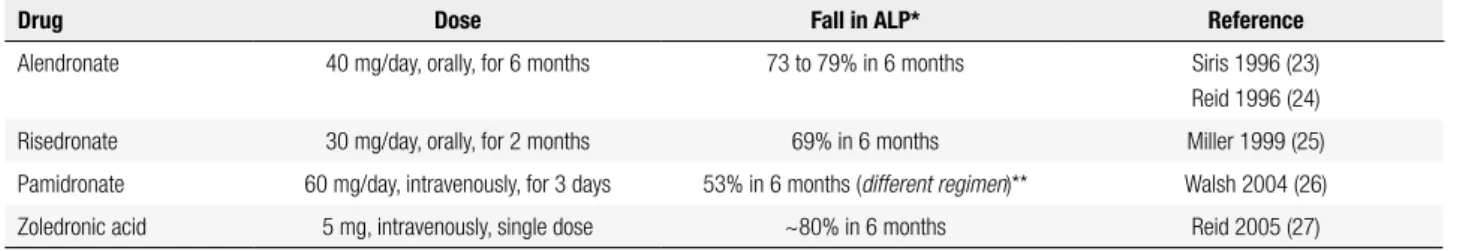
Table 2. Bisphosphonates with proven eficacy for antiresorptive treatment of PDB
Drug Dose Fall in ALP* Reference
Alendronate 40 mg/day, orally, for 6 months 73 to 79% in 6 months Siris 1996 (23)
Reid 1996 (24)
Risedronate 30 mg/day, orally, for 2 months 69% in 6 months Miller 1999 (25)
Pamidronate 60 mg/day, intravenously, for 3 days 53% in 6 months (different regimen)** Walsh 2004 (26)
Zoledronic acid 5 mg, intravenously, single dose ~80% in 6 months Reid 2005 (27)
Cop
yright
© ABE&M t
odos os dir
eit
os r
eser
vados
.
CONCLUSION
Paget’s disease of bone is a chronic progressive disorder of bone metabolism that may go undetected for many years. In Brazil, it is considered to be rare but precise estimates of nationwide incidence do not exist. Indeed, reports from south and northeast of the country support the idea that PDB might be more frequent than assumed in areas with clusters of European ancestry. It is impor-tant, thus, that endocrinologists are alert to its clinical signs and promptly recognize and diagnose PDB before it reaches more advanced stages with irreversible compli-cations. Most cases respond very well to treatment with bisphosphonates in such a way that complications and deterioration of quality of life can be prevented in most cases. It is expected that, as the etiopathogenesis of PDB is better understood, new treatment options might be-come available and further improve patient care.
Acknowledgements: Bruno Ferraz-de-Souza holds a Young In-vestigator grant from the São Paulo Research Foundation (Fa-pesp; 2011/12696-4).
Disclosure: no potential conlict of interest relevant to this article was reported.
REFERENCES
1. Siris ES, Ottman R, Flaster E, Kelsey JL. Familial aggregation of Paget’s disease of bone. J Bone Miner Res. 1991;6(5):495-500. 2. Seton M, Choi HK, Hansen MF, Sebaldt RJ, Cooper C. Analysis of
environmental factors in familial versus sporadic Paget’s disease of bone--the New England Registry for Paget’s Disease of Bone. J Bone Miner Res. 2003;18(8):1519-24.
3. Delmas PD, Meunier PJ. The management of Paget’s disease of bone. N Engl J Med. 1997;336(8):558-66.
4. Ralston SH, Langston AL, Reid IR. Pathogenesis and management of Paget’s disease of bone. Lancet. 2008;372(9633):155-63. 5. Ralston SH. Clinical practice. Paget’s disease of bone. N Engl J
Med. 2013;368(7):644-50.
6. Albagha OM, Wani SE, Visconti MR, Alonso N, Goodman K, Brandi ML, et al. Genome-wide association identiies three new susceptibility loci for Paget’s disease of bone. Nat Genet. 2011;43(7):685-9.
7. Laurin N, Brown JP, Morissette J, Raymond V. Recurrent mutation of the gene encoding sequestosome 1 (SQSTM1/p62) in Paget disease of bone. Am J Hum Genet. 2002;70(6):1582-8.
8. Visconti MR, Langston AL, Alonso N, Goodman K, Selby PL, Fraser WD, et al. Mutations of SQSTM1 are associated with severity and clinical outcome in paget disease of bone. J Bone Miner Res. 2010;25(11):2368-73.
9. Britton C, Walsh J. Paget disease of bone - an update. Aust Fam Physician. 2012;41(3):100-3.
10. Helfrich MH, Hocking LJ. Genetics and aetiology of Pagetic disorders of bone. Arch Biochem Biophys. 2008;473(2):172-82. 11. van Staa TP, Selby P, Leufkens HG, Lyles K, Sprafka JM, Cooper
C. Incidence and natural history of Paget’s disease of bone in England and Wales. J Bone Miner Res. 2002;17(3):465-71. 12. Cooper C, Harvey NC, Dennison EM, van Staa TP. Update on the
epidemiology of Paget’s disease of bone. J Bone Miner Res. 2006;21 Suppl 2:P3-8.
13. Reis RL, Poncell MF, Diniz ET, Bandeira F. Epidemiology of Paget’s disease of bone in the city of Recife, Brazil. Rheumatol Int. 2012;32(10):3087-91.
14. Gennari L, Di Stefano M, Merlotti D, Giordano N, Martini G, Tamone C, et al. Prevalence of Paget’s disease of bone in Italy. J Bone Miner Res. 2005;20(10):1845-50.
15. Werner de Castro GR, Heiden GI, Zimmermann AF, Morato EF, Neves FS, Toscano MA, et al. Paget’s disease of bone: analysis of 134 cases from an island in Southern Brazil: another cluster of Paget’s disease of bone in South America. Rheumatol Int. 2012;32(3):627-31.
16. Griz L, Caldas G, Bandeira C, Assuncao V, Bandeira F. Paget’s disease of bone. Arq Bras Endocrinol Metabol. 2006;50(4):814-22. 17. Siris ES, Lyles KW, Singer FR, Meunier PJ. Medical management
of Paget’s disease of bone: indications for treatment and review of current therapies. J Bone Miner Res. 2006;21 Suppl 2:P94-8. 18. Singer FR. Paget disease: when to treat and when not to treat. Nat
Rev Rheumatol. 2009;5(9):483-9.
19. Bone HG. Nonmalignant complications of Paget’s disease. J Bone Miner Res. 2006;21 Suppl 2:P64-8.
20. Rubin DJ, Levin RM. Neurologic complications of Paget disease of bone. Endocr Pract. 2009;15(2):158-66.
21. Siris ES, Feldman F. Natural history of untreated Paget’s disease of the tibia. J Bone Miner Res. 1997;12(4):691-2.
22. Delmas PD. Biochemical markers of bone turnover in Paget’s disease of bone. J Bone Miner Res. 1999;14 Suppl 2:66-9. 23. Siris E, Weinstein RS, Altman R, Conte JM, Favus M, Lombardi A,
et al. Comparative study of alendronate versus etidronate for the treatment of Paget’s disease of bone. J Clin Endocrinol Metab. 1996;81(3):961-7.
24. Reid IR, Nicholson GC, Weinstein RS, Hosking DJ, Cundy T, Kotowicz MA, et al. Biochemical and radiologic improvement in Paget’s disease of bone treated with alendronate: a randomized, placebo-controlled trial. Am J Med. 1996;101(4):341-8.
25. Miller PD, Brown JP, Siris ES, Hoseyni MS, Axelrod DW, Bekker PJ. A randomized, double-blind comparison of risedronate and etidronate in the treatment of Paget’s disease of bone. Paget’s Risedronate/ Etidronate Study Group. Am J Med. 1999;106(5):513-20.
26. Walsh JP, Ward LC, Stewart GO, Will RK, Criddle RA, Prince RL, et al. A randomized clinical trial comparing oral alendronate and intravenous pamidronate for the treatment of Paget’s disease of bone. Bone. 2004;34(4):747-54.
27. Reid IR, Miller P, Lyles K, Fraser W, Brown JP, Saidi Y, et al. Comparison of a single infusion of zoledronic acid with risedronate for Paget’s disease. N Engl J Med. 2005;353(9):898-908.
28. Reid IR, Lyles K, Su G, Brown JP, Walsh JP, del Pino-Montes J, et al. A single infusion of zoledronic acid produces sustained remissions in Paget disease: data to 6.5 years. J Bone Miner Res. 2011;26(9):2261-70. 29. Pazianas M, Compston J, Huang CL. Atrial ibrillation and
bisphosphonate therapy. J Bone Miner Res. 2010;25(1):2-10. 30. Rosen CJ, Brown S. Severe hypocalcemia after intravenous
bisphosphonate therapy in occult vitamin D deiciency. N Engl J Med. 2003;348(15):1503-4.
31. Whitson HE, Lobaugh B, Lyles KW. Severe hypocalcemia following bisphosphonate treatment in a patient with Paget’s disease of bone. Bone. 2006;39(4):954-8.
32. Ferraz-de-Souza B, Martin RM, Correa PH. Symptomatic intracranial hypertension and prolonged hypocalcemia following treatment of Paget’s disease of the skull with zoledronic acid. J Bone Miner Metab. 2013;31(3):360-5.
33. Schwarz P, Rasmussen AQ, Kvist TM, Andersen UB, Jorgensen NR. Paget’s disease of the bone after treatment with Denosumab: a case report. Bone. 2012;50(5):1023-5.
34. Lyles KW, Siris ES, Singer FR, Meunier PJ. A clinical approach to diagnosis and management of Paget’s disease of bone. J Bone Miner Res. 2001;16(8):1379-87.
35. Langston AL, Campbell MK, Fraser WD, MacLennan GS, Selby PL, Ralston SH. Randomized trial of intensive bisphosphonate treatment versus symptomatic management in Paget’s disease of bone. J Bone Miner Res. 2010;25(1):20-31.