Sao Paulo Med J. 2015; 133(2):131-4 131
SHORT COMMUNICATION
DOI: 10.1590/1516-3180.2014.8470910Pulmonary emphysema induced by methylphenidate:
experimental study
Enisema pulmonar induzido por metilfenidato: estudo experimental
Gabriel Victor Guimarães Rapello
I, Andréia Antoniolli
II, Daniel Martins Pereira
III, Gilberto Facco
IV,
Paulo Manuel Pêgo-Fernandes
V, Rogério Pazetti
VIHealth and Development Program of the Central-Western Region, Universidade Federal de Mato Grosso do Sul (UFMS), Campo Grande,
Mato Grosso do Sul, Brazil
ABSTRACT
CONTEXT AND OBJECTIVE: Methylphenidate is the most widely used drug for treating attention deicit hyperactivity disorder. However, it has important side efects, such as abdominal pain, insomnia, anorexia and loss of appetite, and also some cases of early severe emphysema after drug abuse have been reported. Our aim was to investigate the development of pulmonary emphysema in rats that were subjected to dif-ferent doses of methylphenidate.
DESIGN AND SETTING: Experimental study carried out at the laboratory of a public university.
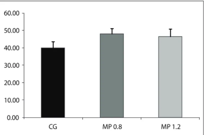
METHODS: Eighteen male Wistar rats were divided into three groups: control (0.9% saline solution); MP 0.8 (methylphenidate, 0.8 mg/kg); MP 1.2 (methylphenidate, 1.2 mg/kg). After 90 days of daily gavage, the ani-mals were sacriiced and lung tissue samples were prepared for analysis on the mean alveolar diameter (Lm). RESULTS: The Lm was greater in MP 0.8 (47.91 ± 3.13; P < 0.01) and MP 1.2 (46.36 ± 4.39; P < 0.05) than in the control group (40.00 ± 3.48).
CONCLUSION: Methylphenidate caused an increase in the alveolar diameter of rats, which was compatible with human pulmonary emphysema.
RESUMO
CONTEXTO E OBJETIVO: O metilfenidato é o medicamento mais utilizado para o tratamento de déicit de atenção e hiperatividade. No entanto, tem efeitos colaterais importantes, tais como dor abdominal, insônia, anorexia, perda de apetite, bem como alguns casos de enisema precoce grave após abuso da droga. Nosso objetivo foi investigar o desenvolvimento de enisema pulmonar em ratos submetidos a diferentes doses de metilfenidato.
TIPO DE ESTUDO E LOCAL: Trata-se de estudo experimental realizado em laboratório de uma universi-dade pública.
MÉTODOS: Dezoito ratos Wistar machos foram divididos em três grupos: Controle (solução salina 0,9%); MP 0.8 (metilfenidato 0,8 mg/kg); MP 1.2 (metilfenidato 1,2 mg/kg). Depois de 90 dias de gavagem diária, os animais sofreram eutanásia e amostras de tecido pulmonar foram preparadas para análise do diâmetro alveolar médio (Lm).
RESULTADOS: Lm foi maior nos grupos 0,8 MP (47,91 ± 3,13, P < 0,01) e MP 1.2 (46,36 ± 4,39, P < 0,05) em comparação com o grupo controle (40,00 ± 3,48).
CONCLUSÃO: O metilfenidato causou aumento no diâmetro alveolar de ratos, o que é compatível com enisema pulmonar humano.
IMSc. Physiotherapist, Hospital Regional de Mato
Grosso do Sul, Campo Grande, Mato Grosso do Sul, Brazil.
IIPhD. Associate Professor, Faculdade de
Medicina (FAMED), Universidade Federal de Mato Grosso do Sul (UFMS), Campo Grande, Mato Grosso do Sul, Brazil.
IIIMSc. Physiotherapist, Hospital Regional de
Mato Grosso do Sul, Campo Grande, Mato Grosso do Sul, Brazil, College Professor, Universidade Anhanguera-Uniderp, Campo Grande, Mato Grosso do Sul, Brazil.
IVMSc. College Professor, Universidade
Anhanguera-Uniderp, Campo Grande, Mato Grosso do Sul, Brazil.
VMD, PhD. Full Professor, Instituto do Coração
(InCor), Faculdade de Medicina da Universidade de São Paulo, São Paulo, Brazil.
VIPhD. Scientiic Researcher, Laboratory of
Thoracic Surgery Research, Faculdade de Medicina da Universidade de São Paulo (FMUSP), São Paulo, Brazil.
KEY WORDS: Methylphenidate.
Attention deicit disorder with hyperactivity. Pulmonary emphysema.
Rats. Lung.
PALAVRAS-CHAVE: Metilfenidato.
Transtorno do déicit de atenção com hiperatividade.
Enisema pulmonar. Ratos.
SHORT COMMUNICATION | Rapello GVG, Antoniolli A, Pereira DM, Facco G, Pêgo-Fernandes PM, Pazetti R
132 Sao Paulo Med J. 2015; 133(2):131-4 INTRODUCTION
Attention deicit hyperactivity disorder (ADHD) is one of the most common psychiatric disorders of childhood and may be accompanied by hyperactive behavior followed by attention prob-lems.1 he prevalence of ADHD is estimated to be between 3% and
6.3% among school-age children in diferent geographical areas,2
including Brazil, and its prevalence is greater among males.3 he
treatment consists of psychological and/or psychiatric interven-tion together with prescripinterven-tion of stimulant drugs. he associainterven-tion of these two therapies is clinically efective and relatively inexpen-sive.4 Methylphenidate is the most widely used psychostimulant
drug for ADHD treatment.2,5,6 It improves attention, concentration
and full cognitive function.7 he most common adverse efects
are abdominal pain, insomnia, anorexia and loss of appetite.8
However, the side efects from longer exposure periods have been insuiciently studied,9 and there are no conclusive data. Sherman10
found a syndrome of pulmonary vascular sclerosis among abusive (intravenous) users of methylphenidate, and reported the cases of six patients with early severe emphysema. An experimental study on rats showed that there was a high concentration of methylphe-nidate in lung tissue ater intraperitoneal injection.11
We hypothesized that chronic use of methylphenidate could be related to early pulmonary emphysema through destruction of the alveolar architecture.
OBJECTIVE
Our aim was to investigate the relationship between methylphe-nidate and pulmonary emphysema in a rodent model.
METHODS
his experimental study was approved by our institutional eth-ics committee on animal use (protocol number 205/2009), and all procedures complied with international standards for ani-mal experimentation. Eighteen ani-male Wistar rats from the aniani-mal house of a university were divided into three groups (n = 6): con-trol (0.9% saline solution, 1 ml/kg), MP 0.8 (methylphenidate, 0.8 mg/kg) and MP 1.2 (methylphenidate, 1.2 mg/kg). Based on data from the literature and from our clinical practice, we chose these doses to represent a therapeutic dose (0.8 mg/kg) and an overdose (1.2 mg/kg).12,13
he animals received either saline or methylphenidate daily by means of gavage for 90 days. Ater this period, the animals were sacriiced by means of a lethal intraperitoneal injection of thiopental sodium (100 mg/kg). he lungs were excised and ixed using intratracheal instillation of 4% paraformaldehyde (at a constant pressure of 20 cmH2O). hus, the trachea was tied of and the lungs were stored in paraformaldehyde solution. Ater 24 hours, lung samples (5 µm slices) were obtained and subjected to processing for histological analysis on slides stained with hematoxylin-eosin.
he mean linear intercept (Lm) was microscopically deter-mined by using an eyepiece with Weibel reticule (50 straight lines and 100 points; 200 x magniication), in 15 ields per slide.14 he
Lm was obtained through the following relationship: Lm = Ltot/ Li, where Ltot was the total length of the reticule straight lines and Li was the number of intercepts between alveolar septa and reticule straight lines.
Lm values were expressed as mean ± standard deviation. he Shapiro-Wilk test was used to investigate whether the data pre-sented normal distribution. Comparisons between groups was made using analysis of variance (ANOVA) with the Tukey post-test. A P value less than 0.05 was considered signiicant.
RESULTS
he Lm was greater in all the methylphenidate-treated animals (MP 0.8 = 47.91 ± 3.13, P < 0.01; MP 1.2 = 46.36 ± 4.39, P < 0.05) than in those of the control group (40.00 ± 3.48) (Figure 1). here was no diference between the MP groups. he Lm analysis is illustrated in Figure 2, which shows the alveolar destruction in methylphenidate-treated animals.
DISCUSSION
In the present study, we used Lm analysis, the gold-standard method for making histological diagnoses of emphysematous pulmonary disease, and observed that there was notable destruc-tion of pulmonary parenchyma in both of the methylphenidate-treated groups.
Until now, the correlation between methylphenidate and pulmonary emphysema10 seems not to have aroused any
atten-tion within the scientiic community. his might be explained by the lack of methodological information about intake and exposure to the drug among users. One group of authors has shown interest in the systemic efects of methylpheni-date.11 hey tested 37 μmol/kg of methylphenidate on rats and
Figure 1. Lmof animals treated either with saline solution (CG, control group) or diferent doses of methylphenidate (MP 0.8 and MP 1.2).
0.00 10.00 20.00 30.00 40.00 50.00 60.00
Pulmonary emphysema induced by methylphenidate: experimental study | SHORT COMMUNICATION
Sao Paulo Med J. 2015; 133(2):131-4 133 observed that there was higher concentration in lung tissue
than in heart, liver and brain tissue. However, no controlled studies have been conducted, thus showing the need to investi-gate these undesirable side efects.
he mechanism through which emphysematous lesions appear is unclear. In a case presented by Sherman et al.,10 a link
between respiratory changes and intravenous injection of talc (magnesium trisilicate), which is present in methylphenidate pills, was suggested. Relevant information such as length of expo-sure and dose has not been elucidated.
Although we did not show that the dose was a determining factor for the changes observed, some studies have tested doses that difered from ours. A double-blind study12 in which the
objec-tive was to evaluate the side efects of diferent doses of methyl-phenidate classiied the doses of 0.3 mg/kg and 0.5 mg/kg as low and high dose, respectively. In contrast, a meta-analysis by Spencer et al.13 found that the doses used in adult patients were smaller
than those used in children (0.5 mg/kg/day versus 1.0 mg/kg/day, respectively).
Because of the lack of consensus in the literature on the ideal dosage of methylphenidate, and based upon the maximum daily dose (60 mg),15 we decided to test 0.8 mg/kg/day as a
therapeu-tic dose in rats. Furthermore, we considered that application of 1.2 mg/kg/day would be an overdose. With regard to the length of exposure in the proposed protocol, we chose to analyze the Lm ater a long period of drug exposure (90 days), considering that drug treatment approaches to ADHD are long-term and may extend until adulthood.16,17
Despite the signiicance of these results, it is important to highlight that a single variable was analyzed (Lm). Further research needs to be developed in order to address other impor-tant variables such as duration of drug exposure, doses and administration routes. In addition, other analytical methodolo-gies should be used such as elastic iber analysis, clinical imag-ing examinations and studies on respiratory mechanics, in order to explain the correlation between methylphenidate and pulmo-nary emphysema.
CONCLUSION
In this study, administration of methylphenidate caused destruc-tion of the alveolar septa in the lung parenchyma in Wistar rats, which was histologically compatible with pulmonary emphysema.
REFERENCES
1. Kay J, Tasman A. Psiquiatria – ciência comportamental e fundamentos
clínicos. São Paulo: Manole; 2000.
2. Ball C. Attention-deficit hyperactivity disorder and the use of
methylphenidate. The Psychiatric Bulletin. 2001;25(8):301-4. Available
from: http://pb.rcpsych.org/content/25/8/301.full.pdf+html. Accessed
in 2014 (Aug 1).
3. Burguess IC. Service innovations: attention-deicit hyperactivity
disorder – development of a multi-professional integrated care pathway.
The Psychiatric Bulletin. 2002;26(4):148-51. Available from: http://
pb.rcpsych.org/content/26/4/148.full.pdf+html. Accessed in 2014 (Aug 1). Figure 2. Photomicrographs of lung parenchyma of rats for
determination of mean alveolar diameter (Lm) using a Weibel reticule. (A) control group: saline solution; (B) methylphenidate 0.8 mg/kg; and (C) methylphenidate 1.2 mg/kg. Note the areas of alveolar space enlargement (stars) due to disruption of alveolar septa, with formation of “drumsticks” (arrows). Hematoxylin-eosin staining, 200 x magniication.
A
B
SHORT COMMUNICATION | Rapello GVG, Antoniolli A, Pereira DM, Facco G, Pêgo-Fernandes PM, Pazetti R
134 Sao Paulo Med J. 2015; 133(2):131-4
4. Ishimatsu M, Kidani Y, Tsuda A, Akasu T. Efects of methylphenidate
on the membrane potential and current in neurons of the rat locus
coeruleus. J Neurophysiol. 2002;87(3):1206-12.
5. Faraone SV, Biederman J, Mick E. The age-dependent decline of
attention deicit hyperactivity disorder: a meta-analysis of follow-up
studies. Psychol Med. 2006;36(2):159-65.
6. Janicak PG, Davis JM, Preskorn SH, Junior FJA. Princípios e práticas em
psicofarmacoterapia. Rio de Janeiro: Medsi; 1996.
7. Efron D, Jarman F, Barker M. Side efects of methylphenidate and
dexamphetamine in children with attention deicit hyperactivity
disorder: a double-blind, crossover trial. Pediatrics. 1997;100(4):662-6.
8. Golinko BE. Side efects of dextroamphetamine and methylphenidate
in hyperactive children--a brief review. Prog Neuropsychopharmacol
Biol Psychiatry. 1984;8(1):1-8.
9. Pastura G, Mattos P. Efeitos colaterais do metilfenidato [Side efects of
methylphenidate]. Rev Psiquiatr Clín (São Paulo). 2004;31(2):100-4.
10. Sherman CB, Hudson LD, Pierson DJ. Severe precocious emphysema
in intravenous methylphenidate (Ritalin) abusers. Chest.
1987;92(6):1085-7.
11. Thai DL, Yurasits LN, Rudolph GR, Perel JM. Comparative
pharmacokinetics and tissue distribution of the d-enantiomers of
para-substituted methylphenidate analogs. Drug Metab Dispos.
1999;27(6):645-50.
12. Barkley RA, McMurray MB, Edelbrock CS, Robbins K. Side efects of
methylphenidate in children with attention deicit hyperactivity
disorder: a systemic, placebo-controlled evaluation. Pediatrics.
1990;86(2):184-92.
13. Spencer T, Biederman J, Wilens T, et al. Pharmacotherapy of
attention-deicit hyperactivity disorder across the life cycle. J Am Acad Child
Adolesc Psychiatry. 1996;35(4):409-32.
14. Kuraki T, Ishibashi M, Takayama M, Shiraishi M, Yoshida M. A novel oral
neutrophil elastase inhibitor (ONO-6818) inhibits human neutrophil
elastase-induced emphysema in rats. Am J Respir Crit Care Med.
2002;166(4):496-500.
15. Ritalina (Cloridato de Metilfenidato): comprimidos [bula de remédios].
Responsável técnico: Marcos A. J. Brazil: Novartis; 2011. Available
from: http://www.anvisa.gov.br/datavisa/ila_bula/frmVisualizarBula.
asp?pNuTransacao=10716102013&pIdAnexo=1909485. Accessed in
2014 (Aug 19).
16. Spencer T, Bierdeman J, Wilens TE, Faraone SV. Adults with
attention-deicit/hyperactivity disorder: a controversial diagnosis. J Clin
Psychiatry. 1998;59 Suppl 7:59-68.
17. Shekim WO, Asarnow RF, Hess E, Zaucha K, Wheeller N. A clinical
and demographic proile of a sample of adults with attention
deicit hyperactivity disorder, residual state. Compr Psychiatry.
1990;31(5):416-25.
Sources of funding: None Conlict of interest: None
Date of irst submission: February 22, 2014 Last received: August 25, 2014
Accepted: September 10, 2014
Address for correspondence: Gabriel Victor Guimarães Rapello
Universidade Federal de Mato Grosso do Sul
Av. Costa e Silva, s/no
Universitário — Campo Grande (MS) — Brasil
CEP 79103-151
Tel. (+55 67) 3345-3000