http://dx.doi.org/10.1590/s2175-97902017000116081
A
r
*Correspondence: M. Aragón. Departamento de Farmacia. Universidad
Na-cional de Colombia. Carrera 30 número 45-03, Oicina 312, Bogotá, Colombia.
N. Tel.: +57(1)3165000 Ext. 14630. E-mail: [email protected]
In vitro
intestinal permeability studies, pharmacokinetics and tissue
distribution of 6-methylcoumarin after oral and intraperitoneal
administration in Wistar rats
Paola Andrea Cárdenas
1, Jadel Müller Kratz
2, Aura Hernández
1, Geison Modesti Costa
3, Luis
Fernando Ospina
4, Yolima Baena
1, Cláudia Maria Oliveira Simões
2, Álvaro Jimenez-Kairuz
5,
Marcela Aragon
1*1Grupo Sistemas de Liberación Modiicada de Moléculas Biológicamente Activas, Departamento de Farmacia, Universidad
Nacional de Colombia, Bogotá, Colombia, 2Grupo de Análise e Desenvolvimento de Fármacos de Origem Natural,
Departamento de Ciências Farmacêuticas, Universidade Federal de Santa Catarina - UFSC, SC, Florianópolis, Brasil,3Grupo
de Estudio y Aprovechamiento de Productos Naturales Marinos y Frutas de Colombia Departamento de Química, Universidad Nacional de Colombia, Bogotá, Colombia, 4Grupo de Principios Bioactivos de Plantas Medicinales, Departamento de
Farmacia, Universidad Nacional de Colombia, Bogotá, Colombia, 5Unidad de Investigación y Desarrollo en Tecnología
Farmacéutica (UNITEFA), CONICET and Departamento de Farmacia, Facultad de Ciencias Químicas, Universidad Nacional de Córdoba. Ciudad Universitaria, Córdoba, Argentina
6-Methylcoumarin (6MC) is a semisynthetic coumarin with important in vitro and in vivo anti-inlammatory activity. In order to continue the pre-clinical characterization of this molecule, in vitro
intestinal permeability, plasma proile and tissue distribution after oral administration in rats were studied. The permeability of 6MC was evaluated by the Caco-2 cellular model in both the apical-basal (A-B) and basal-apical (B-A) directions. The pharmacokinetics and biodistribution were evaluated in rats after oral and intraperitoneal administration at doses of 200 mg/kg. Transport experiments with Caco-2 cells showed that 6MC presented high permeability at all concentrations evaluated. This inding suggested that 6MC could be transported across the gut wall by passive difusion. The plasma concentration-time curve showed that the maximum concentration (Cmax) was 17.13 ± 2.90 µg/mL at maximum time (Tmax) of 30 min for the oral route and Cmax 26.18 ± 2.47 µg/mL at 6.0 min for the intraperitoneal administration, with elimination constant of (Ke) 0.0070 min
-1 and a short life half time of (T
1/2) lower that 120 min. The distribution study showed that 6MC has high accumulation in the liver, and widespread distribution in all the organs evaluated.
Uniterms: 6-Methylcoumarin/Pharmacokinetics/rats. 6-Methylcoumarin/distribution. Intestinal permeability/study. Intestinal permeability/study/In vitro.
INTRODUCTION
The oral route is the most common route for administration of new drugs, as it is considered the safest
and most convenient. However, it also has limitations, as
the drug must be absorbed from the site of absorption to
the systemic circulation, and then distributed to the target
organs, in order to produce its pharmacological effect (Hedaya, 2007).
According to the Biopharmaceutics Classiication System (BCS) of the US Food and Drug Administration, drugs are classified based on two intrinsic properties
that control their oral absorption: aqueous solubility and intestinal permeability (Amidon et al., 1995). Knowledge
of these drug properties not only assists in the classiication of a drug in the BSC, but also guides the selection of
candidate drugs during the drug development process
(Griin, O´Dricoll, 2008).
parameter that defines the amount of material that can dissolve in a given solvent at equilibrium, it is one of
the most critical and widely studied physical-chemical
attributes of candidate drugs (Wei-Qin, 2008).
Permeability, meanwhile, is the property that determines the speed at which a dissolved drug passes through the intestinal wall and reaches the systemic
circulation. Permeability is considered one of most important features in the absorption of drugs. This is a complex kinetic process dependent on several physiological, physiochemical properties of the drug, and on the biophysiochemical properties of a gastrointestinal barrier membrane. Diferent mechanisms of permeation
through biological barriers have been described, the most
important ones being passive diffusion (transcellular and paracellular), active uptake, and efflux transport (Fagerholm, 2007; Matsson et al., 2005).
Likewise, knowledge of the pharmacokinetic proiles allows us to characterize compounds in terms of their bioavailability and desirable action of duration. Four pharmacokinetic (PK) parameters are the most useful in characterizing the in vivo disposition of a compound: (i)
clearance (Cl, units of volume/time), a measure of the ability of the body to eliminate a compound; (ii) volume of distribution (Vd, units of volume), the apparent volume/ space in the body that contains the compound; (iii) half-life (t1/2, units of time), the time taken for a compound to decrease to half of its initial concentration in the luid
or tissue in which it is measured in (e.g., plasma), and
(iv) bioavailability (F, unitless, often expressed as %), the fraction of a compound that reaches the systemic circulation following non-intravenous administration (Fan, De Lannoy, 2014). Meanwhile, biodistribution studies describe the transit of a drug through the organism,
the anatomic sites reached by the drug when it is in the systemic circulation, and the sites where it can accumulate
(Hedaya, 2007).
On the other hand, some simple and complex
coumarins have shown different biological activities, such as antibacterial, effects on the cardiovascular system, efects on the central nervous system, antioxidant activities, cytotoxicity, and anti-inlammatory properties (Grazul, Budzisz, 2009; Kidane et al., 2004; Beillerot et al., 2008; Anand, Singh, Singh, 2012; Sashidhara et al.,
2011; Kang et al., 2009; Li et al., 2011; Hoult, Paydt,
Paya, 1996). Furthermore, 6-methylcoumarin (6MC, Figure 1), another simple coumarin, has shown important
anti-inlammatory activity in in vivo and in vitro models
(Cárdenas et al., 2014).
A l t h o u g h 6 M C h a s s h o w n r e m a r k a b l e pharmacological efect, knowledge of its permeability,
concentration-time and distribution is essential for understanding its biopharmaceutical profile. Thus, the aim of this work was to study the in vitro permeability and in vivo pharmacokinetics after oral and intraperitoneal administration in Wistar rats, in order to complete the
biopharmaceutical characterization of 6MC.
MATERIAL AND METHODS
Chemical reagents
6-Methylcoumarin (6MC, Sigma; St. Louis, MO, USA); trichloroacetic acid (Analytical grade, Merck, Darmstadt, Germany); acetic acid, methanol (HPLC grade, Merck, Darmstadt, Germany), and water HPLC grade were prepared by the MilliQ Plus system (Millipore Co.,
France).
Animals
Twelve-week-old Male Wistar rats were obtained from the animal house of the Pharmacy Department of the Universidad Nacional de Colombia. The assays
were carried out in accordance with the international and
local ethical guidelines on the use and care of laboratory
animals. The local Research Ethics Committee (Act
03/2012 Faculty of Science) approved this study.
Drug analysis
For all the analyses, an Agilent 1100 Series HPLC
chromatograph was used. For the permeability studies, a
Phenomenex reversed phase column (Macclesield, UK) Bondclone C-18 (150 x 3.9 mm; 10 µm) was used. The mobile phase was composed of A) water: methanol: acetic acid (95:5:1 v/v) and B): methanol: acetic acid (100:1 v/v); ratio 80:20 (A:B), under isocratic low 1.0 mL/min. The analytical wavelength was 321 nm, and samples of 10 µL were injected.
follows: 0 to 14 min: 0 % B to 50 % B; 14 to 23 min: 50 % B; 23 to 24 min: 50 % B to 0 % B; 24 to 32 min: 0 % B. UV detection was performed at 321 nm. The injection volume
was 50 µL. The data were processed using the software
program Value Solution Chemstation® (ChemStation for LC 3D systems Rev. B.03.02 [341]) (Hernández, Ospina, Aragón, 2014).
Caco-2 permeability studies
Cell culture
Caco-2 cells (ATCC:HTB-37) were cultured in
high-glucose DMEM (Gibco, USA) supplemented
with 10% fetal bovine serum and 1% non-essential amino acids (Gibco, USA), at 37 °C in a humidified 5% CO2 atmosphere, until the cells reached 80-90% conluence. For the transport experiments, 100,000 cells
(passages 113–115) were harvested and seeded on each polycarbonate insert (0.6 cm2, 0.4 μm pore size; Millipore,
USA), and allowed to grow and diferentiate for 21–28
days prior to the experiments, as described previously by
Kratz et al. (2012).
Transport experiments
The determination of in vitro intestinal permeability
of 6MC was carried out under sink conditions in a series of pH-gradient bidirectional transport experiments with Caco-2 cells. Before the experiments, cell monolayers were rinsed with Hank’s balanced salt solution (HBSS) and equilibrated for 30 min at 37 °C. The integrity of the monolayers was assessed before and after the
experiments, by transepithelial electrical resistance (TEER) measurement. Only monolayers with TEER
values above 200 Ωcm2 were considered.
A stock solution of 6MC (10 mM DMSO) was diluted to final concentration of 10, 25, 50 or 100 µM in HBSS pH 6.5 (apical transport buffer) or pH 7.4 (basolateral transport bufer). Bidirectional experiments (apical-to-basolateral [AB] and basolateral-to-apical [BA]) were initiated by adding 6MC solutions to the donor compartment, and fresh buffer to the acceptor compartment. Caco-2 cell monolayers were incubated for
1 h at 37 °C under constant stirring (150 rpm). Receiver compartments were sampled at 0, 15, 30, 45 and 60 min,
refilled with an equivalent amount of transport buffer, and samples were submitted to analysis by HPLC/UV. Apparent permeability coeicients (Papp) were calculated
from the equation
where ΔQ/Δt is the steady-state lux (mol/s), C0 is the initial
concentration in the donor chamber at each time interval
(mol/mL), and A is the surface area of the filter (cm2).
Carbamazepine (CBZ) (50 µM) and hydrochlorothiazide (HCT) (200 µM) were used as controls. The data are presented as means ± SD of six independent monolayers.
Pharmacokinetic study
Male Wistar rats (12 weeks, 250 ± 10 g), with 5 animals for each administration route, were administrated,
by oral gavage (p.o.) or intraperitoneal (i.p.) route, with
6MC at 200 mg/Kg, suspended in saline solution (NaCl 0.9 %) and tween-80. 200 mg/kg was chosen as doses, since
in previous assays (data not shown) the biopharmaceutics parameter, Tmax, Cmax and AUC proved be linear in a
range of dose of 100 to 400 mg/kg.
Blood samples, 400 µl, were collected from the retro-orbital sinus, at 2,6,10,15,20,25,30,45,60,120, 360 and 480 min after oral administration. After each sampling, the blood samples were centrifuged at 6000 rpm for 10 min, at 4 °C to separate the plasma, and then 200 µL of plasma was homogenized with 200 µL of trichloroacetic acid (20 %), and centrifuged at 13000 rpm for 10 min. The supernatant was recovered for 6MC quantiication using the HPLC-DAD method previously described. The
maximal observed plasma concentration (Cmax) and corresponding sampling time (Tmax) were determined by
visual inspection of the data.
The apparent elimination rate constant (Ke) was
estimated by linear regression of the log-transformed
plasma concentrations during the terminal log-linear
decline phase. The apparent terminal elimination half-life
time (t1/2) was calculated as ln2/Ke. The area under the
6MC plasma concentration-time curve from time zero to the last quantiiable point (AUC0-t) was calculated using the linear trapezoidal rule. The AUC extrapolated to ininity
(AUC0-α) was calculated as the sum of AUC0-t, and the last
quantifiable point was divided by the elimination rate
constant. The apparent oral clearance (Cl/f) was calculated as the dose divided by AUC0-α. The apparent volume of
distribution (V/F) was calculated as the apparent oral clearance divided by the elimination constant.
Tissue distribution study
sampling time). The organs (liver, heart, lung, kidney and
spleen) were removed and washed with saline solution.
The organs were then homogenized with a same volume of trichloroacetic acid (20 %) in a Polytron PT-10 – 35 (Kinematica, Newark, NJ, USA), and centrifuged at 13000 rpm for 10 min. The supernatanat was recovered for 6MC quantiication.
Identification of the main 6-methylcoumarin metabolite
To determine possible metabolites of 6MC after oral administration, the plasma samples were analyzed by LC-ESI/MS. The system consisted of a Shimadzu HPLC equipped with two isocratic pumps, on-line degasser, a Rhodyne manual injector, a UV detector and a mass spectrometer (Shimadzu LCMS-2010EV). The
chromatographic parameters were the same as those used
in the HPLC-DAD analysis. LabSolution® V.3.0 software was used for the data acquisition and processing. Full scan mass spectra were recorded at between m/z 50 and 500 in positive mode. Nitrogen was used as nebulizer gas at 1 L/ min, capillary voltage was 4.500 V and detector voltage was 1.500 V. The collision dissolution line (CDL) and QarrayRF voltage were both 150 V. CDL and the Heat Block temperature as set at 250°C. Standard samples of coumarin and 6MC were analyzed following the same
methodology.
Statistical analysis
All results were expressed as the arithmetic mean of
the values obtained, ± the respective standard deviation.
Simple comparisons between groups were performed using the Student’s t-test, with a conidence level of 95%.
RESULTS
Permeability
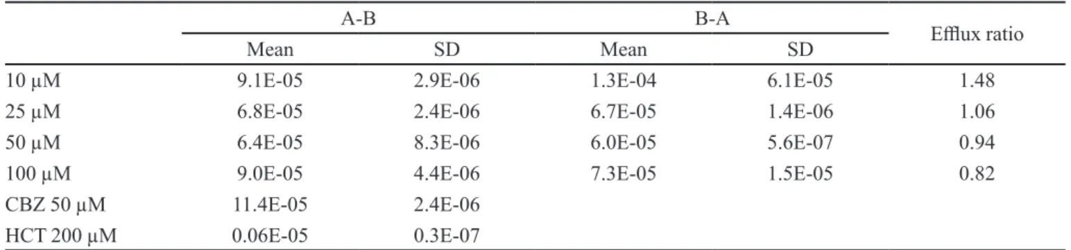
Table I shows that 6MC presented a high Papp in the absorptive and secretory directions in Caco -2 cells. Given the inding of Papp values over 10-5, it is suggested that 6MC is highly permeable in both directions. The elux
ratio (Papp BA/ Papp AB) of less than 2 in all cases indicates
a passive difusion mechanism and the absence of elux.
For all the concentrations evaluated, 6MC had high mass
balance values, with average recovery of more than 70% in both the AB and BA directions.
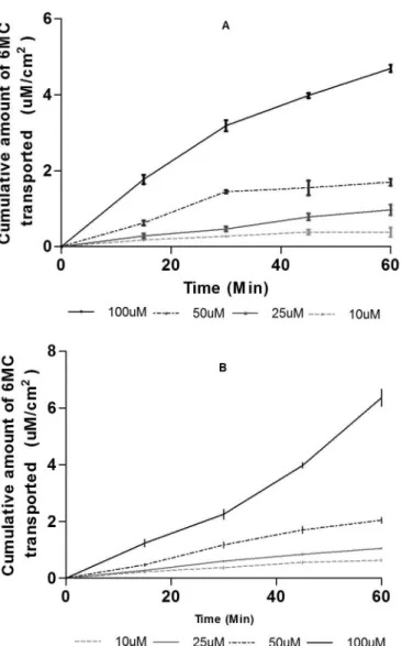
The permeation of 6MC was performed in both A- B and B-A in the Caco-2 cellular model. Figure 2 shows the cumulative amount permeated (µM/cm2) in the AB and
BA directions. The relation of the concentration versus the
cumulative amount permeated showed a linear relationship proportional to concentration (in both directions), with
determinant coeicients of 0.9903 and 0.9853 for AB and BA direction, respectively. This means that there is no saturation of the acceptor medium in any concentration
and time during the test time.
Pharmacokinetic
The plasma samples were analyzed using an HPLC-DAD validated method (Hernández, Ospina, Aragón, 2014). The mean plasma concentration versus time proiles of 6MC are shown in Figure 3. The pharmacokinetic parameters calculated for 6MC in plasma are summarized in Table II.
Plasma pharmacokinetics of 6-methylcoumarin in rats following oral administration. Data are expressed as mean ± SD of ive animals. Dose administered 200 mg/kg.
TABLE I - Comparable Papp (cm/s) values for 6MC obtained in the absorptive (A-B), secretory (B-A) directions, and elux ratio
(PappB-A/ Papp A-B)
A-B B-A
Elux ratio
Mean SD Mean SD
10 µM 9.1E-05 2.9E-06 1.3E-04 6.1E-05 1.48
25 µM 6.8E-05 2.4E-06 6.7E-05 1.4E-06 1.06
50 µM 6.4E-05 8.3E-06 6.0E-05 5.6E-07 0.94
100 µM 9.0E-05 4.4E-06 7.3E-05 1.5E-05 0.82
CBZ 50 µM 11.4E-05 2.4E-06
HCT 200 µM 0.06E-05 0.3E-07
Maximum concentration (Cmax), Maximun
time (Tmax), Area Under Curve from time zero to the last quantifiable point (AUC0-480), Area Under Curve extrapolated to infinite (AUC0-α), Elimination constant
(Ke), Absortion constant (Ka), elimination half –time
(t1/2), Apparent oral clearance (Cl/F), Apparent volume of distribution (V/F). Cl/f and Vd were calculated taking into
account the soluble 6MC in the volume administered. The
data are generated using the mean values from individual
rats ± SD (n=5).
As shown in Table II, the Cmax, Tmax, and ABC 0-480 and ABC0- α values for intraperitoneal administration
are higher than those obtained after oral administration. This fact could be explained by that fact that in the
intraperitoneal route, absorption occurs through the mesenteric vessels, draining to the portal vein, and thereby accessing the bloodstream in less time than by the oral route (Turner et al., 2011), making this a more rapid absorption route (Morton et al., 2001).
FIGURE 2 - Transport of 6-methylcoumarin across Caco-2 cells. Cumulative amount of 6-methylcoumarin transported across Caco-2 cells in the Apical to Basolateral (A) or Basolateral to Apical direction (B). Data are expressed as means ± SD of six
independent monolayers.
TABLE II - Plasma pharmacokinetics parameters of 6-methylcoumarin after oral and intraperitoneal administration in Wistar rats,
dose 200 mg/kg
Oral Intraperitoneal
AUC0-480(µg/mL*min) 977.2 ± 276.5 2177.0 ± 331.6
AUC0- α(µg/mL*min) 1015.4 ± 296.1 2234.9 ± 354.1
C max (µg/mL) 17.13 ± 2.90 26.18 ± 2.47
Tmax (min) 30 6.0
ke (min-1) 0.0064 ± 0.0009 0.0076 ± 0.0007
Ka (min-1) 0.3311 ± 0.0489
t ½ (min) 109.8 ± 15.0 92.3 ± 8.7
Cl/f (mL/min) 0.8 ± 0.3 0.4 ± 0.07
Vd (mL) 134.9 ± 30.4 48.8 ± 3.9
F 0.45
F I G U R E 3 - Plasma concentration - time profile of 6-methylcoumarin after oral and intraperitoneal administration
Regarding the values obtained of ke, t1/2 and the Cl/f, no signiicant diference was found that could be attributed to the administration route. This fact is expected, since these are intrinsic parameters of the drug.
Besides 6MC, another majority compound was detected in the plasma after oral administration of 6MC.
This compound could be related to gastrointestinal
metabolism of 6MC, since it was not found in the blood samples analyzed after i.p. administration. Data from the LC-ESI/MS were used for preliminary identiication of the main metabolite of 6-MC. The retention times (Rt), UV λmax values and the molecular ions in positive mode
are shown in Table III.
Distribution Tissue
Using a validated HPLC-DAD method, the concentration of 6MC after oral administration was determined in liver, heart, lung, kidney and spleen
(Figure 4).
In all the organs, 6MC concentrations were found to be in the range of between 0.35 ± 0.14 and 4.18 ± 0.03 µg per g of tissue. At 15 min, the highest concentrations were found in plasma and liver, and the lowest in kidneys,
heart and lungs. At 30 min, the concentration increased proportionally in all the organs sampled. At 60 min, the
distribution changed; in the kidney, the concentration of
6MC increased significantly, while the concentrations
in the other organs decreased. At this same time point,
the concentrations (µg/mL) were as follows; heart: 2.54 ± 0.10; plasma: 2.48 ± 0.62; spleen: 1.89 ± 0.15, lung: 0.59 ± 0.10 lung, kidney 3.40 ± 0.31, liver 1.45 ± 0.10. In addition, the 6MC fraction present in the evaluated organs was very low, since the total 6MC found in all the organs tested at 30 and 60 minutes was 0.104% and the 0.046% of the 6MC administered, respectively.
DISCUSSION
Ideal drug candidates should have adequate
aqueous solubility and permeability in order to achieve
effective concentration in the target tissue (Kratz et al., 2012). In order to evaluate the in vitro intestinal
permeability, transport experiments were performed on Caco-2 cells. These cells are able to fully polarize into diferentiated monolayers, with well-established tight
junctions and brush border membrane. They can also
express several membrane transporters and metabolizing enzymes, allowing the measurement of functional permeability, both through passive difusion and active transport (Kratz et al., 2012). The apparent permeability of 6MC was high, with values of magnitude of 10-5.
This value is comparable with the Papp of other similar
compounds such as umbelliferone, scopoletine, and coumarin, in which high permeability and no elux were reported (Galkin, Fallarero, Vuorela, 2009).
The high intestinal permeability of a drug indicates
that its transport across the gut wall does not represent
a relevant restriction for its oral absorption. The linear increase in the amount of 6MC permeated, indicating that
its transport across the gut wall, is probably mediated by
passive difusion, and the low elux values in the diferent concentrations evaluated, indicating that elux does not represent an interference for absorption (Galkin, Fallarero, Vuorela, 2009).
The BCS considers that a drug has high aqueous solubility if its maximum dose is completed dissolved in 250 ml of pH 1-7.5 bufered aqueous solution (Wei-Qin, 2008). According to this deinition, it is suggested that 6MC has low solubility, since the dose of 6MC that TABLE III - The main coumarinic compounds identifiedbyLC-ESI/MS analysis in plasma after oral administration of 6-methylcoumarin in Wistar rats
Peak Rt (min) λmáx (nm) [M+H]+(m/z) Compound
1 12.5 278 and 321 147 Coumarin
2 14.6 278 and 321 161 6-methylcoumarin
FIGURE 4 - Biodistribution proile of 6-methylcoumarin after oral administration in Wistar rats. Data expressed mean ± D.S
showed anti-inflammatory activity in rats is 200 mg/ kg,(Cárdenas et al., 2014) and its solubility is 0.57 ± 0.03 µg/mL in water and bufered solutions of pH 1.2, 6.8 and 7.4 (Cárdenas et al., 2013). As consequence, 6MC can be classiied as a class II compound (Low solubility/High permeability) accord to the BCS, when the absorption
across the gut wall is limited mainly by solubility.
The concentration-time proiles (Fig 3) showed fast
absorption, but low plasma concentration in relation to the
dose administered (10.25 ± 0.72 y 26.18 ± 2.47 µg/mL oral and intraperitoneal via respectively). This fact may
be related to the high clearance rate (CL/F) and possibly,
a high rate of metabolism after administration of 6MC.
Regarding other coumarins orally administered in rats, 6MC have a similar t1/2 to that reported for
daphnoterine and daphnetin (t1/2of 93 ± 26 and ± 262.23 96.31 min respectively) (Wei et al., 2015; Lin et al., 2005; Zhang et al., 2014). For coumarin, half-lives (t1/2) of
between 60-240 h were found in humans and other species, such as rats (Lake, 1999).
The absolute bioavailability is defined as the fraction of administered dose that reaches the systemic circulation, compared to the fraction of intravenously
administered dose, while the relative bioavailability is the
fraction of administered dose that reaches the systemic
circulation compared to an administration route other
than the intravenous one (Haidar, Kwon, Lionberger, 2008). Due to the low solubility of 6MC, its intravenous administration was not possible; therefore, the value reported for bioavailability in this study corresponds
to the intraperitoneal route, a parenteral route with rapid absorption (Turner et al., 2011). The relative
bioavailability found for 6MC was 0.45 (45%) which is higher than that reported for coumarin administered orally in humans, which showed rapid absorption from the gastrointestinal tract and was rapidly metabolized by the first-pass effect, resulting in only 2-6 % found
intact in the systemic circulation (Felter et al., 2006).
Similarly, other authors have described the behavior of high metabolization and low plasma concentrations of coumarin, indicating an absolute bioavailability of only 1.5 % (Hoult, Paydt, Paya, 1996).
As shown in Figure 4, in all the organs tested at
60 min, the 6MC concentration decreased to half the initial concentration after 30 min. This fact suggests that 6MC is rapidly eliminated from the systemic
circulation (t1/2 less than 2 hour and high apparent clearance) or extensively metabolized. Previous studies
on the in vivo metabolism of coumarins reported that coumarins are metabolized mainly into two kinds of
compounds: its hydroxyl derivatives, and its smaller
acid derivatives, such as ortho-hydroxyphenyllactic acid, hydroxyphenylacetic acid and
ortho-hydroxyphenylpropionic acid (Lake, 1999).
In this study, some peaks presented pseudo-molecular ions related to known coumarin metabolites. Although the highest peak observed (Rt 14.6 min) was the non-metabolized 6MC, metabolites could be observed and tentatively identiied, such as coumarin (Rt = 12.5). These results suggest that demethylation is probably the primary biotransformation for 6MC in order to increase the polarity of the molecule. This demethylation reaction is reported for other compounds such as lobeglitazone (Song et al., 2014), and for the
Ostol, a coumarinic compound where the demethylation
has been described as a phase I metabolic reaction of
(Yuan et al., 2009). Another peak (Rt = 11.8, [M+H]+ (m/z) 657.3) was observed, but was not possible to propose a structure, because the molecular weight is not
comparable with the common metabolites of coumarin
structure. The conjugation with glutathione (GSH) is
common for coumarin (Lake, 1999).
The biodistribution after oral administration in the studied organs evidenced a high accumulation of 6MC in the plasma and in the most irrigated organs; similar behavior was found for 6MC biodistribution after intraperitoneal administration (Hernández, Ospina, Aragón, 2014). High levels of accumulation were found in the liver and kidney, which suggests that these organs are involved in the processes of metabolism, elimination and excretion of 6MC. The high accumulation in all organs
evaluated is in concordance with the high Vd found both administration routes (p.o and i.p), indicating that 6MC is widely distributed throughout the body.
CONCLUSION
Some biopharmaceutical properties of 6MC
were determined in order to increase the preclinical
characterization of this promising drug. It was found that 6MC is highly permeable and according to the BCS, may be considered a compound class II. The biopharmaceutical and pharmacokinetic parameters were determined 6MC after oral and intraperitoneal administration in Wistar rats. It was found that the compound has a rapid removal times, as relected in its short half-life of 110 min under constant removal of 0.0070 min -1. A large volume of
distribution was also observed in the biodistribution study, indicating extensive distribution. Coumarin was
ACKNOWLEDGEMENTS
The authors thank the VRI and the DIB of the Universidad Nacional de Colombia (UNC) for their financial support. We also thank the Pharmaceutical Department of the UNC for providing the equipment and laboratories. JMK and CMOS thank the Brazilian funding agencies CAPES (PNPD 2207/2009, MEC) and CNPq (MCTI) for their research fellowships.
CONFLICT OF INTEREST
The author(s) declare(s) that they have no conlict of interest to disclose.
REFERENCES
AMIDON, G.L.; LENNERNÄS, H.; SHAH, VP.; CRISON, J.R. A theoretical basis for a biopharmaceutic drug classiication: the correlation of in vitro drug product dissolution and in
vivo bioavailability. Pharm. Res., v.12, n.3, p.413-420,
1995.
ANAND, P.; SINGH, B.; SINGH, N. A review on coumarins as acetylcholinesterase inhibitors for Alzheimer’s disease.
Bioorg. Med. Chem., v.20, n.3, p.1175-1180, 2012.
BEILLEROT, A.; DOMÍNGUEZ, J.C.; KIRSCH, G.; BAGREL, D. Synthesis and protective efects of coumarin derivatives
against oxidative stress induced by doxorubicin. Bioorg.
Med. Chem. Lett., v.18, n.3, p.1102-1105, 2008.
CÁRDENAS, P.A.; BARRERA, J.; HERNÁNDEZ, A.; OSPINA, L.F.; NOVOA, D.M. Efect of
6-Methylcoumarin-loaded polycaprolactone microparticles on Carrageenan
Paw edema in rats. Lat.Am. J. Pharm., v.33, n.4, p.550-556,
2014.
CÁRDENAS, P.A.; BAENA, Y.; ARAGÓN, D.M.; JIMÉNEZ- KAIRUZ, A.; MARTÍNEZ, F. Solution thermodynamics of
6-Methylcoumarin in aqueous media at several pH values.
Lat. Am. J. Pharm., v.32, n.6, p.793-801, 2013.
FAGERHOLM, U. Prediction of human
pharmacokinetics--gastrointestinal absorption. J. Pharm. Pharmacol., v.59,
n.7, p.905-916, 2007.
FAN, J.; DE LANNOY, I.A.M. Pharmacokinetics. Biochem. Pharmacol., v.87, n.1, p.93-120, 2014.
FELTER, S.P.; VASALLO, J.; CARLTON, B.; DASTON, G. A safety assessment of coumarin taking into account species-speciicity of toxicokinetics. Food Chem. Toxicol., v.44, n.4,
p.462-475, 2006.
GALKIN, A.; FALLARERO, A.; VUORELA, P.M. Coumarins permeability in Caco-2 cell model. J. Pharm. Pharmacol.,
v.61, n.2, p.177-184, 2009.
GRAZUL, M.; BUDZISZ, E. Biological activity of metal ions complexes of chromones, coumarins and lavones. Coord. Chem. Rev., v.253, n.21, p.2588-2598, 2009.
GRIFFIN, B.; O´DRICOLL, C. Models of the small intestine.
In: EHRHARDT, C.; JIN-KIM, K.(Eds) Drug absortions
studies. New York: American Association of Pharmaceutical
Scientists: Springer Science, 2008. p.34-65.
HAIDAR, S.H.; KWON, H.; LIONBERGER, R.; YU, L. Bioavailability and bioequivalence. In: KRISHNA, R.; YU, L. (eds.) Biopharmaceutics applications in drug development. New York: Springer, 2008. p.262-289.
HEDAYA, M. Basic pharmacokinetics. Boca Raton: Taylor and
Francis Group, 2007. p.5.
HERNÁNDEZ, A.R.; OSPINA, L.F.; ARAGÓN, D.M. Biodistribution study of free and microencapsulated 6-methylcoumarin in Wistar rats by HPLC. Biomed. Chromatogr., v.29, n.2, p.176-181, 2014.
HOULT, J.R.S.; PAYDT, M.; PAYA, M. Pharmacological and biochemical actions of simple coumarins: Natural products
with therapeutic potential. Gen. Pharmacol., v.27, n.4,
p.713-722, 1996.
JAMBHEKAR, S.S.; BREEN, P.J. Drug dissolution: signiicance of physicochemical properties and physiological conditions.
Drug Discov. Today, v.18, n.23, p.1173-1184, 2013.
KANG, K.H.; KONG, C.S.; SEO, Y.; KIM, M.; KIM, S. Anti-inlammatory efect of coumarins isolated from Corydalis heterocarpa in HT-29 human colon carcinoma cells. Food Chem. Toxicol., v.47, n.8, p.2129-2134, 2009.
KIDANE, A.G.; SALANCINSKI, H.; TIWARI, A.; BRUCKDORFER, K.; SEIFALIAN, A. Anticoagulant
and antiplatelet agents: their clinical and device
applications together with usages to engineer surfaces.
KRATZ, J.M.; TEIXEIRA, M.R.; FERRONATO, K.; TEIXEIRA, H.F.; KOESTER, S.; SIMOES, C.M. Preparation, characterization, and in vitro intestinal
permeability evaluation of
thalidomide-hydroxypropyl-beta-cyclodextrin complexes. AAPS PharmSciTech., v.13,
n.1, p.118-124, 2012.
LAKE, B.G. Coumarin metabolism, toxicity and carcinogenicity: relevance for human risk assessment. Food Chem. Toxicol.,
v.37, n.4, p.423–453, 1999.
LI, Z.P.; HU, J.M.; SUN, M.N.; JI, H.J.; ZHAO, M.; WU, D.H.; LI, G.Y.; LUI, G.; CHEN, N.H. Efect of compound IMMLG5521, a novel coumarin derivative, on
carrageenan-induced pleurisy in rats. Eur. J. Pharmacol., v.661, n.1/3,
p.118–123, 2011.
LIN, L.-C.; YANG, K.I.; CHEN, Y.F.; WANG, S.C.; TSAI, T.H. Measurement of daphnoretin in plasma of freely moving rat
by liquid chromatography. J. Chromatogr. A., v.1073, n.1/2,
p.285-289, 2005.
MATSSON, P.; BERGSTRÖM, C.A.; NAGAHARA, N.; TAVELIN, S.; NORINDER, U.; ARTURSSON, P. Exploring the role of different drug transport routes in
permeability screening. J. Med. Chem., v.48, n.2,
p.604-613, 2005.
MORTON, D.B.; JENNINGS, M.; BUCKWELL, A.; EWBANK, R.; GODFREY, C.; HOLGATE, B.; INGLIS, I.; JAMES, R.; PAGE, C.; SHARMAN, I.; VERSCHOYLE, R.; WESTALL, L.; WILSON, B. Reining procedures for the administration of substances. Lab. Anim., v.35, p.1-41,
2001.
SASHIDHARA, K.V.; KUMAR, A.; CHATTERJEE, M.; RAO, K.; SINGH, S.; VERMA, A.; PALIT, G. Discovery and synthesis of novel 3-phenylcoumarin derivatives as
antidepressant agents. Bioorg. Med. Chem. Lett., v.21, n.7,
p.1937-1941, 2011.
SONG, M.; LEE, D.; KIM, S.; BAE, J.; LEE, J.; GONG, Y.; LEE, T.; LEE, S. Identification of metabolites of N-(5-Benzoyl-2-(4-(2-Methoxyphenyl)piperazin-1-yl) thiazol-4-yl)pivalamide including CYP3A4-Mediated
C-Demethylation in human liver microsomes with
high-resolution/high-accuracy tandem mass. Drug Metab.
Dispos., v.42, n.8, p.1252-1260, 2014.
TURNER, P.V.; BRABB, T.; PEKOW, C.; VASBINDER, M. Administration of substances to laboratory animals: routes of administration and factors to consider. J. Am. Assoc. Lab. Anim. Sci., v.50, n.5, p.600-613, 2011.
WEI, L.; WANG, X.; ZHANG, P.; SUN, Y.; JIA, L.; ZHAO, J.; DONG, S.; SUN, L. An UPLC-MS/MS method for simultaneous quantitation of two coumarins and two flavonoids in rat plasma and its application to a pharmacokinetic study of Wikstroemia indica extract. J. Chromatogr. B: Analyt. Technol. Biomed. Life Sci., v.1008, n.1, p.139-145, 2016.
WEI-QIN, T. Molecular and physicochemical propierties impacting oral absortion of drugs. In: KRISHNA, R.; YU, L.
(Eds.). Biopharmaceutics applications in drug development.
New York: Springer, 2008. p.36.
YUAN, Z.; XU, H.; WANG, K.; ZHAO, Z.; HU, M. Determination of osthol and its metabolites in a phase I reaction system and the Caco-2 cell model by HPLC-UV and LC-MS/MS. J. Pharm. Biomed. Anal., v.49, n.5,
p.1226-1232, 2009.
ZHANG, W.; DI, L.; LI, J.; SHAN, J.; KANG, A.; QIAN, S.; CHEN, L. The efects of Glycyrrhizae uralenis and its major bioactive components on pharmacokinetics of daphnetin
in Cortex daphnes in rats. J. Ethnopharmacol., v.154, n.3,
p.584-592, 2014.
Received for publication on 04nd May 2016