Biological Restorations as an
Alternative to Reconstructing
Posterior Teeth: A Case Report
NLG Albuquerque
JS Mendonc¸a
CSR Fonteles
JC Pereira
SL Santiago
Clinical Relevance
Biological restoration using tooth fragments offers a viable restorative option for the clinician because it restores tooth function and esthetics with the use of a very conservative and cost-effective approach.
SUMMARY
This article reports on a three-year follow-up of two biological restorations performed on a 15-year-old female patient. After clinical eval-uation, tooth fragments from extracted perma-nent molars were obtained from a Human Teeth Bank and were autoclaved, adjusted to
the prepared cavity, and bonded to the re-maining tooth structure with dual resin ce-ment. The technical aspects are described and the benefits and disadvantages of biological restorations as an alternative treatment for rehabilitation of severely destroyed perma-nent molars are discussed.
INTRODUCTION
Reconstructions of posterior teeth are still a chal-lenge for restorative dentistry because of the absence of sufficiently resistant restorative materials with favorable biological properties compatible with den-tal tissues.1Currently, many different materials and techniques, such as resin composite as a direct or indirect restoration or porcelain, have been used to rehabilitate function and esthetics. Often, however, the use of clinical judgment and creativity is essential to modify existing techniques or even to create new ones. Therefore, deciduous and perma-nent teeth have been reused as an alternative to anatomically restore the lost structure.1-4
Since Buonocore’s first introduction of the acid-etch technique in 1955,5 biological restoration tech-niques of tooth fragments became a possibility. In 1978, Tenery6stated that the use of tooth structure
Nadine LG Albuquerque, DDS, Post-Graduate Program in Dentistry, Faculty of Pharmacy, Dentistry and Nursing, Federal University of Ceara´, Fortaleza, Ceara´, Brazil
Juliano Sartori Mendonc¸a, PhD, Department of Restorative Dentistry, Faculty of Pharmacy, Dentistry and Nursing, Federal University of Ceara´, Fortaleza, Ceara´,Brazil
Cristiane SR Fonteles, PhD, Department of Clinical Dentistry, Faculty of Pharmacy, Dentistry and Nursing, Federal University of Ceara´, Fortaleza, Ceara´, Brazil
Jose´ Carlos Pereira, PhD, Department of Operative Dentistry, Endodontics and Dental Materials, Bauru School of Dentist-ry, University of Sa˜o Paulo, Bauru, Sa˜o Paulo, Brazil
*Se´rgio Lima Santiago, PhD, Department of Restorative Dentistry, Faculty of Pharmacy, Dentistry and Nursing, Federal University of Ceara´, Fortaleza, Ceara´, Brazil
*Corresponding author: Rua Monsenhor Furtado, s/n, Rodolfo Teo´filo, 60430-355 Fortaleza-CE, Brazil; e-mail: [email protected]
as a restorative material should always be consid-ered as the first treatment alternative. In addition, several authors2,7,8 have suggested the use of natural tooth fragments as an efficient method for restoring fractured anterior teeth.
The biological restoration technique comprises the use of adhesives, composites, resin cements, and human teeth, frequently those procured from a Human Teeth Bank (HTB). Thus, sufficient infor-mation, such as origin and preparation of the dental fragment, should be provided to patients and/or legal guardians in order to obtain informed consent.1,9,10 This article describes a three-year follow-up in the clinical case of a 15-year-old female patient in whom the biological restoration technique with tooth fragments obtained from a HTB was the treatment elected to restore two permanent molars subjected to extensive amalgam restorations.
CASE REPORT
A healthy 15-year-old female patient presented to the Dental Clinic of the Bauru School of Dentistry, University of Sa˜o Paulo (Brazil), seeking dental treatment. Clinical examination showed the absence of active caries lesions and two extensive amalgam restorations without marginal adaptation on the lower left (#19) and upper right (#3) first molar teeth. Treatment plan alternatives for the replace-ment of these restorations included 1) direct or indirect composite resin material, 2) porcelain inlay, or 3) biological restoration. These teeth presented no clinical signs or symptoms of pulp inflammation/ degradation; hence, normal responses were observed to cold and percussion tests. In addition, the patient reported no sensitivity or spontaneous or induced pain associated with these teeth. The patient and legal guardians were informed of the advantages and disadvantages of each treatment option and elected the biological restoration technique as their first choice of treatment.
The mandibular left first molar was the first tooth to be restored (Figure 1). Following the administra-tion of local anesthesia, the area to be restored was isolated with rubber dam and retentive areas were eliminated (Figure 2). An arch impression with an irreversible hydrocolloid material (Jeltrate Plus, Dentsply Ind. e Com. Ltda, Petro´polis, RJ, Brazil) was performed in order to obtain a plaster cast. The mesiodistal, cervico-occlusal, and buccolingual di-mensions of the tooth were measured to facilitate the selection of an extracted tooth from the HTB with coronal length, height, and width that best fit the prepared tooth (Figure 3). The tooth was also
matched by color during selection. The patient was released with a temporary restoration until the next session.
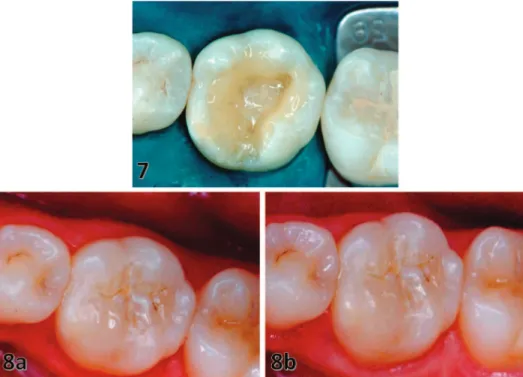
The selected dental specimen was decoronated and the coronal fragment adjusted with diamond points at high speed under air/water spray coolant until it fit the cavity. Articulating paper was interposed between the fragment and the cavity in the plaster cast to demarcate the areas that needed further adjustment. The extracted tooth had been previously sterilized by autoclaving, in accordance with biosecurity stan-dards. At the second visit, prophylaxis was completed and the adaptation of the fragment to the tooth was checked (Figure 4). Acid-etching with a 37% phos-phoric acid gel (3M/ ESPE, St Paul, MN, USA) was extended approximately 2 mm beyond the margin of the cavity for 15 seconds; the fragment was acid-etched for 15 seconds and was subsequently washed (Figure 5). Single Bond (3M/ESPE) adhesive system was applied in two consecutive layers on the tooth and fragment, and each side was light-cured for 20 seconds using a visible light-curing device (XL 3000, 3M/ESPE) (Figure 6). The fragment was bonded with a dual-cure, resin-based cement, shade A2 (Rely X ARC, 3M/ESPE) (Figure 7) and light-cured for 60 seconds. Small imperfections were corrected with light-curing composite resin (Filtek Z250, 3M/ESPE) and the occlusion checked with articulating paper. Figure 8a illustrates the final clinical aspects of the restoration and the three-year follow-up (Figure 8b).
After seven days, the patient was readmitted to perform the treatment in the maxillary right first molar (Figure 9), following the same standards used for the above procedure. Since cavity preparation was deeper, the cavity floor was protected with a calcium hydroxide cement layer (Dycal, Dentsply Ind. Com. Ltda), and a resin-modified glass ionomer cement base was used to replace dentin tissue (Vitrebond, 3M/ESPE). The steps for selection of a tooth compat-ible with the remaining tooth structure, cutting, and adaptation of the fragment to the plaster cast were the same as described previously. The prepared cavity received a temporary restoration, and at the second visit, the same steps described above were followed. Once treatment was concluded, oral hygiene instruc-tions were provided and the need for periodic evaluations was emphasized. Figure 10a illustrates the final aspects of the restoration and the three-year follow-up (Figure 10b).
DISCUSSION
reconstruction of the dental margins, 2) minimal need for dental restorative material, 3) durability and preservation of the remaining tooth structure, 4) resilience comparable to that of the original tooth, and 5) excellent esthetic results compared to com-posite resins and stainless-steel crowns, providing good translucency.1,2,7,8 In addition, this biological restoration allows maintenance of pulp vitality11and has low cost.12In spite of being simple, the technique requires professional expertise to adequately pre-pare and adapt the natural crowns to the cavity.13 Disadvantages of the biological restoration tech-nique include 1) the difficulty in selecting the fragment to adequately meet the natural tooth color, especially in cases involving partial destruction of the crown, and 2) the possibility of nonacceptance by the patient, as the technique involves a carrier of tooth fragment from another individual.1 It is important to inform the patient or his/her parents (legal guardians) that prior to clinical use, tooth
fragments are submitted to a rigorous sterilization process in an autoclave at 1218C for 15 minutes, ensuring all biosecurity standards.14The advantag-es and disadvantagadvantag-es of the technique and treatment alternatives must also be provided to enable patients to choose in an informed manner what they understand as the most appropriate treatment option in each case.
Although over the course of many years amalgam has been referenced as the material of choice to restore posterior teeth in different parts of the world, in spite of its relatively low and long-term cost effectiveness,15,16 esthetic limitation remains a dis-advantage. Likewise, gold inlays are still indicated for larger restorations that need support to with-stand intense masticatory stress, presenting unsur-passable longevity with minimal w ear on antagonists, and in contrast to amalgam, this material is not susceptible to corrosion.17 Direct Figure 1. Extensive amalgam resto-ration without marginal adaptation in the mandibular left first molar (#19). Figure 2. The area to be restored was isolated with rubber dam, and retentive areas were eliminated. Figure 3. The mesiodistal, cervico-occlusal, and buccolingual dimen-sions of the tooth were measured to facilitate the selection of an extracted tooth from the HTB with coronal length, height, and width that best fit the prepared tooth.
Figure 4. Checking the adaptation of the fragment.
composite resin restorations present the following advantages: 1) these restorations involve a low cost compared to other esthetic materials; and 2) these restorations involve single-session procedures and do not requirement temporary restorations, reducing chair time and dispensing a second session for cementation. In the present clinical case, the patient
expressed a desire for an esthetic restoration. Hence, treatment options discussed with the patient and her parents included direct composite resin restorations, porcelain inlay, and biological restoration. Since no synthetic material is capable of replicating the esthetic characteristics or color stability of natural teeth,18composite resin restorations are less esthetic
Figure 9. Extensive amalgam resto-ration without marginal adaptation in the maxillary right first molar (#3). Figure 10. Final clinical aspects of restoration (a) and three-year follow-up (b).
Figure 7. The fragment was bonded with a dual-cured, resin-based ce-ment.
compared to the biological restoration technique. In spite of a necessary laboratory stage, this technique requires a relatively short clinical time compared to other esthetic restorative procedures and offers superior physical properties compared to composite resins.17 Porcelain inlay is a more expensive tech-nique that may require greater tooth wear to provide an adequate dental preparation for indirect restora-tion. In young patients, it is desirable to preserve dental structure in order to avoid or postpone the progression toward endodontic treatment and porce-lain-metal crown restorations or future tooth loss and implant rehabilitation, justifying the treatment alternative chosen for our 15-year-old patient. The biological restoration technique is a more conserva-tive clinical approach, one that offers greater durability, better cost-effectiveness, and shorter chair time, which in turn allows natural results in terms of anatomic shape, surface shine, smoothness, and translucence of the enamel, when compared to other choices of treatment.
The first report in the literature of the use of fragments of extracted teeth as dental restorative materials was published in 1964 by Chosak and Eidelman,19 and the expression ‘‘biological restora-tion’’ was first coined by Santos and Bianchi20 in 1991. Busato and others2described the technique of using human teeth from Tooth Banks in large restorations, emphasizing the greater resistance of teeth restored with tooth fragments, as compared to restorations with composite resin materials. The author presented a two-year evaluation of a clinical case, showing that the biological restoration tech-nique has extraordinary clinical potential and social impact. Tavano and others8 presented the esthetic and functional rehabilitation of an upper left central incisor (#9) through homogeneous bonding of a dental fragment. Biological restoration was used to restore this incisor because the patient did not have the original tooth fragment itself. After a one-year follow-up, the results obtained were highly satisfac-tory. In 2010, Correˆa-Faria and others1 reported a clinical case performed by means of biological restoration using homogeneous fragment bonding associated with biological posts to reconstruct an extensively fractured central maxillary incisor and after one year demonstrated excellent results. Carvalho and others21 described a clinical case demonstrating the quality and functionality of a biological restoration performed to reestablish func-tion and esthetics to a left maxillary first premolar (#24) that presented fracture of the entire buccal region. A 12-month follow-up indicated a stable
restoration. In addition, this technique has also been described as an alternative to the reconstruction of extensively destroyed deciduous teeth.13,22 Sanches and others13 reported on two young children, aged four and five years, in whom biological restorations using tooth fragments were placed in primary molars with severely damaged crowns due to extensive carious lesions. The restorations were bonded to the remaining tooth structure with either adhesive system (case 1) or dual-cure, resin-based cement (Enforce, Dentsply Ind. Com. Ltda) (case 2) over a calcium hydroxide layer and a glass ionomer cement base. Periodical clinical and radiographic controls were carried out and the restored teeth were followed for four and three years, respectively, until exfoliation. Thus, biological restoration technique using tooth fragments has a practical clinical applicability and may present as an interesting treatment alternative when treating pediatric pa-tients.
Currently, with the existence of HTBs and the characteristics of the adhesive materials, rehabilita-tion of extensively destroyed teeth with this tech-nique became possible and quite feasible.1,8,10 Therefore, there is a need to organize the function-ality of HTBs standardizing autoclave sterilization for 40 minutes according to the American Dental Association23 and the Centers for Disease Control and Prevention (CDC).24 This method does not alter the physical properties of the dentinal tissues and does not compromise the goals and/or results of the application of these teeth in therapeutics.23,24 In addition, the proper storage with saline solution, water, and disinfectants, as recommended by the CDC, is essential to maintaining the chemical, physical, and mechanical properties of these teeth.24 All of these precautions must be followed carefully, thereby eliminating the possibility of transmission of pathogenic microorganisms.
as long as standard of care and treatment options are carefully considered in all cases.
CONCLUSION
In conclusion, the restoration technique using biological fragments of teeth is a feasible restorative option for adolescents, showing excellent clinical applicability, in addition to great cost effectiveness, for the restoration of permanent molars with crowns containing extensive amalgam restorations required for replacement due to secondary caries or marginal failure.
Conflict of Interest
The authors have no proprietary, financial, or other personal interest of any nature or kind in any product, service, and/or company that is presented in this article.
(Accepted 23 June 2014)
REFERENCES
1. Correˆa-Faria P, Alcaˆntara CEP, Caldas-Diniz MV, Botel-ho AM, & Tavano KT (2010) ‘‘Biological restoration’’: Root canal and coronal reconstructionJournal of Esthetic and Restorative Dentistry22(3)168-178.
2. Busato ALS, Loguercio AD, Barbosa NA, Sanseverino MCS, Macedo RP, & Baldissera RA (1998) Biological restorations using tooth fragmentsAmerican Journal of Dentistry11(1)46-48.
3. Ramires-Romito ACD, Wanderley MT, Oliveira MDM, Imparato JCP, & Coˆrrea MSNP (2000) Biologic restora-tion of primary anterior teethQuintessence International
31(6)405-411.
4. Terry DA (2003) Adhesive reattachment of a tooth fragment: The biological restorationPractical Procedures & Aesthetic Dentistry15(5)403-409.
5. Buonocore MG (1955) A simple method of increasing the adhesion of acrylic filling material to enamel surfaces Journal of Dental Research34(6)849-853.
6. Tenery TN (1978) The fractured tooth reunited using the acid etch bonding techniquesTexas Dental Journal96(8)
16-17.
7. Nogueira Filho GR, Machion L, Teixeira FB, Pimenta LA, & Sallum EA (2002) Reattachment of an autoge-nous tooth fragment in a fracture with biologic width violation: A case report Quintessence International
33(3)181-184.
8. Tavano KT, Botelho AM, Motta TP, & Paes TM (2009) Biological restoration: Total crown anteriorDental Trau-matology25(5)535-540.
9. Demarco FF, Moura FRR, Tarquinio SBC, & Lima FG (2008) Reattachment using a fragment from an extracted tooth to treat complicated coronal fracture—Case report Dental Traumatology24(2)257-261.
10. Yilmaz Y, Zehir C, Eyboglu O, & Belduz N (2008) Evaluation of success in the reattachment of coronal fracturesDental Traumatology24(2)151-158.
11. Chu FCS, Yim TM, & Wei SHY (2000) Clinical consider-ations for reattachment of tooth fragmentsQuintessence International31(6)385-391.
12. Ehrmann EH (1989) Restoration of a fractured incisor with exposed pulp using original tooth fragment: Report of a caseJournal of American Dental Association118(2)
118-183.
13. Sanches K, de Carvalho FK, Nelson-Filho P, Assed S, Silva FW, & de Queiroz AM (2007) Biological restoration as a treatment option for primary molars with extensive coronal destruction—Report two cases Brazilian Dental Journal18(3)248-252.
14. Imparato JCP (2003) Banks of Human Teeth Maio: Curitiba, Brazil [in Portuguese].
15. Alcaraz MGR, Veitz-Keenan A, Sahrmann P, Schmidlin PR, Davis D, & Iheozor-Ejiofor Z (2014) Direct composite resin fillings versus amalgam fillings for permanent or adult posterior teeth Cochrane Database Systematic Review 31(3)Article Number: CD005620. DOI: 10.1002/ 14651858.CD005620.pub2.
16. Forss H, & Widstrom E (2004) Reasons for restorative therapy and the longevity of restorations in adultsActa Odontologica Scandinavica62(2)82-86.
17. Hopp CD, & Land MF (2013) Considerations for ceramic inlays in posterior teeth: A review Clinical Cosmetic Investigational Dentistry18(5)21-32.
18. Grewal N, & Seth R (2008) Comparative in vivo evaluation of restoring severely mutilated primary anterior teeth with biological post and crown preparation and reinforced composite restorationsJournal of Indian Society Pedodontics Preventive Dentistry26(4)141-148.
19. Chosack A, & Eidelman E (1964) Rehabilitation of a fractured incisor using the patient’s natural crown—Case reportJournal of Dentistry for Children3119-21.
20. Santos JFF, & Bianchi J (1991) Restoration of severely damaged teeth with resin bonding systems: Case reports Quintessence International22(8)611-615.
21. Carvalho MF, Botelho AM, Tavano KT, & Fernandes VC (2013) Biological restoration: A 4/5 crownJournal of the Indian Society of Pedodontics and Preventive Dentistry
31(4)282-285.
22. Barcelos R, Neves AA, Primo L, & de Souza IP (2003) Biological restoration as an alternative treatment for primary posterior teeth Journal of Clinical Pediatric Dentistry27(4)305-310.
23. American Dental Association (2003) Handling extracted teeth; Retrieved online January 1, 2012 from: http://www. ada.org/sections/professionalResources/pdfs/