Xylazine Activates Adenosine
Monophosphate-Activated Protein Kinase
Pathway in the Central Nervous System of
Rats
Xing-Xing Shi1, Bai-Shuang Yin2, Peng Yang1, Hao Chen1, Xin Li1, Li-Xue Su1, Hong-Gang Fan1, Hong-Bin Wang1*
1Department of Veterinary Surgery, College of Veterinary Medicine, Northeast Agricultural University, Harbin, Heilongjiang Province, People’s Republic of China,2Department of Veterinary Medicine, Jilin Agriculture Science and Technology College, Jilin, Jilin Province, People’s Republic of China
Abstract
Xylazine is a potent analgesic extensively used in veterinary and animal experimentation.
Evidence exists that the analgesic effect can be inhibited using adenosine 5’
-monophos-phate activated protein kinase (AMPK) inhibitors. Considering this idea, the aim of this study was to investigate whether the AMPK signaling pathway is involved in the central analgesic mechanism of xylazine in the rat. Xylazine was administrated via the intraperito-neal route. Sprague-Dawley rats were sacrificed and the cerebral cortex, cerebellum, hippo-campus, thalamus and brainstem were collected for determination of liver kinase B1 (LKB1)
and AMPKαmRNA expression using quantitative real-time polymerase chain reaction
(qPCR), and phosphorylated LKB1 and AMPKαlevels using western blot. The results of our
study showed that compared with the control group, xylazine induced significant increases in AMPK activity in the cerebral cortex, hippocampus, thalamus and cerebellum after rats
received xylazine (P<0.01). Increased AMPK activities were accompanied with increased
phosphorylation levels of LKB1 in corresponding regions of rats. The protein levels of
phos-phorylated LKB1 and AMPKαin these regions returned or tended to return to control group
levels. However, in the brainstem, phosphorylated LKB1 and AMPKαprotein levels were
decreased by xylazine compared with the control (P<0.05). In conclusion, our data
indi-cates that xylazine alters the activities of LKB1 and AMPK in the central nervous system of rats, which suggests that xylazine affects the regulatory signaling pathway of the analgesic mechanism in the rat brain.
Introduction
Xylazine is exclusively used as a sedative, analgesic, and muscle relaxant in veterinary medicine, and is marketed as Rompun, Anased, Sedazine, Megaxilor, Paxman and Chanazine [1,2]. OPEN ACCESS
Citation:Shi X-X, Yin B-S, Yang P, Chen H, Li X, Su L-X, et al. (2016) Xylazine Activates Adenosine Monophosphate-Activated Protein Kinase Pathway in the Central Nervous System of Rats. PLoS ONE 11 (4): e0153169. doi:10.1371/journal.pone.0153169
Editor:Renping Zhou, Rutgers University, UNITED STATES
Received:February 5, 2016
Accepted:March 24, 2016
Published:April 6, 2016
Copyright:© 2016 Shi et al. This is an open access article distributed under the terms of theCreative Commons Attribution License, which permits unrestricted use, distribution, and reproduction in any medium, provided the original author and source are credited.
Data Availability Statement:All relevant data are within the paper and its Supporting Information files.
Xylazine (N-(2,6-dimethylphenyl)-5,6-dihydro-4H-1,3-thiazin-2-amine) is an effective seda-tive and analgesic and has potential to use during surgical operations for pain relief to reduce discomfort and stress [1]. In animal experiments, xylazine is a component of the most common injectable anesthetic, ketamine-xylazine, which is used in rats, mice, hamsters, and guinea pigs [3]. Intravenous administration results in deep dose-dependent sedation, characterized by somnolence and low head carriage in horses [4]. Xylazine is a strongα2-adrenergic agonist whose effects are mediated via stimulation of centralα2-receptors.α2-adrenergic stimulation decreases the release of norepinephrine and dopamine in the central nervous system (CNS) resulting in sedation, muscle relaxation, and decreased perception of painful stimuli. Moreover, its actions may also be involved in cholinergic, serotonergic, dopaminergic,α1-adrenergic, his-taminergic, or opiate mechanisms [5]. Xylazine is absorbed, metabolized, and eliminated rap-idly. It diffuses extensively and penetrates the blood brain barrier, as expected due to the uncharged, lipophilic nature of the compound [6]. When xylazine and otherα2-adrenergic receptor agonists are administered, they distribute throughout the body within 30 to 40 min-utes. The sedative and analgesic effects of xylazine inhibit the transmission of neural impulses in the CNS [7]. As an agonist, xylazine leads to a decrease in neurotransmission of norepineph-rine and dopamine [6].
In recent years, there has been increasing interest in the study of the molecular and cellular mechanisms underlying general anaesthesia. Among the different mechanisms, research on the regulation of pain signaling by adenosine 5’-monophosphate activated protein kinase (AMPK) has become a main research focus [8]. The heterotrimeric protein AMPK plays a critical regu-latory role in cellular energy homeostasis and organismal metabolism [9]. This serine/threo-nine kinase is formed by anαcatalytic subunit and two regulatory subunits,βandγ[10,11]. In mammals, the catalyticαsubunit of AMPK has two isoforms,α1 andα2 [12]. AMPKα1 and AMPKα2 can be activated in response to pharmacological agents in a Liver kinase B1 (LKB1)-dependent manner [13]. LKB1 is a tumor suppressor gene mutated in the inherited cancer dis-order Peutz-Jeghers syndrome [14]. In addition to nucleotide binding, phosphorylation of Thr172 at AMPK is required for its activation, and several groups have demonstrated that LKB1 is constitutively active and phosphorylates AMPK at Thr172 of theαsubunit [15,16]. Genetic studies of tissue-specific deletion of LKB1 have revealed that LKB1 mediates the majority of AMPK activation in nearly every tissue type examined to date [17,18]. AMPK can also be phosphorylated in response to calcium flux via calcium/calmodulin-dependent kinase 2 (CAMKK2) kinase [19,20].
When activated by LKB1, AMPK elicits its effects by regulating the activities of key meta-bolic enzymes via suppression of the mammalian target of rapamycin complex 1 pathway [21, 22]. Recent evidence suggests that peripheral pain plasticity is promoted and potentially main-tained via changes in translation control that are mediated by mammalian target of rapamycin complex 1 and mitogen-activated protein kinase. Due to its significant sensor role in modulat-ing pathways in catabolic processes, AMPK activation is correlated with peripheral nerve injury- and incision-induced pain and the blockade of the development of chronic pain follow-ing surgery [8]. AMPK gene expression levels have been studied in electroacupuncture-induced analgesia [23]. Moreover, AMPK plays a unique role for drug development in the kinase area for pain because it is pharmacologically manipulated via activation rather than inhibition [8]. Altogether, the physiology, pharmacology, and therapeutic opportunities surrounding AMPK make it an attractive target for novel intervention for pain.
To the best of our knowledge, there are no data available on the gene expression patterns or protein levels of AMPK and its upstream regulator LKB1 in different brain regions of xylazine-treated rats. We measured the mRNA and protein levels of LKB1 and AMPKαin the cerebral cortex, cerebellum, hippocampus, thalamus and brainstem of rats following intraperitoneal Competing Interests:The authors have declared
injection of xylazine. Hence, to elucidate the mechanisms involved in intraperitoneal xylazine administration, the goal of the present study was to determine whether the LKB1-AMPK path-way is involved in xylazine-induced sedation in the CNS which would lead us to further under-standing in the field of veterinary anaesthesia and analgesia.
Materials and Methods
Ethics statement
All animal work was conducted according to the guidelines for the care and use of experimental animals established by the Ministry of Science and Technology of the People's Republic of China (Approval number: 2006–398), and was approved by the Laboratory Animal Manage-ment Committee of Northeast Agricultural University. All efforts were made to minimize ani-mal stress/distress.
Animals and experimental protocol
Healthy male Sprague-Dawley rats (n = 30) weighing approximately 160–180 g were obtained from the Animal Center of Harbin Medical University (Harbin, China). Animals were housed at constant room temperature and maintained under a 12/12-h light-dark cycle. Rats were allowed free access to commercial pellets (Animal Center of Harbin Medical University, Har-bin, China) for at least one week. All experiments were performed in rats that had been deprived of food for 24 h, but free access to water. After acclimatization, the rats were ran-domly assigned to control or xylazine groups. Six rats received intraperitoneal injection of saline (0.5 mL, control group) and were sacrificed 10 min later. Twenty-four rats in the xyla-zine group were further subdivided into four groups. After receiving an injection of xylaxyla-zine (5.2 mg/kg diluted in 0.5 mL saline), the rats were sacrificed 10 min (Xyl1 group), 20 min (Xyl2 group), 40 min (Xyl3 group) or 60 min (Xyl4 group) respectively. Six rats from each group were euthanized by cervical dislocation. A midline incision on the scalp was made to fully expose the sutures on the dorsal surface of the skull. The occipital, parietal and temporal skull plates were quickly removed with the use of bone rongeurs. Then the brains were immediately removed and placed in ice-cold slurry of 0.9% (w/v) NaCl. Five brain structures were dissected under a microscope: cerebral cortex, cerebellum, hippocampus, thalamus and brainstem. Dis-sected tissues were immediately frozen in liquid nitrogen and stored at−80°C for pending
analysis.
Quantitative real-time polymerase chain reaction (qPCR)
Total RNA was isolated from the brain tissues using TransZol reagent (TransGen Biotech, Bei-jing, China) following the manufacturer’s instructions, and the quality was assessed by spectro-photometric absorbance at 260/280 nm. First-strand complementary DNA (cDNA) synthesis was performed with 0.5μg of total RNA using the ReverTra Ace qPCR RT Master Mix with a
amplification by qPCR were synthesized by Sangon Biotech (Shanghai, China). All other reagents used in this study were of analytic grade.
Protein isolation and western blot analysis
Frozen tissues were homogenized in ice-cold radioimmunoprecipitation assay lysis buffer (Beyotime Biotechnology, Nanjing, China) containing protease and phosphatase inhibitors (Sangon Biotech) for 5 min, and then incubated on ice for 30 min. The homogenate was then centrifuged at 14,000 × g for 10 min at 4°C. After the supernatant was collected, protein con-centrations of the supernatants were determined with the bicinchoninic acid protein assay kit (Beyotime Biotechnology) according to the manufacturer’s protocol. The supernatants used as protein samples were boiled at 100°C for 5 min with 5 × SDS sample buffer (Beyotime Biotech-nology), which were equivalent to 50μg of protein. Samples were subjected to sodium dodecyl
sulfate polyacrylamide gel electrophoresis by using 10% (w/v) gel, followed by transfer onto nitrocellulose membranes using the Bio-Rad Wet Trans-Blot apparatus (Bio-Rad, Hercules, USA). Non-specific binding sites were blocked by incubation with 5% (w/v) non-fat dry milk freshly prepared in Tris-buffered saline containing 0.05% (v/v) Tween-20 (TBST) for 2 h at room temperature. The nitrocellulose membranes were then incubated with primary antibod-ies overnight at 4°C. After three washes with TBST, the membranes were incubated with appropriate secondary antibodies conjugated to horseradish peroxidase for 2 h at room tem-perature. After washing four times in TBST, the signals were visualized using enhanced chemi-luminescence (ECL) detection reagents (Advansta, Menlo Park, USA). The bands were scanned using a Tanon 5200 Imaging System (Tanon Science & Technology Co., Shanghai, China) with a 16-bit camera, and quantified by densitometry. The primary antibodies were used at the following dilutions, and obtained from the indicated sources: anti-p-AMPKα
(Thr172) diluted 1:3000 (Cell Signaling Technology, Beverly, USA), anti-AMPKαdiluted 1:3000 (Cell Signaling Technology), anti-p-LKB1 (Ser428) diluted 1:3000 (Cell Signaling Tech-nology), anti-LKB1 diluted 1:3000 (Cell Signaling TechTech-nology), and anti-β-actin diluted 1:5000 (Zhongshan Goldenbridge Biotech, Beijing, China). Secondary horseradish peroxidase-conju-gated antibodies (Zhongshan Goldenbridge Biotech) were used at a dilution of 1:5000.
Statistical analysis
All data are presented as the mean ± standard error of the mean (SEM). The mean mRNA expression ratio in the control group was designated as one. Statistically significant differences Table 1. Sequences of primers used for the quantitative real-time PCR.
Gene GenBank number Primer sequences (50-30) Product size (bp)
LKB1 NM_001108069 Forward: AGCCAAGAGGTTCTCCATCC 114
Reverse: CAGCGGTCCTTAGTGTCTGG
AMPKα1 NM_019142 Forward: GAAGTCAAAGCCGACCCAAT 116
Reverse: AGGGTTCTTCCTTCGCACAC
AMPKα2 NM_023991 Forward: ATGATGAGGTGGTGGAGCAG 117
Reverse: GTGAATGGTTCTCGGCTGTG
β-actin NM_031144 Forward: AGGGAAATCGTGCGTGACAT 163
Reverse: CCTCGGGGCATCGGAA
LKB1, liver kinase B1; AMPKα1, adenosine 5’-monophosphate-activated protein kinaseα1; AMPKα2, adenosine 5’-monophosphate-activated protein kinaseα2.
among the means were determined using one-way analysis of variance (ANOVA) followed by Tukey's post hoc tests. All calculations were performed using PASW Statistics 17 (SPSS Inc., Chicago, USA). A p value of<0.05 was considered statistically significant.
Results
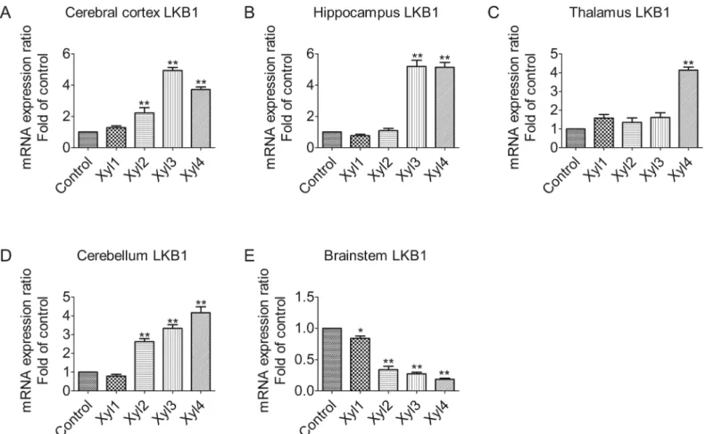
Xylazine alters mRNA levels of LKB1 in different brain regions of rats
Quantitative real-time PCR analysis revealed that treatment with 5.2 mg/kg of xylazine altered the expression of LKB1 mRNA in different rat brain regions (Fig 1). However, the magnitude of the alteration varied with time. In the cerebral cortex, the level of LKB1 increased slightly at 10 min (Xyl1, 1.3-fold increase, P>0.05) after drug administration compared with the controlgroup, peaked at 40 min (Xyl3, 4.9-fold increase, P<0.01), and then decreased rapidly, but
remained elevated at 60 min (3.7-fold increase, P<0.01,Fig 1A). In the hippocampus, the
level of LKB1 began to increase markedly at 40 min (Xyl3, 5.2-fold increase, P<0.01,Fig 1B).
Similarly, thalamic levels of LKB1 mRNA increased at 60 min (Xyl4, 4.1-fold increase, P<0.01,Fig 1C). The level of LKB1 decreased slightly at 10 min (Xyl1, 0.8-fold increase,
P>0.05) and then increased markedly (Xyl2, 2.6-fold increase, P<0.01) at 20 min in the
cere-bellum, and this elevation became more marked as time increased (Xyl4, 4.2-fold increase, Fig 1. Effect of xylazine administration on the mRNA levels of LKB1 in rats.(A) Cerebral cortex, (B) Hippocampus, (C) Thalamus, (D) Cerebellum and (E) Brainstem. Rats received saline (0.5 mL) or xylazine (5.2 mg/kg) intraperitoneally and then were sacrificed 10, 10, 20, 40 or 60 min later for control, Xyl1, Xyl2, Xyl3 or Xyl4, respectively. Total RNA was isolated and subjected to real-time PCR analysis. Each value of the expression levels of LKB1 was normalized to the expression levels ofβ-actin, and the control value was set to one. Data are presented as the means±SEM, n = 6.*P<0.05,**P<0.01 vs control.
P<0.01,Fig 1D). Unlike the previous four regions, there was a continuous downward
ten-dency of LKB1 mRNA levels in the brainstem. A marked decrease in mRNA expression was observed at 60 min (Xyl4, 0.2-fold decrease, P<0.01,Fig 1E).
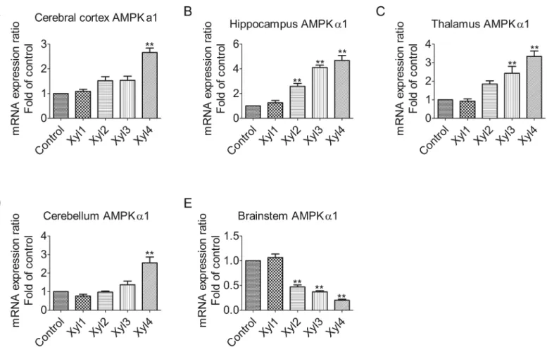
Xylazine alters the expression of AMPK
α
1 mRNA levels in different brain
regions of rats
Changes in AMPKα1 mRNA levels in the rat brain were determined by qPCR (Fig 2). The mRNA levels for AMPKα1 were raised after rats were intraperitoneally injected with xylazine in the cerebral cortex. A significant elevation was observed at 60 min (Xyl4, 2.7-fold increase, P<0.01,Fig 2A). The most obvious increase in mRNA expression was observed at 60 min
(Xyl4, 4.7-fold increase, P<0.01) in the hippocampus (Fig 2B). Whereas, in the thalamus, the
level of AMPKα1 mRNA began to increase significantly at 40 min (Xyl3, 2.4-fold increase, P<0.01). A further increase was seen at 60 min (Xyl4 3.3-fold increase, P<0.01,Fig 2C). As
shown inFig 2D, however, no significant differences were observed among the control, Xyl1, Xyl2 and Xyl3 groups for the cerebellum, but Xyl4 showed increased (2.6-fold increase, P<0.01) levels at 60 min. In contrast, xylazine treatment caused a marked downregulation of
Fig 2. Effect of xylazine administration on the mRNA levels of AMPKα1 in rats.(A) Cerebral cortex, (B) Hippocampus, (C) Thalamus, (D) Cerebellum and (E) Brainstem. Rats received saline (0.5 mL) or xylazine (5.2 mg/kg) intraperitoneally and then were sacrificed 10, 10, 20, 40 or 60 min later for control, Xyl1, Xyl2, Xyl3 or Xyl4, respectively. Total RNA was isolated and subjected to real-time PCR analysis. Each value of the expression levels of AMPKα1 was normalized to the expression levels ofβ-actin, and the control value was set to one. Data are presented as the means±SEM, n = 6.*P<0.05,**P<0.01 vs control.
AMPKα1 mRNA expression at 20 min (Xyl2, 0.5-fold decrease, P<0.01,Fig 2E) in the
brainstem.
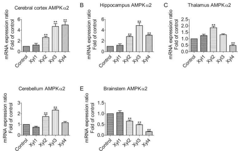
Xylazine alters the expression of AMPK
α
2 mRNA levels in different brain
regions of rats
We examined the expression pattern of AMPKα2 among different brain regions (Fig 3). The qPCR analysis showed that the relative expression levels of AMPKα2 tended to increase with time in the cerebral cortex. The level of AMPKα2 mRNA was markedly elevated at 60 min (Xyl4, 4.9-fold increase, P<0.01,Fig 3A). In the hippocampus, AMPKα2 mRNA was signifi-cantly upregulated at 40 min (Xyl3, 4.8-fold increase, P<0.01), but declined at 60 min (Xyl4,
3.1-fold increase, P<0.01,Fig 3B). Xylazine increased mRNA levels at 20 min (Xyl2, 1.8-fold
increase, P<0.01), with a subsequent decline at 60 min (Xyl4, 0.5-fold decrease, P<0.01,
Fig 3C) in the thalamus. In the cerebellum, the levels of AMPKα2 increased markedly at 20 min (Xyl2, 1.7-fold increase, P<0.01) and reverted to control levels (Xyl4, 1.2-fold increase,
P>0.05,Fig 3D). Consistent with the results of AMPKα1 in the brainstem, the progressive attenuation of AMPKα2 mRNA was observed at 20 min (Xyl2, 0.7-fold decrease, P<0.01,
Fig 3E).
Fig 3. Effect of xylazine administration on the mRNA levels of AMPKα2 in rats.(A) Cerebral cortex, (B) Hippocampus, (C) Thalamus, (D) Cerebellum and (E) Brainstem. Rats received saline (0.5 mL) or xylazine (5.2 mg/kg) intraperitoneally and then were sacrificed 10, 10, 20, 40 or 60 min later for control, Xyl1, Xyl2, Xyl3 or Xyl4, respectively. Total RNA was isolated and subjected to real-time PCR analysis. Each value of the expression levels of AMPKα2 was normalized to the expression levels ofβ-actin, and the control value was set to one. Data are presented as the means±SEM, n = 6.*P<0.05,**P<0.01 vs control.
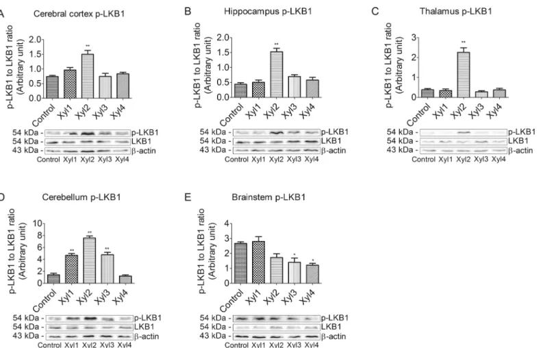
Effect of xylazine on phosphorylated LKB1 levels in different brain
regions of rats
To investigate whether the LKB1/AMPK signaling pathway was involved in analgesia, we first examined the phosphorylation of LKB1 at Ser428 using western blot (Fig 4). In the cerebral cortex, compared with the control group, the phosphorylation of LKB1 was significantly increased in the Xyl2 group at 20 min after drug administration and recovered at 40 min (P<0.01,Fig 4A). The change in LKB1 expression in the hippocampus or thalamus was
simi-lar to that in the cerebral cortex. However, the phosphorylated LKB1 levels increased at 20 min and decreased again at 30 min, while no significant changes were seen among the other groups in the hippocampus or thalamus (Fig 4B and 4C). The phosphorylation of LKB1 began to increase markedly at 10 min after xylazine administration (P<0.01) and recovered at 60 min
in the cerebellum of rats (Fig 4D). In the brainstem, phosphorylated LKB1 levels increased slightly at 10 min compared with the control group, and then began to decrease at 40 min (P<0.05,Fig 4E).
Fig 4. Effect of xylazine administration on the levels of phosphorylated LKB1 in rats.(A) Cerebral cortex, (B) Hippocampus, (C) Thalamus, (D) Cerebellum and (E) Brainstem. Rats received saline (0.5 mL) or xylazine (5.2 mg/kg) intraperitoneally and then were sacrificed 10, 10, 20, 40 or 60 min later for control, Xyl1, Xyl2, Xyl3 or Xyl4, respectively. Western blot analyses were performed with anti-LKB1 and anti-phospho-LKB1 (Ser428). Data for
densitometry represent the mean±SEM obtained from six independent series of Western blotting for each animal group and time point after the procedure.*
P<0.05,**P<0.01 vs control. Representative blots are shown below graph.
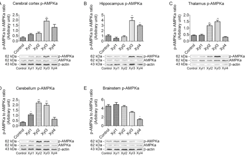
Effect of xylazine on phosphorylated AMPK
α
levels in different brain
regions of rats
We also examined the phosphorylation of AMPKαat Thr172. The brain tissues from rats that were intraperitoneally injected with saline or xylazine were subjected to western blot analysis using the anti-phospho-AMPKα(Thr172) and anti AMPKαantibodies (Fig 5). In the cerebral cortex, phosphorylated AMPKαwas slightly increased and peaked in the Xyl3 group at 40 min (P<0.01)
after drug administration, and then decreased. Levels remained elevated in the Xyl4 group (Fig 5A). Phosphorylation levels of AMPKαbegan to increase in the Xyl3 group and decline in the Xyl4 group in the hippocampus (Fig 5B). Thalamic levels of phosphorylated AMPKαincreased at 40 min and then returned to control levels at 60 min (Fig 5C). In the cerebellum, phosphorylated AMPKαlevels peaked at 20 min in Xyl2 rats and recovered in Xyl4 rats (Fig 5D). Levels of phos-phorylated AMPKαdid not significantly change in Xyl1 or Xyl2 rats compared with control. Phos-phorylation levels decreased in the brainstem at 60 min in the Xyl4 group (P<0.05,Fig 5E).
Discussion
It was proposed that adrenoceptors and cholinoceptors, which have properties in common with classicalα-adrenoceptors, were involved in the anti-nociceptive action of xylazine [25].
The anti-nociceptive effect of systemic, intrathecal or intracerebroventricular administration of xylazine has been reported [26]. It was shown that the spinal and systemic antinociceptive effects of xylazine are dependent on anα2-adrenoceptor-mediated, and not on an opioid-mediated mechanism [27]. At an individual level, conditioned pain modulation was related to blood oxygenation level-dependent responses in human secondary somatosensory cortices [28]. Moreover, the xylazine-induced reduction in cerebral blood flow could explain the reduced brain oxygenation observed in ketamine-xylazine anesthetized rats [29]. It has also been demonstrated that xylazine inhibits the release of cytokines and chemokines and thus contributes to the down-regulation of inflammatory pain and hyperalgesia [30]. However, the role analgesics play in regulating signal transduction has not been fully elucidated. The present work investigated this hypothesis at both the gene and protein level.
Gene expression and regulation is an important part in signal transduction. By using spa-tially and temporally regulated transcription factors in a concentration-dependent fashion, genes are able to be expressed in a precise, temporally and spatially controlled manner [31]. Therefore, we measured the mRNA levels of LKB1, AMPKα1, and AMPKα2 following treat-ment with xylazine in time and space. AMPK activity in neurons is regulated mainly via LKB1 in mice and rats [32]. LKB1 is a serine/threonine protein kinase that is highly expressed in the neurons of rats [33]. It is reported that mRNA levels of LKB1 and AMPKαdecrease signifi-cantly in response to fasting, and that refeeding normalizes this effect [34]. The results of our study showed that, compared with the sedative state, xylazine induced a significant decrease in the mRNA levels of LKB1 in the brainstem at 40 min after rats received xylazine, whereas a sig-nificant increase was observed in the cerebral cortex, hippocampus, thalamus, and cerebellum, suggesting that a region-dependent regulation mechanism may modulate LKB1 gene expres-sion at the transcriptional level in rats exposed to xylazine. However, it was showed that the rapid increase of AMPKαgene expression was necessary, but not sufficient to induce tissue plasminogen activator [35], which may affect synaptic plasticity in glucose-deprived rat pri-mary astrocytes. Moreover, xylazine restrained the excitement of the nervous system via increasing LKB1-AMPK gene expression in the cerebral cortex, hippocampus, thalamus and cerebellum, but decreased LKB1-AMPK gene expression in the brainstem. Furthermore, besta-tin improved the peripheral anbesta-tinociceptive effect of xylazine, suggesbesta-ting the mobilization of endogenous opioid peptides since the presence of mRNA or protein have been observed in the synthesis of opioid peptides [36]. Although the mechanisms by which LKB1 or AMPK regulate downstream protein translation in the rat CNS is not clear, the data from the present study sug-gests that the AMPK signaling pathway may be one of the key regulators involved in the xyla-zine-induced analgesic effect in the CNS of rats. These results indicate that xylazine may play an important role in influencing gene expression, and thus, more efforts should be made at the molecular level for future research.
the phosphorylation of Ser-428-LKB1 in the rat [32,40]. Taken together, these findings suggest that it is possible that AMPK activation is a consequence of anα2-adrenergic receptor-medi-ated effect and may involve LKB1. The present results provide evidence that the transcriptional activation of the LKB1-AMPK signaling pathway needs not only phosphorylation of LKB1 at Ser428, but also phosphorylation of AMPKαat Thr172 in the rat brain following treatment with xylazine. Furthermore, other upstream kinases need to be considered in the activation of AMPK in the brain in sedated rats. In this study, we examined the regulation of AMPK by phosphorylation in rats and found that phosphorylation at Thr172 of AMPK increased with a concurrent increase in the phosphorylation of Ser428 of LKB1 in the rat cerebral cortex, hippo-campus, thalamus and cerebellum after an intraperitoneal xylazine injection, whereas the phos-phorylation of Thr-172-AMPKα, as well as the phosphorylation of Ser-428-LKB1, began to decrease at 20 min in the Xyl2 rat brainstem. The similar trend in LKB1 activity and AMPK activity indicated that a LKB1-AMPK signaling pathway may exist in the brain structures of rats. However, the most marked increase in AMPK phosphorylation was observed at 40 min in the Xyl3 cerebral cortex, hippocampus and thalamus, while the phosphorylation of LKB1 reached a maximal level at 20 min in Xyl2 rats in the corresponding regions. These results, which indicate that changes in p-LKB1 are not always closely associated with parallel alter-ations in p-AMPK, suggest that LKB1 is not the sole upstream regulator of AMPK activation [34,41]. These studies suggest that AMPK kinase activity in the rat corresponds to LKB1, but it does not rule out the possibility that other AMPK activities exist in different tissues.
Conclusion
In summary, our study showed that xylazine administration altered mRNA expression and protein phosphorylation levels of AMPK signaling molecules, suggesting that the LKB1-AMPK pathway plays a role in the sedative and tranquilizing effects in the CNS caused by xylazine treatment. These results are the first to suggest that the central analgesic effect of xylazine is associated with the LKB1-AMPK signal transduction pathway. The results of this work con-tribute to a greater understanding of the central analgesia mechanisms of a drug widely used in veterinary anaesthesia and analgesia. However, more work needs to be done to elucidate the relationship between xylazine and the signal transduction pathways.
Supporting Information
S1 Table. Effect of xylazine administration on the mRNA levels of LKB1 in rats.Rats received saline (0.5 mL) or xylazine (5.2 mg/kg) intraperitoneally and then were sacrificed 10, 10, 20, 40 or 60 min later for control, Xyl1, Xyl2, Xyl3 or Xyl4, respectively. Total RNA was iso-lated and subjected to real-time PCR analysis. The relative expression levels of mRNA were analyzed using the 2−ΔΔCtmethod. Each value of the expression levels of LKB1 was normalized to the expression levels ofβ-actin. The mean mRNA expression ratio in the control group was designated as one. Statistical analyses were performed using one-way ANOVA followed by Tukey's post hoc tests.
(DOC)
S2 Table. Effect of xylazine administration on the mRNA levels of AMPKα1 in rats.Rats
was designated as one. Statistical analyses were performed using one-way ANOVA followed by Tukey's post hoc tests.
(DOC)
S3 Table. Effect of xylazine administration on the mRNA levels of AMPKα2 in rats.Rats
received saline (0.5 mL) or xylazine (5.2 mg/kg) intraperitoneally and then were sacrificed 10, 10, 20, 40 or 60 min later for control, Xyl1, Xyl2, Xyl3 or Xyl4, respectively. Total RNA was iso-lated and subjected to real-time PCR analysis. The relative expression levels of mRNA were analyzed using the 2−ΔΔCtmethod. Each value of the expression levels of AMPKα2 was normal-ized to the expression levels ofβ-actin. The mean mRNA expression ratio in the control group was designated as one. Statistical analyses were performed using one-way ANOVA followed by Tukey's post hoc tests.
(DOC)
S4 Table. Effect of xylazine administration on the levels of phosphorylated LKB1 in rats. Rats received saline (0.5 mL) or xylazine (5.2 mg/kg) intraperitoneally and then were sacrificed 10, 10, 20, 40 or 60 min later for control, Xyl1, Xyl2, Xyl3 or Xyl4, respectively. Western blot analyses were performed with anti-LKB1 and anti-phospho-LKB1 (Ser428). Data for densi-tometry were obtained from six independent series of Western blotting for each animal group and time point after the procedure. Densitometric analysis of p- LKB1 to LKB1 is represented as an arbitrary unit, normalized byβ-actin. Statistical analyses were performed using one-way ANOVA followed by Tukey's post hoc tests.
(DOC)
S5 Table. Effect of xylazine administration on the levels of phosphorylated AMPKαin rats.
Rats received saline (0.5 mL) or xylazine (5.2 mg/kg) intraperitoneally and then were sacrificed 10, 10, 20, 40 or 60 min later for control, Xyl1, Xyl2, Xyl3 or Xyl4, respectively. Western blot analyses were performed with anti-AMPKαand anti-phosphor-AMPKα(Thr172). Data for densitometry were obtained from six independent series of Western blotting for each animal group and time point after the procedure. Densitometric analysis of p-AMPKαto AMPKαis represented as an arbitrary unit, normalized byβ-actin. Statistical analyses were performed using one-way ANOVA followed by Tukey's post hoc tests.
(DOC)
Acknowledgments
I am greatly indebted to my supervisor, Professor Hong-Bin Wang, for his guidance on my manuscript and the experiment. And I would like to express my gratitude to all those who have helped me with my work.
Author Contributions
Conceived and designed the experiments: XXS HGF HBW. Performed the experiments: XXS PY HC XL LXS. Analyzed the data: XXS PY HC. Contributed reagents/materials/analysis tools: HBW BSY. Wrote the paper: XXS BSY HBW. Performed samples preparation: XMD HL GB JB HFL.
References
1. Doran GS, Bradbury LA. Quantitation of the anaesthetic xylazine in ovine plasma by LC–MS/MS. J Chroma-togr B. 2015; 997:81–4. doi:http://dx.doi.org/10.1016/j.jchromb.2015.06.005
3. Veilleux-Lemieux D, Castel A, Carrier D, Beaudry F, Vachon P. Pharmacokinetics of ketamine and xylazine in young and old Sprague-Dawley rats. J Am Assoc Lab Anim Sci. 2013; 52(5):567–70. Epub 2013/09/18. PMID:24041212; PubMed Central PMCID: PMCPmc3784662.
4. Daunt DA, Steffey EP. Alpha-2 adrenergic agonists as analgesics in horses. Vet Clin North Am Equine Pract. 2002; 18(1):39–46, vi. doi:10.1016/s0749-0739(02)00004-4PMID:12064181.
5. Hoffmann U, Meister CM, Golle K, Zschiesche M. Severe intoxication with the veterinary tranquilizer xylazine in humans. J Anal Toxicol. 2001; 25(4):245–9. Epub 2001/06/02. PMID:11386637.
6. Ruiz-Colon K, Chavez-Arias C, Diaz-Alcala JE, Martinez MA. Xylazine intoxication in humans and its impor-tance as an emerging adulterant in abused drugs: A comprehensive review of the literature. Forensic Sci Int. 2014; 240:1–8. Epub 2014/04/29. doi:10.1016/j.forsciint.2014.03.015PMID:24769343.
7. Delehant TM, Denhart JW, Lloyd WE, Powell JD. Pharmacokinetics of xylazine, 2,6-dimethylaniline, and tola-zoline in tissues from yearling cattle and milk from mature dairy cows after sedation with xylazine hydrochlo-ride and reversal with tolazoline hydrochlohydrochlo-ride. Vet Ther. 2003; 4(2):128–34. PMID:14506588.
8. Price TJ, Dussor G. AMPK: An emerging target for modification of injury-induced pain plasticity. Neurosci Lett. 2013; 557 Pt A:9–18. doi:10.1016/j.neulet.2013.06.060PMID:23831352.
9. Hardie DG, Carling D, Carlson M. The AMP-activated/SNF1 protein kinase subfamily: metabolic sensors of the eukaryotic cell? Annu Rev Biochem. 1998; 67:821–55. doi:10.1146/annurev.biochem.67.1.821PMID: 9759505.
10. Mackenzie RW, Elliott BT. Akt/PKB activation and insulin signaling: a novel insulin signaling pathway in the treatment of type 2 diabetes. Diabetes Metab Syndr Obes. 2014; 7:55–64. Epub 2014/03/13. doi:10.2147/ dmso.s48260PMID:24611020; PubMed Central PMCID: PMCPmc3928478.
11. Steinberg GR, Kemp BE. AMPK in Health and Disease. Physiol Rev. 2009; 89(3):1025–78. doi:10.1152/ physrev.00011.2008PMID:19584320.
12. Hardie DG. AMP-activated/SNF1 protein kinases: conserved guardians of cellular energy. Nat Rev Mol Cell Biol. 2007; 8(10):774–85. doi:10.1038/nrm2249PMID:17712357.
13. Shaw RJ, Lamia KA, Vasquez D, Koo S-H, Bardeesy N, Depinho RA, et al. The kinase LKB1 mediates glu-cose homeostasis in liver and therapeutic effects of metformin. Science. 2005; 310(5754):1642–6. doi:10. 1126/science.1120781PMID:16308421.
14. Shackelford DB, Shaw RJ. The LKB1-AMPK pathway: metabolism and growth control in tumour suppression. Nat Rev Cancer. 2009; 9(8):563–75. doi:10.1038/nrc2676PMID:19629071.
15. Hawley SA, Boudeau J, Reid JL, Mustard KJ, Udd L, Makela TP, et al. Complexes between the LKB1 tumor suppressor, STRAD alpha/beta and MO25 alpha/beta are upstream kinases in the AMP-activated protein kinase cascade. J Biol. 2003; 2(4):28. Epub 2003/09/27. doi:10.1186/1475-4924-2-28PMID:14511394; PubMed Central PMCID: PMCPmc333410.
16. Woods A, Johnstone SR, Dickerson K, Leiper FC, Fryer LGD, Neumann D, et al. LKB1 is the upstream kinase in the AMP-activated protein kinase cascade. Curr Biol. 2003; 13(22):2004–8. doi:10.1016/j.cub.2003.10.031 PMID:14614828.
17. Anderson KA, Ribar TJ, Lin F, Noeldner PK, Green MF, Muehlbauer MJ, et al. Hypothalamic CaMKK2 contrib-utes to the regulation of energy balance. Cell Metab. 2008; 7(5):377–88. doi:10.1016/j.cmet.2008.02.011 PMID:18460329.
18. Tamas P, Hawley SA, Clarke RG, Mustard KJ, Green K, Hardie DG, et al. Regulation of the energy sensor AMP-activated protein kinase by antigen receptor and Ca2+ in T lymphocytes. J Exp Med. 2006; 203 (7):1665–70. Epub 2006/07/05. doi:10.1084/jem.20052469PMID:16818670; PubMed Central PMCID: PMCPmc2118355.
19. Fogarty S, Hawley SA, Green KA, Saner N, Mustard KJ, Hardie DG. Calmodulin-dependent protein kinase kinase-beta activates AMPK without forming a stable complex: synergistic effects of Ca2+ and AMP. Biochem J. 2010; 426(1):109–18. Epub 2009/12/05. doi:10.1042/bj20091372PMID:19958286; PubMed Central PMCID: PMCPmc2830670.
20. Hurley RL, Anderson KA, Franzone JM, Kemp BE, Means AR, Witters LA. The Ca2+/calmodulin-dependent protein kinase kinases are AMP-activated protein kinase kinases. J Biol Chem. 2005; 280(32):29060–6. doi: 10.1074/jbc.m503824200PMID:15980064.
21. Gwinn DM, Shackelford DB, Egan DF, Mihaylova MM, Mery A, Vasquez DS, et al. AMPK phosphorylation of raptor mediates a metabolic checkpoint. Molecular cell. 2008; 30(2):214–26. doi:10.1016/j.molcel.2008.03. 003PMID:18439900.
22. Kahn BB, Alquier T, Carling D, Hardie DG. AMP-activated protein kinase: ancient energy gauge provides clues to modern understanding of metabolism. Cell Metab. 2005; 1(1):15–25. doi:10.1016/j.cmet.2004.12. 003PMID:16054041.
24. Livak KJ, Schmittgen TD. Analysis of Relative Gene Expression Data Using Real-Time Quantitative PCR and the 2−ΔΔCT Method. Methods. 2001; 25(4):402–8. doi:10.1006/meth.2001.1262PMID:11846609. 25. Schmitt H, Le Douarec JC, Petillot N. Antagonism of the antinociceptive action of xylazine, an
alpha-sympa-thomimetic agent, by adrenoceptor and cholinoceptor blocking agents. Neuropharmacology. 1974; 13 (5):295–303. Epub 1974/05/01. PMID:4412037.
26. Romero TR, Pacheco Dda F, Duarte ID. Xylazine induced central antinociception mediated by endogenous opioids and mu-opioid receptor, but not delta-or kappa-opioid receptors. Brain Res. 2013; 1506:58–63. Epub 2013/03/15. doi:10.1016/j.brainres.2013.02.030PMID:23485547.
27. Goodchild CS, Guo Z, Davies A, Gent JP. Antinociceptive actions of intrathecal xylazine: interactions with spi-nal cord opioid pathways. Br J Anaesth. 1996; 76(4):544–51. doi:10.1093/bja/76.4.544PMID:8652328.
28. Bogdanov VB, Vigano A, Noirhomme Q, Bogdanova OV, Guy N, Laureys S, et al. Cerebral responses and role of the prefrontal cortex in conditioned pain modulation: an fMRI study in healthy subjects. Behav Brain Res. 2015; 281:187–98. Epub 2014/12/03. doi:10.1016/j.bbr.2014.11.028PMID:25461267.
29. Lei H, Grinberg O, Nwaigwe CI, Hou HG, Williams H, Swartz HM, et al. The effects of ketamine-xylazine anes-thesia on cerebral blood flow and oxygenation observed using nuclear magnetic resonance perfusion imaging and electron paramagnetic resonance oximetry. Brain Res. 2001; 913(2):174–9. Epub 2001/09/11. PMID: 11549383.
30. Seo J-P, Son W-G, Gang S, Lee I. Sedative and analgesic effects of intravenous xylazine and tramadol on horses. J Vet Sci. 2011; 12(3):281–6. doi:10.4142/jvs.2011.12.3.281PMID:21897102.
31. Sunkin SM. Towards the integration of spatially and temporally resolved murine gene expression databases. Trends Genet. 2006; 22(4):211–7. Epub 2006/02/28. doi:10.1016/j.tig.2006.02.006PMID:16499990.
32. Park HG, Yi H, Kim SH, Yu HS, Ahn YM, Lee YH, et al. The effect of cyclosporine A on the phosphorylation of the AMPK pathway in the rat hippocampus. Prog Neuropsychopharmacol Biol Psychiatry. 2011; 35(8):1933– 7. Epub 2011/10/04. doi:10.1016/j.pnpbp.2011.09.008PMID:21963396.
33. Lu J, Cao Y, Cheng K, Xu B, Wang T, Yang Q, et al. Berberine regulates neurite outgrowth through AMPK-dependent pathways by lowering energy status. Exp Cell Res. 2015; 334(2):194–206. Epub 2015/04/19. doi: 10.1016/j.yexcr.2015.04.006PMID:25889370.
34. Hu X, Liu L, Song Z, Sheikhahmadi A, Wang Y, Buyse J. Effects of feed deprivation on the AMPK signaling pathway in skeletal muscle of broiler chickens. Comp Biochem Physiol B Biochem Mol Biol. 2016; 191:146– 54. Epub 2015/10/27. doi:10.1016/j.cbpb.2015.10.007PMID:26497445.
35. Cho KS, Joo SH, Choi CS, Kim KC, Ko HM, Park JH, et al. Glucose deprivation reversibly down-regulates tis-sue plasminogen activator via proteasomal degradation in rat primary astrocytes. Life Sci. 2013; 92(17– 19):929–37. Epub 2013/04/09. doi:10.1016/j.lfs.2013.03.011PMID:23562854.
36. Romero TRL, de Castro Perez A, de Francischi JN, Gama Duarte ID. Probable involvement of alpha(2C)-adrenoceptor subtype and endogenous opioid peptides in the peripheral antinociceptive effect induced by xylazine. Eur J Pharmacol. 2009; 608(1–3):23–7. doi:10.1016/j.ejphar.2009.02.019PMID:19236861.
37. Pradet-Balade B, Boulme F, Beug H, Mullner EW, Garcia-Sanz JA. Translation control: bridging the gap between genomics and proteomics? Trends Biochem Sci. 2001; 26(4):225–9. Epub 2001/04/11. PMID: 11295554.
38. Woods A, Dickerson K, Heath R, Hong SP, Momcilovic M, Johnstone SR, et al. Ca2+/calmodulin-dependent protein kinase kinase-beta acts upstream of AMP-activated protein kinase in mammalian cells. Cell Metab. 2005; 2(1):21–33. Epub 2005/08/02. doi:10.1016/j.cmet.2005.06.005PMID:16054096.
39. de Leeuw van Weenen JE, Parlevliet ET, Maechler P, Havekes LM, Romijn JA, Ouwens DM, et al. The dopa-mine receptor D2 agonist bromocriptine inhibits glucose-stimulated insulin secretion by direct activation of the alpha2-adrenergic receptors in beta cells. Biochem Pharmacol. 2010; 79(12):1827–36. Epub 2010/02/09. doi: 10.1016/j.bcp.2010.01.029PMID:20138024.
40. Liangpunsakul S, Wou SE, Wineinger KD, Zeng Y, Cyganek I, Jayaram HN, et al. Effects of WY-14,643 on the phosphorylation and activation of AMP-dependent protein kinase. Arch Biochem Biophys. 2009; 485 (1):10–5. Epub 2009/02/25. doi:10.1016/j.abb.2009.02.006PMID:19236843; PubMed Central PMCID: PMCPmc2692688.