Advances in knowledge about induced tooth movement
Part 1: The osteocytes
Alberto Consolaro1
Osteoblasts and clasts were primary targets for the understanding of bone biopathology. In recent years, evidence has shifted attention to the osteocytes. The biology of induced tooth movement and jaw orthopedics should research the role of osteocytes and the specific effects of mediators such as RANKL and sclerostin. The sclerostin represents a regulatory molecule: When more bone is necessary, osteocytes release less sclerostin, when it is necessary to inhibit bone formation, osteocytes release more sclerostin. RANKL is connected to local osteoclastogenesis in order to have more cells capable of reabsorbing the mineralized matrix. New therapeutic ways of controlling the metabolic bone diseases have been targeted at these mediators.
Keywords: Osteocytes. Mechanotransduction. Tooth movement. Sclerostin. RANKL.
Submitted: March 26, 2012 - Revised and accepted: March 31, 2012
» The author reports no commercial, proprietary, or financial interest in the prod-ucts or companies described in this article
Contact address: Alberto Consolaro E-mail: [email protected]
1 Full Professor, Bauru Dental School and Post-graduation courses at Ribeirão Preto
Dental School, University of São Paulo.
How to cite this article: Consolaro A. Advances in knowledge about induced tooth movement. Part 1: The osteocytes. Dental Press J Orthod. 2012 May-June;17(3):14-8. The osteocytes have always been placed in a sec-ond role in the study of the phenomena associated with tooth movement, as well as in bone biology and comprehension of the diseases involving our skel-eton. It was believed that osteocytes were included in the mineralized bone matrix and, thus, did not participate in bone metabolism, the responses to stimuli and aggression.
The dendritic shape of the osteocyte puts it in contact with 40 to 50 cells simultaneously, gener-ating among them a very efficient communicat-ing network, while scavengcommunicat-ing any deformation that the bone may suffer from deflections result-ing from compression and traction. This osteo-cytes communicating network acts as excellent
mechanotransductors and also are centrally in-volved in bone metabolism by releasing mediators that reaches bone surfaces.
As shown in numerous studies over the past five years, there is strong influence of osteocytes in bone remodeling and, by extension and consequence, os-teocytes must actively participate in the biopathology of the induced tooth movement, among which is the biology of orthodontic movement.
THE ORIGIN OF OSTEOCYTES: PRIMARILY MESENCHYMAL CELLS AND, SECONDARILY, DERIVED FROM OSTEOBLASTS!
mediators still in the embryo and fetus. The main mediator of differentiation and synthesizing ac-tivity in this intrauterine phase are the BMPs or osteomorphogenetics proteins. Mediators in the early stage, that determines the form of organs and structures, can be identified as morphogens, such as it is in these osteomorphogenetics proteins. In this osseodifferentiation and synthesis environment, much of the molecules of these mediators are even-tually included in the bone extracellular matrix to be mineralized later. Thus, it can be assured that any mineralized bone matrix has, naturally, osteo-morphogenetic proteins in its composition.
Once the skeleton is formed and adulthood is established, osteoblasts and osteocytes remain in bone environment. Many osteoprogenitor cells, pre-osteoblasts and tissue stem cells, formerly known as undifferentiated mesenchymal cells re-main on bone surfaces. In the bone marrow, con-tained and protected by trabeculae and cortical, there are many tissue stem cells, which can origi-nate almost infinitely new bone cells.
Osteoblasts on the surfaces of the trabecular and cortical bone, are polyhedral cells arranged side by side, like a real fence, railing, or palisade. Its polyhe-dral format allows, on one of its surfaces, bone matrix production, and, in the other surface, expose receptors to mediators located on adjacent connective tissue or bone marrow tissue. At the same time, laterally, osteo-blasts contact and interact with other osteoosteo-blasts to form a true cell layer covering bone surfaces.
In certain conditions the osteoblasts synthesize the bone matrix and mineralize it; in other conditions, as in inflamed and stressed areas, the mediators can induce osteoblasts and move the bone surface, remain on the periphery and command the clasts activity in the context of a osteo-remodeling unit or BMU.
In this bone matrix deposition many osteoblasts eventually end up included in gaps called osteo-plasts (Figs 1, 2 and 3). It was believed for many years that these cells would be trapped, almost by a passive mechanism, as if they had lost the moment to depart, and got involved in the newly deposited matrix. The passive role of osteocytes was proved untrue. On the contrary, these cells seem to per-form a central role in controlling bone remodeling and opposite reactions to certain stimuli.
THE LOCATION AND SHAPE OF OSTEOCYTES
Osteocytes comprise 90-95% of bone cells in an adult.15 These cells are included in the mineralized
bone matrix (Figs 1, 2 and 3) and now, as with os-teoblasts and clasts, we also have greater knowledge about the osteocytes and their functions.
Osteocytes are regularly distributed in the gaps in the bone matrix, also known as osteoplasts, and communicate with each other and with the cells of the bone surface by means of extensions of the canaliculi of 100 to 300nm thickness.3,4,5 They form
a true web with their extensions, one real network comparable to the neural network in the central nervous system (Figs 1, 2 and 3).
Within these tubules, where the cytoplasmic processes of each cell are (Figs 1, 2 and 3), circulates a fluid tissue that carries nutrients and mediators. These canaliculi with its working fluid and its ex-tensions communicate the osteocytes with each other and interconnected with the surface cells of cortical and trabecular bone, in addition to resident cells of the bone marrow.10 This communication can
be cell-cell by means of specialized junctions or me-diators (Figs 1, 2 and 3).
THE BONE MECHANOTRANSDUCTORS: OSTEOCYTES
The osteocytes network form a very sensitive 3D system that uptakes bone deformities. Any change in bone form during skeleton function can be captured by this sensitive network or web of osteocytes, and ex-tensions or mechanotransduction detection system. Exercise can increase bone structure by mechanical stimuli, initially, on this network scavenging strain.
The osteocytes individually pick up signals by mechanical deformation of their cytoskeleton. At the same time, the network in which each osteo-cyte participates, distributed throughout the bone structure, picks up deformations, overloads, deflec-tions and limitadeflec-tions of nutrients. The deformation of the cytoskeleton, the restriction of oxygen and of nutrient stress the osteocytes, which release me-diators to communicate with other osteoblasts and clasts on the bone surface and induce them to reac-tive or adapreac-tive phenomena.
osteocytes in mechanical stress and, thus, it in-creases the production of secreted and circulating mediators through the fluid that circulates in the canaliculi (Figs 1, 2 and 3) and from there to the re-spective periodontal and bone surfaces. Although included in the mineralized bone matrix in their osteoplasts, the osteocytes and its communicating network — by direct contact or mediators — can stimulate or inhibit bone formation and bone re-sorption in the “distant” cortical bone surface (Fig 3). The osteocytes in the bone marrow inside the bone, can influence the higher or lower production of clastic cells and osteoclastogenesis.
The osteocytes, therefore, have a strong influ-ence in the function of bone to adapt its shape ac-cording to the determination of functional demands, changing the mechanical stimuli into biochemical events, a phenomenon known as osteocyte mecha-notransduction.13 The osteocytes also play a role in
regulating the mineral metabolism9 and also induce
changes in the properties of bone matrix around it,12
but these functions were already better known. The skeleton is able to continuously adapt to mechanical loads by the addition of new bone to increase the ability to resist or remove bone in re-sponse to a lighter load or lack of use.6,8 The
osteo-cytes have a high interconnectivity and are consid-ered the bone mechanotransductors.
Osteocytes increases glucose-6-dehydrogenase phosphatase after a few minutes of load,18 a marker
for increased metabolism, as it occurs in cells asso-ciated with bone surface. Seconds after the applied load on the osteocytes, nitric oxide prostaglandins and other molecules such as ATP1 are increased.
Therefore, osteocytes, when facing induced loads, have the ability to release mediators, which stimulate the precursors of clasts or osteoclasto-genesis to differentiate into new clasts increasing the rate of resorption. Among these mediators the M-CSF or stimulating factor of colonies for macro-phages and RANKL should be higlighted.14 It can be
argued that osteocytes can command the activities of the clasts on bone surfaces according to function-al demand. The set or lacunocanfunction-alicular osteocyte system can be seen as a real endocrine body.4
THE OSTEOCYTES AND THE BIOLOGY OF ORTHODONTIC AND ORTHOPEDIC MOVEMENT
In micro-bone lesions that occur daily, osteo-cytes die by apoptosis, such as when the bone tis-sue is dried and heated. The death of osteocytes in areas with 1-2 mm damage, such as microfractures, can generate mediators that stimulate clasts, espe-cially RANKL,7 a group TNF cytokine. Preserving
the osteocytes is to prevent bone reabsorption and clinicians should know this in-formation to take better care of the surgical margins in bone surfaces. In orthodontics many procedures are surgical.
An example of osteocyte preservation can be the divided flap technique in periodontal treatments, which preserves the periosteum attached on the surface. The source of nu-trients in the bone are vessels of the periosteum. Preserving the periosteum means to keep alive the osteocytes so that its death does not induce the thin cortical alveolar bone resorp-tion, leading to an undesirable dehiscence or fenestration.
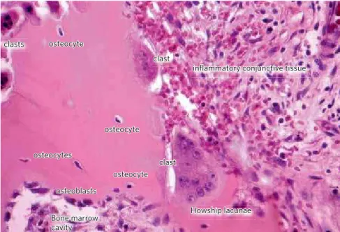
Figure 1 - The osteocyte network participates of the cellular functional control on bone surface, such as the clasts and osteoblasts. The cytoplasmatic prolongations arrive at the canaliculi and make contact with the surface cells or act via mediators (HE; 40X).
clast clasts
clast
infl ammatory conjunctive tissue
Howship lacunae osteocyte
osteocyte
osteocyte osteocytes
osteoblasts
Opening the periosteum inevitably leads to the death of the most superficial osteocytes, for they do not receive nutrients from broken vessels dur-ing this surgical procedure.
When the osteocytes die in bone remodeling tis-sue this area will inevitably be reabsorbed. Thus, the osteocytes should be preserved in the bony walls of the cavity prepared earlier to place the implants, avoiding excessive heat or improper manipulation of surfaces, since the death of osteocytes will lead to increased bone resorption at the site, which can disrupt osseointegration.
Probably some orthopedic facial responses can be explained by bone deformities produced. The re-sponses controlled by the osteocytes can change the shape and size of the bone to adapt to new functional demands. This increasingly requires further studies.
More recently the sclerostin was discovered, a mediator secreted by osteocytes, that circulates the fluid spaces of bone, especially in tubules with cytoplasmic osteocites extensions.16 It represents
a regulatory molecule: If you need more bone, os-teocytes release less sclerostin if you need to inhibit bone formation, osteocytes release more sclerostin. The osteocytes seem to play a central role in bone remodeling.2 On induced tooth movement there are
bone deformations and deflections for each activa-tion devices, especially in the interdental bone crest
and free surfaces. When moving a particular tooth to the lingual or buccal, it is known that on the out-side, bone is deposited on the cortical surface.17
In induced tooth movement with biologically acceptable forces, probably the stimulus released by the network of osteocytes on the farther part of the ligament is of mediators in type and amount required for inducing bone formation, while in the periodontal surface of the alveolar bone, the osteo-cytes stimuli captured by the network lead to bone permeation of mediators that stimulate osteoclas-togenesis and osteoclasia in the region.
In turn, in the tooth movement induced by ex-cessive force, the osteocytes die near the hyalinized ligament along one segment. Subjacent, the surviv-ing osteocytes release mediators, which stimulate the underlying and peripheral osteoclastogenesis, as RANKL, while release more sclerostin to inhibit bone formation at the site. All these phenomena are occurring in the subjacent or adjacent hyalinized periodontal space, i.e., at a distance.
These discoveries in bone biology have led to search for new therapeutic alternatives for the bone metabolic problems. Some substances are death in-hibitors of osteocytes on the skeleton as a whole and so promote less resorption, for example, estrogens and their modulators, bisphosphonates, calcitonin, CD40 ligand and others.2 There are still
anti-scleros-tin to help control bone loss in osteopenia and osteoporosis, the most common manifestations of various metabolic bone diseases.
CONCLUSIONS
The osteocytes form a three-dimensional network with each cell communicating with other 40-50 by numerous cytoplas-mic processes arranged like a real neural network. This com-munication is by cell contact and interaction, but particu-larly by mediators released by osteocytes in different amounts depending on the mechanical stimulus captured. Bone de-formation by compression and
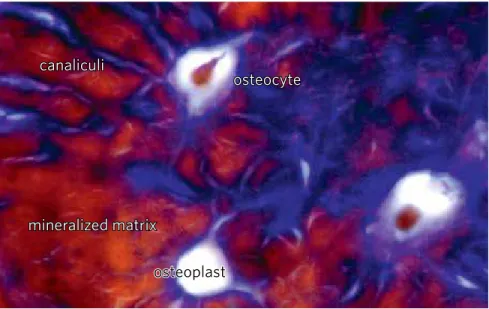
Figure 2 - The osteocytes have many cytoplasmatic prolongations, which intercommunicate with the mineralized matrix with other 20 to 40-50 cells and they detect minimal structural deformations and act as mechanotransducers. They occupy lacunae known as osteoplasts and the prolongations spread out as canaliculi, where mediators circulate in a tissue fluid, which performs ionic exchange with the mineralized extracellular matrix (Mallory, 100X).
mineralized matrix
osteoplast
traction during orthodontic movement stimulates these mechanisms by mediators released by osteo-cytes that virtually controls the formation and re-sorption of bone surfaces.
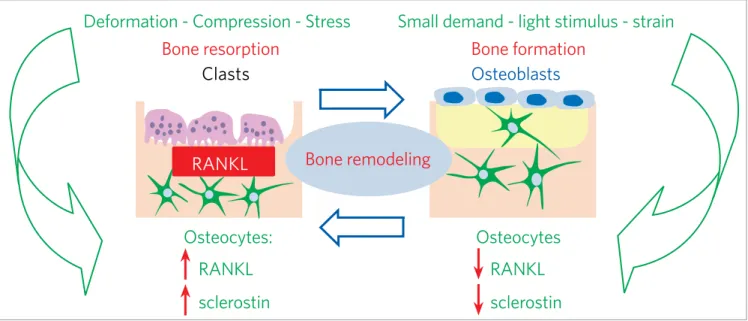
Figure 3 - The osteocytes detect shape and volume changes to increase or decrease the liberation of mediators involved in bone resorption or for-mation. In this manner, bone remodeling responds to the functional demand, modifying and adapting itself structurally (adapted from Nakasima et al,14 2011).
Osteoblasts
Clasts
Deformation - Compression - Stress
Small demand - light stimulus - strain
RANKL
Osteocytes:
RANKL
sclerostin
sclerostin
RANKL
Osteocytes
Bone resorption
Bone formation
Bone remodeling
To study the presence and specific effects of scleros-tin, of RANKL and of osteoprotegerin in the biology of induced tooth movement may represent several insights in Orthodontics and Facial Orthopedics researches.
1. Bakker AD, Soejima K, Klein-Nulend J, Burger EH. The production of nitric oxide and prostaglandin E(2) by primary bone cells is shear stress dependent. J J Biomech. 2001 May;34(5):671-7.
2. Baron R, Hesse E. Update on bone anabolics in osteoporosis treatment: rationale, current status, and perspectives. J Clin Endocrinol Metab. 2012 Feb;97(2):311-25.
3. Bonewald LF. Mechanosensation and transduction in osteocytes. Bonekey Osteovision. 2006 Oct;3(10):7-15.
4. Bonewald LF. Osteocytes as multifunctional cells. J Musculoskelet Neuronal Interact. 2006; 6(4): 331–3.
5. Bonewald LF. The amazing osteocyte. J Bone Miner Res. 2011 Feb;26(2):229-38. 6. Burr DB, Robling AG, Turner CH. Effects of biomechanical stress on bones in animals.
Bone. 2002 May;30(5):781-6.
7. Crockett JC, Rogers MJ, Coxon FP, Hocking LJ, Helfrich MH. Bone remodeling at a glance. J Cell Sci. 2011 Apr;124: 991-8.
8. Ehrlich PJ, Noble BS, Jessop HL, Stevens HY, Mosley JR, Lanyon LE. The effect of in vivo mechanical loading on estrogen receptor alpha expression in rat ulnar osteocytes. J Bone Miner Res. 2002 Sep;17(9):1646-55.
9. Feng JQ, Ward LM, Liu S, Lu Y, Xie Y, Yuan B, et al. Loss of dmp1 causes rickets and osteomalacia and identifies a role for osteocytes in mineral metabolism. Nat Genet. 2006 Nov;38(11):1310-5.
10. Kamioka H, Honjo T, Takano-Yamamoto T. A three-dimensional distribution of osteocyte processes revealed by the combination of confocal laser scanning microscopy and differential interference contrast microscopy. . Bone. 2001 Feb;28(2):145-9. REFERENCES
11. Krstic RV. Human microscopic anatomy. Berlin (DE): Springer-Verlag; 1994.
12. Lane NE, Yao W, Balooch M, Nalla RK, Balooch G, Habelitz S, et al. Glucocorticoid-treated mice have localized changes in trabecular bone material properties and osteocyte lacunar size that are not observed in placebo-treated or estrogen-deficient mice. J Bone Miner Res. 2006 Mar;21(3):466-76.
13. Lanyon LE. Osteocytes, strain detection, bone modeling and remodeling. Calcif Tissue Int. 1993;53 Suppl 1:S102-6; discussion S106-7.
14. Nakashima T, Hayashi M, Fukunaga T, Kurata K, Oh-Hora M, Feng JQ, et al. Evidence for osteocyte regulation of bone homeostasis through RANKL expression. Nat Med. 2011 Sep 11;17(10):1231-4.
15. Parfitt, AM. The cellular basis of bone turnover and bone loss: a rebuttal of the osteocytic resorption—bone flow theory. Clin Orthop Relat Res. 1977;(127):236-47.
16. Poole KE, van Bezooijen RL, Loveridge N, Hamersma H, Papapoulos SE, Löwik CW, et al. Sclerostin is a delayed secreted product of osteocytes that inhibits bone formation. FASEB J. 2005 Nov;19(13):1842-4.
17. Raab-Cullen DM, Thiede MA, Petersen DN, Kimmel DB, Recker RR. Mechanical loading stimulates rapid changes in periosteal gene expression. Calcif Tissue Int. 1994 Dec;55(6):473-8.