Karine Evangelista Martins Arruda1, José Valladares Neto2, Guilherme de Araújo Almeida3
Assessment of the mandibular symphysis of Caucasian Brazilian
adults with well-balanced faces and normal occlusion: The influence
of gender and facial type
original article
Objective: This study aimed to establish cephalometric reference values for mandibular symphysis in adults.
Den-toalveolar, skeletal and soft tissue variables were measured considering the influence of gender and facial type.
Methods: The sample consisted of sixty cephalometric radiographs of white Brazilian adult patients, with a mean
age of 27 years and 6 months, who had not undergone orthodontic treatment and who presented well-balanced faces and normal occlusion. The sample was standardized according to gender (30 males and 30 females) and facial type (20 were dolichofacial, 20 mesofacial and 20 brachyfacial).
Results: The results showed that male and female symphyses are similar, except for symphyseal height, which was
greater in males. In terms of facial type, the dolichofacial group presented narrower symphysis in dentoalveolar and basal areas, with a more accentuated lingual dentoalveolar inclination.
Conclusion: The brachyfacial group showed broader symphysis in the dentoalveolar and basal areas and a greater
buccal dentoalveolar inclination. The projection of the chin was 6.67 mm below the subnasal vertical line and there was no significant difference between the genders or facial types.
Keywords: Mandibular symphysis. Gender. Facial type. Facial balance.
How to cite this article: Arruda KEM, Valladares Neto J, Almeida GA. Assessment of the mandibular symphysis of Caucasian Brazilian adults with well-balanced faces and normal occlusion: The influence of gender and facial type. Dental Press J Orthod. 2012 May-June;17(3):40-50.
Submitted: September 01, 2008 - Revised and accepted: December 30, 2009
» The authors report no commercial, proprietary or financial interest in the products or companies described in this article.
» Patients displayed in this article previously approved the use of their facial and in-traoral photographs.
Contact address: José Valladares Neto R. 132, 113, lote 13 – Setor Sul – Goiânia/GO – Brazil Zip code: 74.093-210 – E-mail: [email protected]
1 MSc in Dental Clinic, FO-UFG. Specialist in Orthodontics, ABO/MG. 2 Assistant Professor of Preventive Orthodontics, FO-UFG. Professor of
Specialization course in Orthodontics, ABO/MG.
3 Associate Professor of Orthodontics, FO-UFU. Coordinator of Specialization
INTRODUCTION
Mandibular symphysis is an anatomical struc-ture of the mandible in which the lower incisors are found including the anterior portion of the chin. Mandibular symphysis contributes to the
compo-sition and balance of facial harmony2,15,25 and must
be considered when deciding on orthodontic
treat-ment in borderline cases.12,20,30
Mandibular symphysis is morphologically di-vided into two regions, the dentoalveolar and basal
symphyses.22 The dentoalveolar symphysis
in-cludes the alveolar process and lower incisors. The long axis of the lower incisors cephalometrically
matches the long axis of the alveolar process22 and
its inclination is influenced by facial type.16,29 This
classical concept dates from the Tweed era and defines the lingual inclination of the alveolar long axis (IMPA) in subjects with a high mandibular plane (FMA), while in subjects with low
mandibu-lar planes, the long axis is more buccally tipped.29
According to this view, the positioning error of the lower incisors could compromise the stability of
orthodontic results and facial esthetics.29
Alveolar bone thickness varies according to
location and facial type.12 Generally, there is a
greater bone thickness at the apex then in the cer-vical region, and towards the lingual surface when
compared to the labial surface.12 This explains the
higher prevalence of bone dehiscence and fenes-tration on the buccal side, and gives rise to peri-odontal concern about the anterior orthodontic
movement of the lower incisors.8
However, studies related to buccal projec-tion3,4,9,10,19,28,30 of lower incisors present conflicting
results, probably due to methodological differences and limitations, and the multifactorial etiology of
periodontal recession.31 However, thin buccal bone
coverage of the root10,12,28 associated to excessive
buccal movement31 and insufficient thickness of the
marginal gingiva have been shown19,31 to be
signifi-cant variables in the development of non-inflam-matory gingival recession.
In terms of cortical bone, the lingual side is thicker than the buccal, and due to the inclination of the lower incisors, there is a closer approximation of the root apex to the lingual cortical. This apex relationship is particularly evidenced in subjects
with vertical growth tendency12 and Class III
mal-occlusion.12,22 since the alveolar bone is very narrow
in this region. Bone in the referred apical region is assumed as non-remodelable anatomical limit and restricts the orthodontic retraction movement,
be-cause it can perforate the lingual cortical.12,20,24
The basal symphysis is part of the main body of the mandibular symphysis with more apical loca-tion, setting the hard menton outline. The menton is considered to be a recent phylogenetic
acquisi-tion ( just over 10,000 years ago), exclusive to Homo
sapiens. The morphological variation of the menton has a strong genetic basis and its occurrence may
have emerged casually14 and, did not add any
biome-chanical advantages for mastication.
The long axis of the basal symphysis differs
cepha-lometrically from that of the alveolar symphysis.22
Tooth movement of the lower incisors cannot influ-ence the shape or position of the basal symphysis. The relationship between the height and width of the mandibular symphysis is one of Björk’s five crite-ria for establishing the mandibular rotation pattern
during growth.1,5,6,27 For long and narrow symphyses,
the tendency of mandibular rotation during growth is predominantly vertical; when short and wide, it is
predominantly horizontal.5 In the vertical pattern,
a mandibular symphysis with a long axis and greater
lingual inclination has also been observed.12,16
The morphology of the mandibular symphysis is
also influenced by the sagittal growth pattern.12,16,22 In
Class III malocclusion, a higher,22 narrower12
symphy-sis with greater anterior projection16 and evident
lin-gual inclination of the long axis has been identified.16,22
In addition, the height and projection of the bas-al symphysis influence the position of the adjacent soft tissue and are significant in terms of aesthetic
and facial harmony.2,15,25 Menton deformities can be
treated satisfactorily using basilar genioplasty. For this procedure, it is necessary to establish norma-tive values for height and anterior projection, that are both influenced by ethnicity and sexual
dimor-phism. These values are usually higher in males.2
Despite its relevance, few studies have focused
on mandibular symphysis17,26 and its standard
Assessment of the mandibular symphysis of Caucasian Brazilian adults with well-balanced faces and normal occlusion: The influence of gender and facial type
original article
to describe the morphology of the mandibular sym-physis in a sample of Brazilian adults with well-bal-anced faces and normal occlusion, individualized in terms of gender and facial type variables.
SUBJECTS AND METHODS
The research project was submitted to the
Re-search Ethics Committee of Universidade Federal
de Uberlândia and approved under the protocol
number 247/07.
Sample selection
The total sample, composed of 60 subjects with well-balanced faces, equally divided between the genders, was prospectively selected from students of the Federal University of Goiás Dental School and complemented with subjects retrospectively selected from patients with minimum morpho-logical occlusion deviations from the researchers’ private clinics. The mean age of participants was 27 years and 6 months. The sample was also evenly distributed between the possible vertical varia-tions in terms of facial type (dolichofacial, mesofa-cial and brachyfamesofa-cial) (Table 1).
The following inclusion criteria had to be ful-filled by all participants: 1) be Brazilian; 2) Cauca-sian; 3) males over 18 and females over 16; 4) ANB between 0° and 4°; 5)well-balanced face; 6) ap-parent facial symmetry (clinically determined); 7) normal occlusion with Class I canine and molar relationship, overjet and overbite up to 3 mm and crowding up to 4 mm; 8) presence of all teeth, ex-cept third molars; 9) no serious medical condition; 10) no history of facial or dental trauma; 11) no pre-vious orthodontic or prosthetic treatment, facial plastic surgery or orthognathic surgery.
In this study, all the subjects showed a well-bal-anced face according to Capelloza’s Pattern I
descrip-tion.7,23 There were no skeletal discrepancies in sagittal
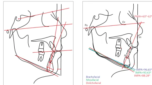
or vertical directions, and the profile was orthogna-thic, in other words, with gentle facial convexity, lips sealed when resting, the proportion of the facial thirds and the upper lip height were equal to half the height of the lower lip. In order to define the facial type, con-cordance between the subjective facial analysis and the angle of the mandibular plane (SN.GoGn) were used as criteria. Subjects were classified as mesofacial when SN.GoGn was between 30° and 34°, brachyfacial when less than 30° and dolichofacial when greater than 34°. For profile evaluation, the menton-neck line (length and angle) was used. Subjects were character-ized as brachyfacial when the line was elongated and the angle more open. For mesofacial subjects, the line was proportional and the angle close to 90°. For doli-chofacial subjects, the line was shortened and the an-gle reduced. For the frontal evaluation, the referential used was the width between gonion landmarks. This reference was comparatively larger for the brachyfa-cial type, balanced for the mesofabrachyfa-cial type and narrow for the dolichofacial type. Cases in which the facial analysis was not compatible with the SN.GoGn angle were excluded from the sample (Fig 1).
Cephalometric method
After the radiographs were taken, the cephalo-gram was performed by a single calibrated examiner. Ultraphan paper, a 0.5 mm propelling pencil, soft white eraser, ruler, protractor, square (Desetec) and lightbox were used. The tracings were performed us-ing predefined points, lines and planes in a dark room using black cardboard to protect the edges of the ra-diographic film. The values obtained were rounded off to 0.5 or the nearest whole number when decimal values were found. Radiographs were excluded when it was impossible to identify anatomical design.
The cephalometric landmarks used were (Fig 2): » Or (orbital): The lowest edge of the infraorbital
margin.
» Po (Porion): Highest edge of the external audi-tory canal.
» Gn (gnathion): Lowest and most anterior edge of the symphysis.
» Me (menton): The lowest edge of the menton symphysis outline.
» Go (gonion): The lowest and most posterior point of the gonial angle.
Brachyfacial Mesofacial Dolichofacial Total
Male 10 10 10 30
Female 10 10 10 30
Total 20 20 20 60
A
B
C
Figure 1 - Extraoral photographs (front and profile) and lateral radiographs with corresponding SN.GoGn values, representative of the female sample. Facial balance was classified into three facial types: A) Brachyifacial, B) mesofacial and C) dolichofacial.
SN.GoGn 26°
SN.GoGn 31.5°
Assessment of the mandibular symphysis of Caucasian Brazilian adults with well-balanced faces and normal occlusion: The influence of gender and facial type
original article
» Pog (pogonion): Most proeminent edge in the symphysis.
» Pog’ (soft pogonion): Most proeminent edge of menton soft tissue.
» Pog’’ (lingual pogonion): Suggested by Nojima
et al22, represents the most posterior point
lo-cated in the external lingual cortical of the man-dibular symphysis.
» Sn (subnasal): Point located at the junction be-tween the upper lip and the base of the nose. » IIi: The uppermost point of the lower incisor
in-cisal edge.
» AIi: Lowest point located at the root apex of the lower incisor.
» Sf: Midpoint between the outer lingual and out-er buccal corticals in the IIiAlipout-erp line, sug-gested by the authors of this study.
» Mi: Point on the mesiobuccal cusp tip of the lower first molar.
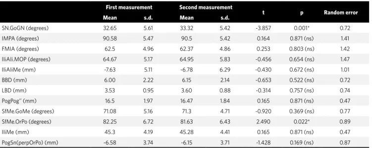
The lines and planes used were (Fig 3): » OrPo: Frankfurt horizontal plane. » GoMe: Mandibular plane.
» IIiAIi: Long axis of the lower incisors also representing the long axis of the alveolar symphysis.
» IIiAIiperp line: Tangent to the apex of the lower incisors perpendicular to their long axis as de-fined by the authors of this study.
» Sn perp Orpo: Line passing through the Sn, per-pendicular to the Frankfurt plane.
» SfMe: Long axis of the basal symphysis.
» IIiMi: Mandibular occlusal plane (MOP),
sug-gested by Arnett et al.2
The angular measurements used were (Fig 3): » SN. GoGn: Mandibular plane inclination in
re-lation to the base of the skull.
» IMPA (GoMe.IIiAIi): Lower incisor inclination in relation to the mandibular plane, also repre-senting the alveolar symphysis inclination. » FMIA (OrPo.IIiAIi): Lower incisor inclination
in relation to Frankfurt plane.
» IIiAIi.MOP: Lower incisor inclination in rela-tion to the mandibular occlusal plane.
» SfMe. GoMe: Inclination of the basal symphysis in relation to the mandibular plane.
» SfMe. Orpo: Inclination of the basal symphysis in relation to the Frankfurt plane.
The linear measurements evaluated were (Fig 3): » IIiAIiMe: Distance from the projection of the
long axis of the lower incisors on the mandibu-lar plane to the Me point.
» BBD: Buccal bone distance, comprising the thickness of the buccal alveolar bone at the apex of the lower incisors, measured from the AIi point to the external buccal cortical point, using the path of the IIiAIiperp line.
» LBD: Lingual bone distance, comprising the thickness of the lingual alveolar bone at the apex of the lower incisors, measured form the AIi point to the external lingual cortical point, using the path of the IIiAIiperp line.
» PogPog’’: Distance between the pogonian and the lingual pogonian points representing the thickness of the basal symphysis, suggested by
Nojima et al.22
» IIiMe: Height of the long axis of the mandibular symphysis.
» Pog’Sn (perpOrPo): Distance from the menton soft tissue to the subnasal line perpendicular to the Frankfurt plane.
Systematic error
In order to evaluate the systematic error, 20 ran-domly selected radiographs used in this study, were remeasured after 30 days. To determine intra-ex-aminer error, the paired t test was applied. Random
error was calculated using Dahlberg’stest13 when
error values greater than 1.5° or 1.0 mm were found. As noted in Table 2, systematic error was statistical-ly significant for SN.GoGn and SfMe.OrPo, but with a slight average difference (0.67° and 0.62°, respec-tively), irrelevant from the clinical point of view. The results revealed a random error less than 1.5° and 1.0 mm, indicating the reliability of the data.
Statistical Analysis
S
Po
Or N
Sn
IIi Mi
Go
AIi
Sf Pog’ Pog
Gn Me Pog’’
Or Po
Sn
IIi
AIi
FMIA=60°-62°
For the statistical treatment of data, the SPSS for Windows (version 16.0) was used, considering a significance level of 5% (a = 0.05).
RESULTS
Composition and characteristics of the sample
The sample consisted of subjects ranging from 18 to 38 years for males and 16 to 35 years for females. All subjects presented well-balanced faces, confirmed by subjective facial analysis and cephalometric measure-ments. The average ANB angle was 2.16±1.63°, indicat-ing harmony in the sagittal position of both maxilla and mandible, and the average SN.GoGn was 32.11±5.46°), which confirmed facial balance in the vertical position. Classification in terms of facial type was clearly estab-lished by SN.GoGn cutoff values (Fig 4).
In this study, the buccolingual inclination of the lower incisors represented the long axis of alveo-lar symphysis. The cephalometric measurements which contributed to this evaluation were IMPA, FMIA, IIiAIi.POM and IIiAIiMe. In general, the lower incisors were implanted perpendicular to the mandibular base (IMPA = 92.78°), buccally in relation to the Frankfurt horizontal plane (FMIA = 61.13°) and lower occlusal plane (IIiAIi.MOP= 63.10°) and the projection of the long axis of these teeth is about 9.51 mm after the Me point (Table 3).
The amount of buccal and lingual bone at the apex of the lower incisor was measured by BBD
and LBD widths, respectively. In this sample, the amount of buccal bone (BBD = 5.12 mm) was thick-er than the amount found for lingual bone (LBD= 3.55 mm) (Table 3). The long axis of the basal and alveolar symphyses was not aligned. The basal symphysis was inclined 22° lingually in terms of the dentoalveolar symphysis in relation to both the mandibular and Frankfurt planes (SfMe.GoMe = 70.33±5,44º and SfMe.OrPo = 83.13±6.50º). The width of the basal symphysis baseline was 15.61 mm (PogPog’’), considered almost twice (BBD LBD = 8.67 mm) that of the dentoalveolar symphysis at the apex of the lower incisors. Sym-physis height (IIiMe) was 44.78±3.79 mm and in terms of soft tissue, the projection of the Pog’ re-mained about 6.7 mm below the vertical subnasal line [Pog’-Sn(perp OrPo)] (Table 3).
Gender
Regarding gender, the results showed no statisti-cally significant difference for most cephalometric measurements. Hence, as a general rule, both male and female mandibular symphyses have a similar morphology, except for a slight inclination of the basal symphysis (SfMe.PoOr) and height (IIiMe).
The basal symphysis inclination in relation to the Frankfurt plane (SfMe.PoOr), was 84.97° for males and 81.28° for females, and this difference was sta-tistically significant at 5% level. However, caution
Figure 2 - Cephalometric landmarks used, em-phasizing the Sf.
Figure 3 - Lines, planes and cephalometric measurements.
Figure 4 - Variations in dentoalveolar symphy-sis inclination means (long axis of the lower incisors, measured using IMPA and FMIA) as a variation of the mandibular plane (FMA).
IMPA=96.65°
IMPA=93.43°
IMPA=88.28°
Brachyfacial
Mesofacial
Assessment of the mandibular symphysis of Caucasian Brazilian adults with well-balanced faces and normal occlusion: The influence of gender and facial type
original article
First measurement Second measurement
t p Random error
Mean s.d. Mean s.d.
SN.GoGN (degrees) 32.65 5.61 33.32 5.42 -3.857 0.001* 0.72
IMPA (degrees) 90.58 5.47 90.5 5.42 0.164 0.871 (ns) 1.41
FMIA (degrees) 62.5 4.96 62.37 4.86 0.253 0.803 (ns) 1.42
IIiAIi.MOP (degrees) 64.67 5.17 64.95 5.83 -0.456 0.654 (ns) 1.47
IIiAIiMe (mm) -7.63 5.11 -6.78 6.29 -0.430 0.672 (ns) 1.01
BBD (mm) 6.00 2.22 6.15 2.14 -0.653 0.522 (ns) 0.72
LBD (mm) 3.53 0.95 3.60 0.88 -0.314 0.757 (ns) 0.74
PogPog’’ (mm) 16.5 1.97 16.47 1.84 0.165 0.871 (ns) 0.47
SfMe.GoMe (degrees) 71.08 5.16 71.3 4.71 -0.920 0.369 (ns) 0.77
SfMe.OrPo (degrees) 82.25 6.72 81.63 6.43 2.490 0.022* 0.89
IIiMe (mm) 45.3 4.19 45.28 4.41 0.165 0.871 (ns) 0.47
PogSn(perpOrPo) (mm) -6.58 3.74 -6.15 3.71 -1.428 0.169 (ns) 0.87
Table 2 - Systematic error values (paired t test) and random error (Dahlberg).
Table 3 - Cephalometric characteristics of the total sample.
Variable Mean s.d. Maximum value Minimum value
SN.GoGn (degrees) 32.11 5.46 42 23
IMPA (degrees) 92.78 6.02 103 79.5
FMIA (degrees) 61.13 5.23 71 46
IIiAIi.MOP (degrees) 63.10 5.43 75 54
IIiAIiMe (mm) -9.51 3.11 -3 -19
BBD (mm) 5.12 1.70 12.5 2
LBD (mm) 3.55 1.07 6 1.5
PogPog” (mm) 15.61 2.13 21.5 11
SfMe.GoMe (degrees) 70.33 5.44 84 51.5
SfMe.OrPo (degrees) 83.13 6.50 96 71
IIiMe (mm) 44.78 3.79 55 39
Pog'Sn(perp OrPo) (mm) -6.66 3.88 1 -14
Total Gender Facial type
M F p Brachyfacial Mesofacial Dolichofacial p
SN.GoGN (degrees) 32.10 (±4.46) 32.91 (±4.43) 31.30 (±6.30) 0.255 26.50 (±2.12)A 31.65 (±1.10)B 38.17(±3.86)C 0.000
IMPA (degrees) 92.78 (±6.02) 93.63 (±5.45) 91.93 (±6.52) 0.278 96.65 (±4.58)A 93.42 (±5.00)A 88.27 (±5.38)B 0.000
FMIA (degrees) 61.12 (±5.23) 60.07 (±4.80) 62.18 (±5.51) 0.118 61.37 (±4.60) 61.00 (±4.68) 61.00 (±6.47) 0.967
IIi.MOP (degrees) 63.10 (±5.42) 63.31 (±5.29) 62.88 (±5.64) 0.760 60.67 (±4.09)A 62.60 (±5.29)AB 66.02 (±5.60)B 0.005
IIiAIiMe (mm) -9.50 (±3.10) -8.83 (±2.86) -10.18 (±3.24) 0.093 -10.37 (±2.07)A -10.07 (±4.17)AB -8.07 (±2.22)B 0.037
BBD (mm) 5.11 (±1.70) 5.27 (±2.04) 4.97 (±1.28) 0.499 5.72 (±2.00)A 5.35 (±1.52)AB 4.27 (±1.20)B 0.017
LBD (mm) 3.55 (±1.06) 3.57 (±1.13) 3.53 (±1.02) 0.905 4.22 (±0.86)A 3.37 (±1.15)B 3.05 (±0.82)B 0.001
PogPog” (mm) 15.60 (±2.12) 15.30 (±2.16) 15.91 (±2.08) 0.265 16.07 (±1.89)A 16.12 (±2.25)A 14.62(±1.96)B 0.038
SfMe.GoMe (degrees) 70.33 (±5.44) 71.45 (±5.98) 69.21 (±4.68) 0.113 71.42 (±4.37) 70.10 (±6.63) 69.47 (±5.17) 0.520
SfMe.OrPo (degrees) 83.12 (±6.50) 81.28 (±6.90) 84.96 (±5.60) 0.027 86.95 (±4.51)A 82.72 (±6.28)AB 79.70 (±6.60)B 0.001
IIiMe (mm) 44.77 (±3.79) 42.58 (±2.13) 46.97 (±3.85) 0.000 43.17 (±3.06)A 44.45 (±3.77)AB 46.70 (±3.79)B 0.010
PogSn(perpOrPo) (mm) -6.65 (±3.87) -6.27 (±3.89) -7.05 (±3.89) 0.439 -5.15 (±3.28) -6.90 (±3.89) -7.92 (±4.07) 0.071
should be exercised when evaluating this finding, because the systematic error was significant for this measurement (Table 4). Mean values for mandibu-lar symphysis height (IIiMe) were 46.97 mm and 42.58 mm, respectively, in both males and females. On average, male mandibular symphysis was 10% higher than female symphysis, and this finding was statistically significant (p < 0.00). Therefore, the height of the mandibular symphysis was consid-ered a distinguishing criterion between the gen-ders (Table 4).
Facial type
Facial type had no correlation with the FMIA, SfMe.GoMe or Pog’Sn (perpOrPo) measurements. IMPA and Pog’Pog’’ measurements were similar for brachyfacial and mesofacial types and LBD mea-surements were similar for mesofacial and dolicho-facial types SfMe.OrPo, BBD, IIiAIiMe, IIiMe and IIi.MOP were statistically different for the extreme facial types (dolichofacial and brachyfacial) but similar for the mesofacial type (Table 4).
DISCUSSION
This study described the cephalometric charac-teristics of the mandibular symphysis of a sample consisted of 60 Brazilian Caucasian adults resi-dents of the central region of the country, with an average age of 27 years and 6 months. Subjects presented well-balanced faces and normal occlu-sion. The measurements analyzed included dento-alveolar, skeletal and soft tissue structures of the mandibular symphysis and the main objective was to evaluate the influence of gender and facial type on the morphology of the symphysis. In this study, the distinction between facial types was made us-ing concordance between facial analysis and the SN.GoGn value. The cutoff value to characterize the mesofacial type was performed with a slight variation (2.0°) from the normative value (32°). Hence, when the facial features were compatible with a SN.GoGn less than 30°, the type was consid-ered well-balanced brachyfacial and dolichofacial when over 34°. From this sample, it can be seen that reading the SN.GoGn angle is quite adequate for evaluation of facial type, just as Tweed suggested
in relation to the FMA angle.29 The data obtained
in this study confirmed certain characteristics of the mandibular symphysis already described in the literature, but it also unprecedentedly showed the influence of certain measurements when drawing up individualized therapeutic targets for Brazilians.
Gender
The similarities between male and female man-dibular symphyses are evident, except in the case of height. The results in general showed significant morphological similarity between the dentoalveo-lar and basal symphyses, both in thickness and in-clination. The absence of sexual dimorphism for the IMPA angle has also been confirmed by other stud-ies17,26 involving normal occlusion.
The expectation of finding a male symphysis sta-tistically more prominent than the female was not confirmed in this study, same findings were
previ-ously reported by Scavone et al25 and Arnett et al.2
The results confirmed that both the width of the basal symphysis and its anterior projection are sim-ilar between the genders. The perception of a more projected mandibular symphysis in males may be explained by a greater vertical tendency and espe-cially by its greater height. On average, the height of the mandibular symphysis in males was 47 mm and 42.5 mm in females. This difference was statistical-ly significant (p = 0.0) and can thus be considered a differentiating factor between the genders.
Facial type
In this study, the sample was based on subjects with skeletally well-balanced faces, but with varia-tions in their mandibular plane angles. In addition to a subjective facial analysis, the subjects were cat-egorized into three distinct facial types: dolichofa-cial, mesofacial and brachyfacial. One of the main objectives of this study was to identify possible variations in the morphology of mandibular sym-physis from the premise of a variation in the facial morphology not involving the extremes.
Dolichofacial types presented features well
de-scribed in the literature,5,6,12,27 which include
Assessment of the mandibular symphysis of Caucasian Brazilian adults with well-balanced faces and normal occlusion: The influence of gender and facial type
original article
point (IIiAIiMe) in the dolichofacial types. These characteristics are typical morphological signs of subjects who are hyperdivergent or also called long faced. This study showed the tendency in the mandibular symphysis morphology in well-bal-anced dolichofacial type subjects and which prob-ably becomes more accentuated as the vertical gap increases. The average thickness of the alveolar symphysis in the region of the apex of the lower
in-cisors found by Handelman,12 in 1996, in patients
with a high mandibular plane was 5.5 mm. This re-sult was lower than the findings of this study for dolichofacial type people with a well-balanced fa-cial pattern (7.32 mm). However, there were meth-odological differences between the studies, such as the inclusion of patients with malocclusion, ex-treme vertical growth patterns and the different criteria for measuring the alveolar symphysis.
After adding the mean values of buccal and lin-gual thickness (BBD + LBD), the dolichofacial type group showed an average of 7.32 mm, while the aver-age for the mesofacial and brachyfacial type groups was 8.72 mm and 9.94 mm, respectively. These val-ues denote that the alveolar symphysis in the apical region of the lower incisors is on average 20% nar-rower in dolichofacial types.
For brachyfacial well-balanced faces, the most striking morphological feature was the greater thick-ness of the bone near the apex of the lower incisors, especially at the lingual region (LBD). In general, the findings of this study are in accordance with the liter-ature in terms of a wider and shorter symphysis, with a greater buccal inclination of the dentoalveolar and basal symphyses for brachyfacial types.
The cephalometric IMPA measurement was in-fluenced by facial type. The mean values were 88.27°, 93.42° and 96.65°, respectively, for the dolichofacial, mesofacial and brachyfacial types. Tweed’s
con-cept,7,29 is summarized as inclining the incisors and
the alveolar portion in the buccal direction as the tendency to grow becomes more horizontal.
In contrast, the FMIA measurement, which evaluates lower incisor inclination in relation to the Frankfurt plane, was less variable with the os-cillation of the mandibular plane. According to the results, this angle ranged between 60° and 62° for most patients (Fig 4).
Clinical implications
For the surgical orthognathic planning in cases of menton deformities, a comparison with norma-tive values is needed. Thus, the extent of the sur-gical movement depends on the pre-sursur-gical mea-surement of the height and anterior symphysis projection of the face. The height of the mandibu-lar symphysis recommended for male and female Caucasian North Americans is 44 mm and 40 mm,
respectively.2 This study found higher mandibular
symphyses, 47 mm and 42.5 mm, respectively. In other words, a 10% greater proportion for males was maintained, just the absolute value increased.
The expression of a higher mandibular symphy-sis and a lesser anterior projection in white Cauca-sian Brazilians contrasts when compared to North Americans. An average position of 6.67 mm below the subnasal line perpendicular to the Frankfurt plane was found, and it is worth noting that no sig-nificant difference was found between the genders.
In North American Caucasians2 the value found was
3.5±1.8 mm for males and 2.6±1.9 mm for females, with a differential methodology in the use of the natural head position. However, the lesser projec-tion of the menton in white Caucasian Brazilians
has also been confirmed by other studies15,25 (Fig 5).
Because of this difference, the use of normative value guideline of samples from North American Cau-casians has been questioned for therapeutic
applica-tion in white Brazilians.25 This statement can be partly
explained by the difference in ethnic origin, as white Brazilian are descendents of people from Mediter-ranean countries, such as Portugal, Italy and Spain, whereas North American Caucasians are mainly of English, Polish, Dutch, Scottish and French origin. Ethnic and individual diversity in human facial con-tours in Caucasians from different countries means
that normative values25 cannot be applied universally.
Another reason to justify this difference is the
crite-rion used for sample selection. Arnett et al2 formed a
sample with photographic models, unlike this study
and others15,25 whose basis for selection was
well-bal-anced faces, not always associated with beauty. Hence, it is essential to individualize orthodontic planning according to the population group being analyzed.
evaluation can establish the extent of safe orthodon-tic movement of the lower incisors, such as
projec-tion and retracprojec-tion.24,28 The possibility or lack of
possibility of this orthodontic movement helps in making decisions for borderline cases undergoing orthodontic treatment with or without tooth extrac-tion or in the treatment of skeletal sagittal discrep-ancies with compensation or with orthognathic
sur-gery.12 Buccal and lingual corticals at the level of the
incisor apex may represent the lower anatomic lim-its for orthodontic movement, since there is no bone
apposition12,20,28. When tooth movement exceeds the
limits imposed by the alveolar symphysis morphol-ogy, there could be a risk of instability or iatrogeni-sis.12,20,30 Hence, severe skeletal discrepancies in
nar-row alveolar symphyses limit orthodontic compen-sation and require orthognathic surgery. This con-cern about mandibular symphysis thickness is par-ticularly acute in dolichofacial types. With the lesser alveolar thickness, subjects with vertical growth are naturally more limited in terms of sagittal orth-odontic movement. An example of this clinical dif-ficulty is the planning of this orthopedic treatment in cases of Class II malocclusion with mandibular deficiency and accentuated vertical growth. Man-dibular growth with clockwise rotation complicates
orthopedic mandibular correction and requires a compensatory projection of the lower incisors in a narrow symphysis. The periodontal prognosis will depend on the quality of local hygiene and mainly on
marginal gingival thickness.3,19,31
Orthodontists have traditionally evaluated lower incisor positioning using angular and linear cepha-lometric measurements. It is important that a mor-phological analysis of the dentoalveolar symphysis be added to this simplistic geometric analysis. For this reason, computed tomography to evaluate buc-cal-lingual bone volume and density in the alveolar region of the symphysis prior to orthodontic
treat-ment has become increasingly common.11,18,19,21,24
Considering these facts and recognizing the un-deniable importance of the mandibular symphysis for orthodontic treatment, this study has empha-sized the need for individualization. It can be con-cluded that even for well-balanced facial patterns, some morphological variations are influenced by gender and facial type.
CONCLUSIONS
Based on these results and in accordance with the methodology used, it was concluded that:
» Mandibular symphysis height was a differ-entiator between the genders and was, on average, 10% higher in males.
» The degree of divergence of the mandibular plane tended to influence the inclination of the dentoalveolar symphysis but not that of the basal symphysis.
» Well-balanced dolichofacial types have a narrower mandibular symphysis in the al-veolar and basal portions and a greater den-toalveolar lingual inclination.
» Well-balanced brachyfacial types have a thicker mandibular symphysis in the alveo-lar and basal portions and a greater dentoal-veolar buccal inclination.
» The soft tissue projection of the chin was on average 6.66 mm below the subnasal vertical line and there was no distinction between the genders and facial types.
Figure 5 - Menton projection and mandibular symphysis height mean values proposed by this study.
-6.66 mm
Assessment of the mandibular symphysis of Caucasian Brazilian adults with well-balanced faces and normal occlusion: The influence of gender and facial type
original article
1. Aki T, Nanda RS, Currier GF, Nanda SK. Assessment of symphysis morphology as a predictor of the direction of mandibular growth. Am J Orthod Dentofacial Orthop. 1994;106:60-9.
2. Arnett GW, Jelic JS, Kim J, Cummings DR, Beress A, Worley CM Jr et al. Soft tissue cephalometric analysis: diagnosis and treatment planning of dentofacial deformity. Am J Orthod Dentofacial Orthop. 1999;116:239-53.
3. Artun J, Krogstad O. Periodontal status of mandibular incisors following excessive proclination. A study in adults with surgically treated mandibular prognathism. Am J Orthod Dentofacial Orthop. 1987;91:225-32.
4. Bimstein E, Crevoisier RA, King DL. Changes in the morphology of the buccal alveolar bone of protruded mandibular permanent incisors secondary of orthodontic alignment. Am J Orthod Dentofacial Orthop. 1990;97:427-30. 5. Björk A. Prediction of mandibular growth rotation. Am J Orthod 1969;55:585-99. 6. von Bremen J, Pancherz H. Association between Björk’s structural signs of
mandibular growth rotation and skeletofacial morphology. Angle Orthod. 2005;75:506-9.
7. Capelloza Filho L. Diagnóstico em Ortodontia. Maringá: Dental Press; 2004. 8. Diedrich P. Problems and risks in the movement of the mandibular anterior teeth.
Fortschr Kieferorthop. 1995;56:148-56.
9. Dorfman HS. Mucogingival changes resulting from mandibular incisor tooth movement. Am J Orthod. 1978;74:286-97.
10. Engelking G, Zachrisson BU. Effects of incisor repositioning on monkey periodontium after expansion through the cortical plate. Am J Orthod. 1982;82:23-32.
11. Fuhrmann R. Three-dimensional interpretation of labiolingual bone width of the lower incisors. Part II. J Orofacial Orthop. 1996;57:168-85.
12. Handelman CS. The anterior alveolus: its importance in limiting orthodontic treatment and its influence on the occurrence of iatrogenic sequelae. Angle Orthod. 1996;66:95-110.
13. Houston WJ. The analysis of errors in orthodontics measurements. Am J Orthod. 1983;83:382-90.
14. Ichim I, Swain M, Kieser JA. Mandibular biomechanics and the development of the human chin. J Dent Res. 2006;85:638-42.
15. Batista KBSL, Paiva JB, Rinoneto J, Queiroz GV, Bozzini MF, Farias B. Avaliações tegumentares, esqueléticas e dentárias do perfil facial. Rev Clin Ortodon Dental Press. 2007;5:95-105.
16. Martins AN. Inclinação da sínfise em relação aos padrões faciais em pacientes leucodermas, sul-brasileiros, portadores de má-oclusão de Classe I, de Classe II (divisão I) e de Classe III de Angle. Ortodontia Paranaense. 1991;12:1-19. 17. Martins DR, Janson GRP, Almeida RR, Pinzan A, Henriques JFC, Freitas MR. Atlas
de crescimento craniofacial. São Paulo (SP): Ed. Santos; 1998. REFERENCES
18. Masumoto T, Hayashi I, Kawamura A, Tanaka K, Kasai K. Relationships among facial type, buccolingual molar inclination and cortical bone thickness of the mandible. Eur J Orthod. 2001;23:15-23.
19. Melsen B, Allais D. Factors of importance for the development of dehiscences during labial movement of mandibular incisors: a retrospective study of adult orthodontic patients. Am J Orthod Dentofacial Orthop. 2005;127:552-61. 20. Mulie RM, Hoeve AT. The limitations of tooth movement within the symphysi,
studied with laminography and standardized occlusal films. J Clin Orthod. 1976;10:882-93.
21. Nauert K, Berg R. Evaluation of labio-lingual bony support of lower incisors in orthodontically untreated adults with the help of computed tomography. J Orofac Orthop. 1999;60:321-34.
22. Nojima K, Nakakawaji K, SakamotoT, Isshiki Y. Relationships between mandibular symphysis morphology and lower incisor inclination in skeletal Class III malocclusion requiring orthognatic surgery. Bull Tokyo Dent. Coll 1998;39:175-81. 23. Reis SAB, Capelozza Filho L, Cardoso MA, Scanavini MA. Características
cefalométricas dos indivíduos Padrão I. R Dental Press Ortodon Ortop Facial. 2005;10:67-78.
24. Sarikaya S, Haydar B, Ciger S, Ariyürek M. Changes in alveolar bone thickness due to retraction of anterior teeth. Am J Orthod Dentofacial Orthop. 2002;122:15-26. 25. Scavone H, Zahn-Silva W, do Valle-Corotti KM, Nahás AC. Soft tissue profile in
white Brazilian adults with normal occlusions and well-balanced faces. Angle Orthod. 2008;78:58-63.
26. Silva OP, Oliveira AG, Oliveira JN, Souza LA, Silva ESO. Padrão cefalométrico de brasileiros, leucodermas, portadores de oclusão “normal”. R Dental Press Ortodon Ortop. 2004;9:59-78.
27. Skieller V, Björk A, Linde-Hansen T. Prediction of mandibular growth rotation evaluated from a longitudinal implant sample. Am J Orthod. 1984;86:359-70. 28. Steiner GG, Pearson JK, Ainamo J. Changes of the marginal periodontium as a
result of labial tooth movement in monkeys. J. Periodontol. 1981;52:314-20. 29. Tweed CH. The Frankfort-mandibular incisor angle (FMIA) in orthodontic
diagnosis, treatment planning and prognosis. Angle Orthod. 1954;24:121-9. 30. Wehrbein H, Bauer W, Diedrich P. Mandibular incisors, alveolar bone and
symphysis after orthodontic treatment. A retrospective study. Am J Orthod Dentofacial Orthop. 1996;110:239-46.