Increased Biodiversity in the Environment
Improves the Humoral Response of Rats
Cinthia Pi1, Emma H. Allott2, Daniel Ren1, Susan Poulton1, S. Y. Ryan Lee1,
Sarah Perkins3, Mary Lou Everett1, Zoie E. Holzknecht1, Shu S. Lin1, William Parker1*
1Department of Surgery, Duke University Medical Center, Durham, NC, United States of America, 2Lineberger Comprehensive Cancer Center, University of North Carolina at Chapel Hill, Chapel Hill, NC, United States of America,3Cardiff School of Biosciences, Biomedical Sciences Building, Museum Avenue, Cardiff, United Kingdom
Abstract
Previous studies have compared the immune systems of wild and of laboratory rodents in an effort to determine how laboratory rodents differ from their naturally occurring relatives. This comparison serves as an indicator of what sorts of changes might exist between mod-ern humans living in Westmod-ern culture compared to our hunter-gatherer ancestors. However, immunological experiments on wild-caught animals are difficult and potentially confounded by increased levels of stress in the captive animals. In this study, the humoral immune re-sponses of laboratory rats in a traditional laboratory environment and in an environment with enriched biodiversity were examined following immunization with a panel of antigens. Biodiversity enrichment included colonization of the laboratory animals with helminths and co-housing the laboratory animals with wild-caught rats. Increased biodiversity did not ap-parently affect the IgE response to peanut antigens following immunization with those anti-gens. However, animals housed in the enriched biodiversity setting demonstrated an increased mean humoral response to T-independent and T-dependent antigens and in-creased levels of“natural”antibodies directed at a xenogeneic protein and at an autologous tissue extract that were not used as immunogens.
Introduction
We and others have previously compared the immune systems of wild rodents with that of lab-oratory rodents [1–4]. The studies provide one way of accessing the effect of laboratory envi-ronments on immune function in rodents. Of potential medical importance, this comparison can serve as a model for comparing the immune systems of humans in a hunter-gatherer envi-ronment with humans in a modern, Westernized envienvi-ronment. The studies have provided a trove of information, uncovering a number of mechanisms by which the immune systems of wild rats are much differently regulated than that of laboratory rats. For example, low levels of
“natural”antibodies were found in laboratory rats compared to wild rats [5]. This finding has implications for the progression of cancer in biome depleted environments, since natural
OPEN ACCESS
Citation:Pi C, Allott EH, Ren D, Poulton S, Lee SYR, Perkins S, et al. (2015) Increased Biodiversity in the Environment Improves the Humoral Response of Rats. PLoS ONE 10(4): e0120255. doi:10.1371/ journal.pone.0120255
Academic Editor:Alash'le G. Abimiku, University of Maryland School of Medicine, UNITED STATES
Received:October 14, 2014
Accepted:January 29, 2015
Published:April 8, 2015
Copyright:© 2015 Pi et al. This is an open access article distributed under the terms of theCreative Commons Attribution License, which permits unrestricted use, distribution, and reproduction in any medium, provided the original author and source are credited.
Data Availability Statement:All relevant data are within the paper.
Funding:This work was supported in part by the Coalition for SafeMinds (http://www.safeminds.org/) to WP. The funders had no role in study design, data collection and analysis, decision to publish, or preparation of the manuscript.
antibodies are important for tumor surveillance [6,7]. However, those studies have some limi-tations inherent in immunological studies utilizing wild caught rodents. Not only are the genet-ics of the wild-caught animals poorly defined, but experiments on the animals involving multiple procedures and captivity are impractical due to the extreme stress induced by captivity and the potential effects of that stress on immune function.
We sought to further define the effects of the laboratory environment on immune function, but rather than using wild-caught rats for comparison with laboratory animals, we utilized lab-oratory animals which had been exposed to a“wild-like”environment. This wild-like environ-ment, with greatly increased biodiversity (biome enriched) compared to the laboratory setting (biome depleted), included inoculation of the animals with helminths, co-housing with wild-caught rats, and the introduction of bedding from unregulated rodent facilities. This approach has considerable advantages over using wild-caught rodents in terms of isolating the variable of biodiversity. In particular, genetic differences between cohorts of animals are eliminated, and variation in factors such as diet, exercise and stress are minimized.
The model we utilized is less than ideal in terms of defining how exactly specific symbionts alter immune function. Indeed, it would be difficult if not impossible to define all of the changes, some of which might be transient, in the biodiversity of the wild-like environment. However, the wild-like model is very useful for examining the general role of biodiversity in immune function and, as stated above, has several advantages over our previous experiments using wild-caught animals.
Because the wild-like environment utilizes domesticated, laboratory rodents rather than wild-caught rodents, experiments involving multiple procedures and long-term captivity are feasible. With this in mind, we evaluated the humoral response of laboratory rats in a tradition-al laboratory setting (biome depleted) and in the wild-like environment (biome enriched). The response to a series of immunizations, including known allergens, T-dependent antigens and T-independent antigens in the two groups of animals was compared.
Methods
Standard laboratory conditions (biome depleted) and
“
biome enriched
”
conditions
All experiments were approved by the Duke University Institutional Animal Care and Use Committee. Male (n = 4) and female (n = 8) Sprague Dawley rats from Harlan Sprague Dawley (Indianapolis, IN, USA) were housed in a standard (hygienic) laboratory setting, except that cages were modified to accommodate the experiment. Specifically, the plastic sides of the tradi-tional cages were replaced with wire so that free exchange between the animals and their envi-ronment could occur. Although this modification was necessary only in the biome enriched environment, the same cage system was used in the traditional laboratory setting (biome de-pleted) to avoid any potential effects of housing from confounding the comparison between the two groups. Cages consisted of a 40.6 cm high box with floor dimensions of 61 cm by 35.5 cm. The sides and top were constructed of 1.27 cm by 2.54 cm galvanized steel mesh, and a drop-in 7.62 cm deep plastic pan was used as the flooring. After acclimatization for 62 days, the animals were bred, yielding 31 female F1rats (male F1animals were not used in the
experi-ment). Each F1female rat was weighed at 4 days of age and again at 23 days of age. The animals
were weaned at 23 days of age. Twenty of the 31 female F1rats were selected at random and
im-munized according to the protocol described below.
that was located in a building not used for the housing of any other laboratory rodents. Hous-ing conditions, includHous-ing temperature, lightHous-ing, cage construction, food and water were identi-cal to those in standard laboratory housing. In this setting, the F0rats were exposed to three
sources of potential biome enrichment:
1. Nine days before arrival of the F0rats, four female wild rats (Rattus Norwegicus) were caught
in live traps in Durham, North Carolina and housed in the facility. Upon arrival, 62 days before breeding, the F0laboratory rats were placed in cages next to the wild rats, and used bedding
from the wild rat cages was introduced weekly into the cages housing the F0laboratory rats.
2. After arrival at the animal facility, the F0rats were exposed to bedding, refreshed weekly,
from rats housed under non-standard (no barrier or regular screening) conditions from a commercial pet supplier.
3. 56 days before breeding, each F0female was fed 3Hymenolepis diminutacysticercoids (the
larval state of the rat tapeworm, a helminth) in a drop of 0.6% saline. The cysticercoids were harvested from meal bugs containing the organisms (Carolina Biological Supply, Burling-ton, NC, USA) using a Hund Wetzlar Wilovert dissecting microscope. This approach re-sulted in confirmed colonization (confirmed using a modified version of the McMaster technique) withHymenolepis diminutaof 2 out of the 3 female F0rats used in this study.
(Of the original 8 F0females, two animals were sacrificed prior to breeding and three others
did not get pregnant during breeding; see below.) Confirmation of the presence or absence of helminth colonization was performed 3 weeks after exposure toHymenolepis diminuta
cysticercoids, and was repeated three times. AlthoughHymenolepis diminutareadily colo-nizes laboratory rats and will survive for the lifespan of those rats [8], it remains unknown why the organisms failed to colonize one of the rats under biome-enriched conditions, and it is unknown how long the helminths survived under those conditions.
At the biome enrichment facility, one F0male and two F0females were sacrificed due to
ap-parent respiratory difficulty and weight loss. Breeding of the 6 remaining F0female rats
re-sulted in 3 pregnant female F0rats and 15 female F1rats. We subsequently (in a study
unrelated to the present work) determined that removal of the commercial bedding improved the rate of effective breeding, suggesting that factors (e.g., scent from older males) present in the commercial bedding may have interfered with the willingness of the animals to breed. The wild rats remained in the facility for the duration of the experiment, although bedding from the wild rats and bedding from other non-clean facilities were not transferred to the cages of the F1laboratory rats after weaning at 23 days of age. Further, F1laboratory rats were not
in-tentionally colonized with helminths;Hymenolepis diminutarequires an intermediate host (an insect containing the cysticercoid that is ingested by the primary host) for completion of its life cycle, so it is unlikely that the organism is an important component of the biome in mammals prior to weaning, before the animals forage. However, the F1 animals may have acquired the helminths naturally after weaning, and the extent of biome enrichment withHymenolepis diminutaor with a wide range of other potential symbionts was not quantified (see Discus-sion). All 15 of the female F1rats in the biome enrichment facility were immunized according
to the protocol described below.
Labeling of keyhole limpet hemocyanin (KLH) and bovine serum albumin
(BSA) with fluorescein isothiocyanate (FITC)
(Sigma-Aldrich, St. Louis, MO, USA) with FITC (Sigma-Aldrich, St. Louis, MO, USA), 1.5mL of 10mg/mL KLH was first dialyzed against 2L of 100mM NaCO3buffer at pH 9.3 in dialysis
tubing (MWCO: 3,500; VWR Scientific, Rancho Dominguez, CA, USA) at 4°C for 24 hrs with one change in dialysis solution during this process. FITC and KLH were then combined at a 20μg:1mg ratio, and incubated for 1.5 h at room temperature. Next, the FITC-KLH conjugate
was dialyzed in 500mL saline solution overnight at 4°C. The buffer was changed, and the sam-ple was dialyzed for 2 hrs at room temperature. The absorbance of purified FITC-KLH was read at 280nm and 495nm, and the labeling ratio (FITC:Protein) was determined based on the ratio of those absorbances. Two batches, with labeling ratios 2.4 and 5.3, were made and pooled. BSA (Roche, Indianapolis, IN, USA) was labeled using the same protocol, with a mo-lecular labeling ratio of 1:4. Both labeled proteins, FITC-KLH and FITC-BSA, were stored at -80°C until use. Labeled proteins were stored so that each aliquot was thawed only once prior to use.
Peanut antigen extraction
A peanut extract was prepared in order to provide an immunogen that would elicit an IgE re-sponse upon injection. For this purpose, a peanut extract was prepared according to the meth-od of deJonge et al. [9] with minor modifications: Raw peanuts were lightly blanched at 100°C for 3 min, pulverized using a BioPulverizer (Biospec Products, Inc. Bartlesville, OK, USA) and mixed with phosphate buffered saline (Roche, Indianapolis, IN, USA) at a 1kg:4L ratio. A ho-mogenizer (OMNI GLH) was used to further homogenize the peanut-PBS mixture. The mix-ture was stirred overnight at 4°C, then centrifuged for 30 min at 100 x g at room temperamix-ture. The aqueous fraction was centrifuged for 30 more min at 100 x g at room temperature to re-move the residual lipids. The remaining aqueous fraction, or“peanut extract”, was collected and stored at -80°C until use. The protein concentration was determined with the Pierce BCA Protein Assay Kit (Thermo Scientific, Rockford, IL, USA) according to manufacturer’s instruc-tions. The extract was stored so that each aliquot was thawed only once prior to use.
Immunization with peanut extract, FITC-KLH, and DNP-Ficoll
Biome depleted and biome enriched F1rats (n = 20 and 15, respectively) were immunized with
peanut extract, FITC-KLH, and DNP-Ficoll (2,4 Dinitrophenyl conjugated to AminoEthylCar-boxyMethyl-Ficoll, a high molecular weight polysaccharide; Biosearch Technologies, Inc., No-vato, CA, USA) according to the following protocol. An immunization“cocktail”was prepared by thawing and mixing peanut extract, FITC-KLH, and DNP-Ficoll at a concentrations of 2mg/mL:1mg/mL:2mg/mL, respectively. The cocktail was aliquoted and stored at -80°C until use. On Day 0, a portion of the cocktail was thawed, combined with Imject Alum (Pierce, Rock-ford, IL, USA) in a 1:1 (v/v) ratio, and mixed for 30 min on an Everlast Rocker at medium set-ting at room temperature. Rats were weighed (Day 0, age 43 days old), and peanut extract, FITC-KLH, and DNP-Ficoll emulsified with alum were injected intraperitoneally at a dose of 2mg/kg, 1mg/kg, and 2mg/kg, respectively. On days 2, 4, 7, and 9, the peanut extract (alone, without other antigens) was injected intraperitoneally at 2 mg/kg (using the Day 0 weight). On day 14, rats were injected with the immunization cocktail in a manner similar to that on Day 0, except that the alum conjugate was not included. On day 28 of the immunization protocol, when the F1rats were 71 days old, the animals were euthanized with CO2and blood was
Measurement of Ig concentrations by ELISA
ELISA assays were run under conditions in which a linear relationship existed between the serum concentration and the absorbance readout. Initial dilution series were run for each anti-body to determine this“linear range”. Assays for total IgE and anti-peanut IgE employed a di-lution of 1:800. All other ELISA assays employed a didi-lution of 1:50. All assays were run in duplicate except for the sera standard on each plate, which was run in quadruplicate.
Relative concentrations of total serum IgE were evaluated by ELISA as follows. Ninety-six-well Nunc Maxisorp plates (Nalge Nunc International, Rochester, NY, USA) were incubated for 1 to 1.5 hours with 50μL/well of PBS containing 7.5μg/mL goat anti-rat IgE (Immunology
Consultant Laboratory, Portland, OR, USA). Plates were washed three times with 200μL/well
of PBS, blocked with 200μL/well of PBS with 0.1%BSA (PBS-BSA) and incubated for 1 hr at
room temperature. Plates were washed with 200μL/well PBS and incubated overnight in
200μL/well PBS at 4°C. Serum samples were thawed and diluted with PBS-BSA and added at
50μL/well. The serum from one rat was selected as a standard and was used on each plate.
After incubating for 3 hrs at 4°C, the wells were washed and alkaline phosphatase-labeled goat anti-rat IgG (whole molecule) (Sigma-Aldrich, St. Louis, MO, USA), was added at 50μL/well.
The plates were incubated for 1 hr at room temperature and washed 6 times with PBS. Plates were developed with 0.5mM MgCl2and NaN3in a 100 mM diethanolamine buffer at pH 9.5.
The absorbance at 405 nm was measured.
Relative concentrations of peanut-specific IgE were determined in a similar manner, except that a) the wells were initially coated with peanut extract diluted in PBS (~10μg/mL), and b)
goat anti-rat IgE and the alkaline phosphatase-labeled rabbit anti goat-IgG whole molecule (Sigma-Aldrich), were used instead of alkaline phosphatase-labeled goat anti-rat IgG.
Relative concentrations of FITC-specific IgG were determined in a similar manner, except that the wells were initially coated with FITC-BSA diluted in PBS (~10μg/mL), and that
alka-line phosphatase-labeled goat anti-rat IgG (γ-chain specific) (Kirkegaard & Perry,
Gaithers-burg, MD, USA) was used.
Relative concentrations of DNP-specific IgM and IgG were determined in a similar manner, except that the wells were initially coated with DNP-human serum albumin (DNP-HSA) (Sigma-Aldrich) diluted in PBS (~10μg/mL), and that alkaline phosphatase-labeled goat
anti-rat IgM (μ) (Kirkegaard & Perry) and alkaline phosphatase-labeled goat anti-rat IgG (γ-chain
specific) were used, respectively.
Relative concentrations of HSA-specific IgM and IgG were determined in a similar manner except that the wells were initially coated with HSA (Sigma-Aldrich) rather than BSA.
Relative concentrations of DNP-specific IgG1, IgG2a, IgG2b, and IgG2c were determined in a similar manner except that a) the wells were initially coated with DNP-Albumin (Sigma-Aldrich) diluted in PBS, and b) the appropriate biotin labeled mouse anti-rat IgG subclass antibody (BD Biosciences) and alkaline phosphatase-labeled Streptavidin (BD Biosciences, Franklin Lakes, NJ, USA) at a 1:500 dilution, were used before adding the diethanolamine buffer developer.
Preparation of rat muscle tissue extracts
suspension at 90 x g for approximately 1 minute, and disposal of the supernatant. After both washes were completed, additional PBS was added to bring the suspension to a total volume of 5.56 mL/g of muscle tissue. This suspension was homogenized using a homogenizer (OMNI GLH) with 2 pulses of approximately 45 seconds each, to lyse cell membranes and expose intra-cellular antigens. The homogenized suspension was spun once at 9,710 x g for 30 minutes at 4°C, and the supernatant, having a protein concentration of about 4.68 mg/mL, determined using a DC Protein Assay Kit (Bio-Rad Laboratories, Hercules, CA, USA) was collected and mixed with 10% glycerol. The muscle tissue extract was aliquotted, flash frozen in liquid nitro-gen, and stored at -80°C.
Binding of Immunoglobulin to muscle antigens as determined by
immunoblotting
The antigen mixture for immunoblotting was prepared by mixing 280μL muscle extract,
108μL Novex NuPAGE LDS sample buffer (4x) (Life Technologies, Carlsbad, CA, USA), and
43μL Novex NuPAGE sample reducing agent (10x) (Life Technologies) containing 500 mM
dithiothreitol. The mixture was vortexed, boiled for 7 minutes at 100°C, vortexed again, and centrifuged for 7 minutes at 15,996 x g. Two hundredμL of the antigen mixture was loaded
onto each 4 to 12% acrylamide gradient preparation gel (Novex NuPAGE 4–12% Bis-Tris ZOOM Gel, Life Technologies). FiveμL of a molecular weight standard (PageRuler Plus,
Thermo Scientific) was loaded into the standard well of each gel. Proteins were separated by electrophoresis at 70V for 3 hours. Antigens were transferred to PVDF membranes for 7 min-utes at 20V using an iBlot gel transfer device (Ethrog Biotechnologies Ltd., Invitrogen) and iBlot mini gel transfer stacks (Life Technologies). PVDF membranes were blocked for 70 min-utes at room temperature using 1.0% bovine serum albumin, 0.1% Tween 20, and 0.01% sodi-um azide in Tris buffered saline (blocking buffer). Each membrane was then cut into 16 strips (excluding the standard strip) approximately 4 mm wide. Fifteen strips were incubated over-night at 4°C with specific rat sera diluted 1/400 in blocking buffer, while 1 strip was incubated overnight in blocking buffer to act as a control for secondary antibody-conjugate binding (see below) to muscle antigens. Rat sera from biome depleted laboratory rats (n = 7) and biome en-riched rats (n = 8) weighing greater than 250 grams were selected at random for the study.
After overnight incubation in rat sera, membrane strips were washed 3 x 10 minutes with Tris buffered saline. Strips were then incubated either for 1 hour at room temperature in alka-line-phosphatase conjugated, affinity purified goat antibody specific for the Fc region of rat IgG (Sigma-Aldrich), diluted 1/1000 in blocking buffer, to detect natural IgG antibody binding or for 1 hour at room temperature in alkaline-phosphatase conjugated, affinity purified goat antibody specific for ratμ-chain (Sigma-Alrich), diluted 1/1000 in blocking buffer, to detect
natural IgM binding. Strips were then washed 3 x 10 minutes with Tris buffered saline and developed with 1-Step NBT-BCIP (nitro blue tetrazolium and 5-bromo-4-chloro-3-indolyl-phosphate) (Thermo Scientific). Strips that had been incubated in goat anti-rat IgG were devel-oped for 28 minutes with fresh developer added after 14 minutes, and strips that had been incubated in goat anti-rat IgM were developed for 7 minutes. Finally, all strips were washed with distilled water twice for 6 minutes each, gently blotted with filter paper (to remove large water droplets), and air dried. Two membranes were utilized in this fashion for analysis of each antigen, one membrane for assessment of IgM binding, and another, run at the same time, for IgG binding.
a rolling disk radius of 100 for strips incubated in goat anti-rat IgM, to adjust for noise caused by natural antibody and anti-IgG or anti-IgM conjugate binding to the blocking buffer. All IgG bands were detected using a sensitivity of 50.5 and a noise filter of 4.4, and all IgM bands were detected using a sensitivity of 23.6 and a noise filter of 4.0. After all bands had been quantified, the amount of binding for each band on the control strip was subtracted from the correspond-ing bands on all the other strips, to account for anti-IgG and anti-IgM conjugate bindcorrespond-ing to muscle-derived antigens.
Statistical analyses
Results were evaluated to determine if the data were normally distributed using the D’Agostino and Pearson omnibus normality test. Isotype concentrations from biome depleted and biome enriched animals were compared using a t-test if the data passed the normality test, and a Mann-Whitney test if the data did not pass the normality test. Multiple subclasses of IgG were evaluated using a 2-way ANOVA, followed by post-hoc t-tests or Mann-Whitney tests, where appropriate. GraphPad Prism Version 5.01 (GraphPad Software, Inc., San Diego, CA) was uti-lized for all statistical calculations.
Results
Body weights of biome depleted and biome enriched rats
All F1rats exhibited normal respiration and social interactions. In addition, body weights of all
F1rats were measured when the rats were 4 days old, 23 days old and again at 63 days old to
gauge the effect of biome depletion and enrichment on the overall health of the animals. As shown inFig. 1, no significant difference was found between the mean body weights of the biome depleted and the biome enriched animals. However, at 23 days of age, two of the biome depleted animals showed a 40% greater body mass than average for either group. Two biome depleted animals of unusually high weight were also present at day 63, although it is unknown if these were the same two animals, and their weights were not as high above the mean at day 63 as on day 23.
Total serum IgE and anti-peanut IgE levels in biome depleted and biome
enriched rats
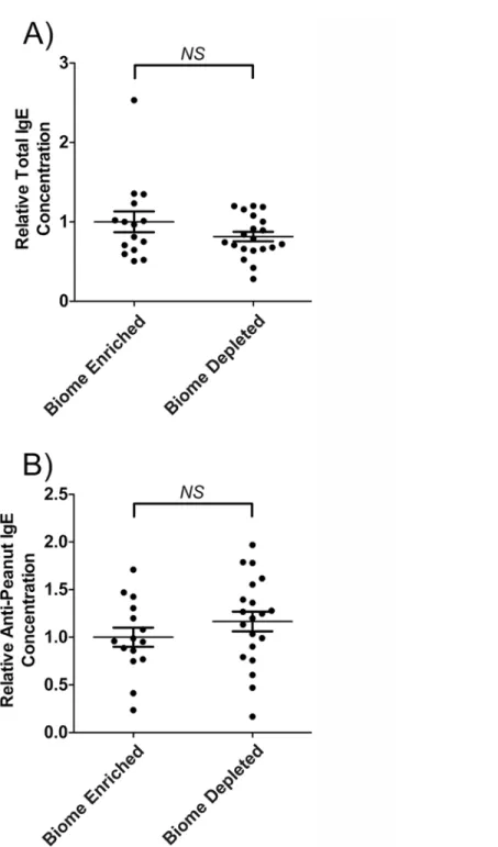
As shown inFig. 2Aand inTable 1, no statistically significant difference was observed between serum IgE levels in biome depleted versus biome enriched rats following immunization. Simi-larly, no statistically significant difference was observed between anti-peanut IgE levels in biome depleted versus biome enriched rats following immunization (Fig. 2B).
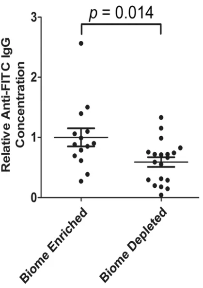
Anti-FITC IgG in the serum of biome depleted and biome enriched rats
Anti-DNP IgM and IgG in the serum of biome depleted and biome
enriched rats
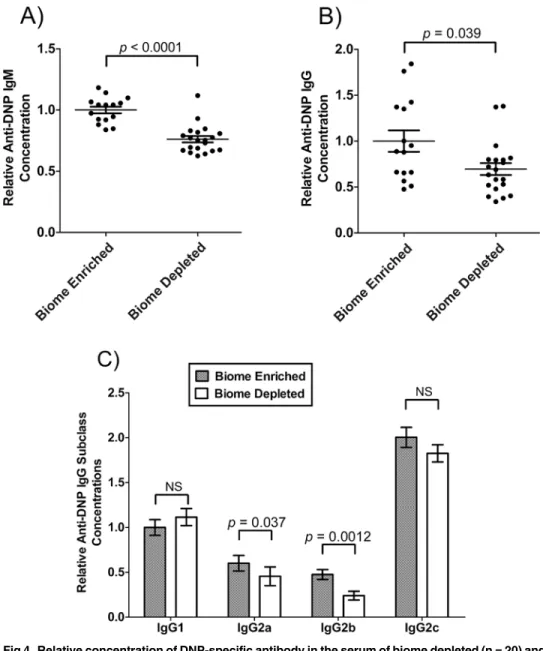
The level of anti-DNP IgM and IgG in serum following immunization of biome depleted and biome enriched animals was assessed as a measure of the immune response against DNP-Fi-coll, a T-independent antigen. The mean concentrations of anti-DNP IgM (Fig. 4A) and anti-DNP IgG (Fig. 4B) were significantly greater in biome enriched animals compared to biome depleted animals (p<0.0001 and p = 0.039, respectively) following immunization.
The relative levels of anti-DNP IgG1, IgG2a, IgG2b, and IgG2c in the serum of biome de-pleted (n = 20) and biome enriched (n = 15) rats following immunization are shown inFig. 4c. Although the 2-way ANOVA showed no significant effect of the state of the biome on the re-sults, post-hoc tests showed significant effects on IgG2a concentrations (p = 0.037) and IgG2b concentrations (p = 0.0012), with biome enriched rats having mean levels approximately 30% greater and 2-fold greater, respectively, than biome depleted rats. Further, mean levels of anti-DNP IgG1 were actually decreased in biome enriched rats. Although this decrease was not statistically significant, it does strongly suggest that biome depletion and enrichment does not affect all aspects of the humoral response equally.
Levels of
“
natural
”
IgM and IgG antibodies in biome depleted and biome
enriched animals
The levels of natural antibodies, antibodies that are present without a known history of sensiti-zation, were evaluated in biome depleted and biome enriched animals. For this purpose, two antigens were selected, one xenogeneic (human serum albumin; HSA) and one autologous (rat muscle tissue extract). Typically, antibodies in normal (non-diseased) rats which bind to these
Fig 1. Body weights of biome depleted and biome enriched rats at (A) 4 days old, (B) 23 days old, and (C) 63 days old.The mean±the standard error is indicated by the horizontal lines. No statistical significance (NS) was observed with comparing data from biome depleted and biome enriched animals using a t-test.
Fig 2. Relative concentration of IgE and of peanut-specific IgE in the serum of biome depleted (n = 20) and biome enriched (n = 15) rats following immunization.The relative concentration of antibody was determined by ELISA as described in the Methods. The means and standard errors are indicated by the horizontal bars, and the data were assessed using a t-test. (A) Following immunization, no statistically significant difference (NS) was observed when comparing total serum IgE levels from biome depleted and biome enriched animals. In addition, (B), following immunization, no statistically significant difference (NS) was observed when comparing anti-peanut IgE levels from biome depleted and biome enriched animals.
antigens are polyreactive (broadly reactive) antibodies that are the first line of defense against foreign antigens or damaged tissue, and do not react strongly with intact tissues[10–15]. Levels of both IgM and IgG natural antibodies were evaluated. Differences observed in natural anti-body levels as a function of biome enrichment were somewhat dependent on isotype, with lev-els of anti-HSA IgG but not anti-HSA IgM being significantly affected by biome enrichment (Fig. 5). The decrease in anti-HSA antibodies associated with biome depletion was particularly notable, with those animals having less than 40% of the level of antibody found in biome en-riched animals (p = 0.0015;Fig. 5).
The repertoire of natural antibodies recognizing autologous antigens (rat muscle tissue ex-tract) in biome enriched and biome depleted animals was evaluated by Western blotting (Figs.
6and7). The results suggest that the concentration of natural antibodies is dramatically in-creased by biome enrichment. The binding of IgM to autologous antigens (Fig. 6) was charac-terized by a moderate (11%) difference in the average number of antigens recognized (Fig. 6B), but given the high density of bands, it is possible that the presence of overlapping bands may have caused an underestimation of the bands recognized, particularly in the lanes with high in-tensity of binding. Biome depleted animals had about 34% less binding of IgM to autologous antigens, on average, and a distribution analysis showed enhanced binding of IgM from biome enriched animals to both high intensity and low intensity bands on the membranes (Fig. 6C). These results suggest that the increase in IgM binding to autologous antigens as a result of biome enrichment may be due to both increased concentrations of antibody and an increased range of specificity of the repertoire. Similar results were obtained when examining the IgG repertoire (Fig. 7), again suggesting that the increase in IgG binding to autologous antigens as a result of biome enrichment may be due to both increased concentrations of antibody and in-creased range of specificity of the repertoire.
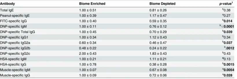
Table 1. Effect of biome depletion on the humoral immune response.
Antibody Biome Enriched Biome Depleted p-value1
Total IgE 1.00±0.51 0.81±0.26 b0.38
Peanut-specific IgE 1.00±0.39 1.17±0.47 a0.27
FITC-specific IgG 1.00±0.40 0.59±0.35 b0.014
DNP-specific IgM 1.00±0.11 0.76±0.12 b<0.0001
DNP-specific Total IgG 1.00±0.45 0.70±0.29 b0.039
DNP-specific IgG1 1.00±0.34 1.12±0.43 b0.34
DNP-specific IgG2a 0.60±0.34 0.46±0.47 b0.037
DNP-specific IgG2b 0.48±0.22 0.24±0.22 b.0012
DNP-specific IgG2c 2.00±0.43 1.83±0.43 b0.43
HSA-specific IgM 1.00±0.21 1.11±0.21 a0.13
HSA-specific IgG 1.00±0.78 0.38±0.28 b0.0015
Muscle-specific IgM 1.00±0.07 0.67±0.08 a0.0054
Muscle-specific IgG 1.00±0.09 0.72±0.06 a0.028
Antibody levels were determined by ELISA, except anti-muscle antibodies, which were determined by immunoblotting. The means and standard deviations are shown.
1The p-values were determined using a t-test for normally distributed data indicated byaor a Mann-Whitney test for non-normally distributed data indicated byb(seeMethods). Signi
ficant values (<0.0500) are shown inbold. Thep-values shown for IgG subclasses are post hoc tests run after a 2-way ANOVA that showed no significant effect of the state of the biome on IgG subclass concentrations.
Discussion
The biome enrichment protocol employed was directed primarily at the biome of parental rats, with the F1 rats receiving less stimulation, as described in the Methods. For example, the F0 rats but not the F1 rats received regular exposure to bedding from the cages of wild-caught rats. This protocol simulated, in part, the fact that newborn F1 rats are not, in the wild, exten-sively exposed to new components of the biome except through contact with the mother. How-ever, F1 rats were housed in the same room as the wild rodents and, until weaning, in the same cages as the F0 rats, so extensive enrichment of the biome in the F1 rats may have occurred. That being said, the immune response of the F1 rats could have been extensively altered by transmission of immune components through the mother’s milk and by epigenetic effects. With this in mind, future studies might initiate biome enrichment after birth to probe the ex-tent to which mother-to-offspring transmission of immune components is important.
Fig 3. Relative concentration of FITC-specific IgG in the serum of biome depleted (n = 20) and biome enriched (n = 15) rats.The relative concentration of antibody was determined by ELISA as described in the Methods. The means and standard errors are shown. No statistical significance (NS) was observed with comparing data from biome depleted and biome enriched animals using a t-test.
The results demonstrate that biome enrichment enhances immune responsiveness under the conditions used in this study, which employed vaccination with adjuvant. Although this study is clearly an experiment involving enrichment of the biodiversity of laboratory animals, the study has implications for humans in a Westernized, biome depleted environment. These studies suggest that biome depletion may be associated with attenuated humoral immune re-sponses to both T-independent and T-dependent antigens. To the extent that biome depletion affects laboratory animals and humans in Western countries in a similar fashion, the implica-tions of this finding are potentially far-reaching. For example, attenuated responses to tumor antigens as a result of biome depletion might underlie, at least in part, the proposed connection between increased rates of cancer and biome depletion[16]. Further, decreased levels of
Fig 4. Relative concentration of DNP-specific antibody in the serum of biome depleted (n = 20) and biome enriched (n = 15) rats.The relative concentration of antibody was determined by ELISA as described in the Methods. Relative levels of (A) IgM, (B) IgG, and (C) subclasses of IgG are shown. The means and standard errors are shown. Thep-values associated with comparing data from biome depleted and biome enriched animals using a t-test are shown. (NS = not significant)
Fig 5. Natural anti-human serum albumin antibody levels in the serum of biome depleted (n = 20) and biome enriched (n = 15) rats.The relative concentration of antibody was determined by ELISA as described in the Methods. Relative levels of (A) IgM and (B) IgG are shown. Binding to human serum albumin (HSA) was used as a measure of reactivity toward a xenogeneic antigen for which the animals lacked previous exposure. The means, standard errors, and thep-values associated with comparing data from biome depleted and biome enriched animals using a t-test are shown. (NS = not significant)
“natural”IgG and IgM observed in biome depleted (laboratory) environments could exacerbate the problem, since the natural antibody repertoire is involved in tumor surveillance [6,7]. In this manner, decreased tumor surveillance in biome depleted environments could promote cancer progression and operate synergistically with biome depletion-associated inflammation, a potential initiator and promoter of carcinogenesis [16,17].
Studies comparing wild and laboratory rats [5] have suggested the importance of biodiversi-ty in the development of the natural antibody repertoire. These studies extend that work con-siderably, emphasizing the potential importance of biome enrichment for effective function of the humoral immune system and giving traction to the idea of biome enrichment for all West-ernized humans. Such enrichment in the clinical setting might conceivably utilize a wide array
Fig 6. Binding of natural anti-rat muscle IgM in the serum of biome depleted and biome enriched rats as evaluated by immunoblotting.(A) Rat muscle extracts were separated by SDS PAGE and probed by immunoblotting as described in the Methods. The analysis was limited to 15 animals (n = 8 biome enriched; lanes E1 through E8, and n = 7 biome depleted; lanes D1 through D7) due to size constraints of the gel. A control strip with no serum is labeled“C”, and indicates reactivity of the anti-IgM conjugate with muscle-derived antigens. (B) The number of bands recognized by natural IgM in individual sera (p = 0.0089) and the total reactivity of natural IgM from each serum sample (p = 0.0093) are shown, with the bars indicating the mean and standard error. (C) The distribution of bands as a function of band size is shown. For this analysis, the average number of bands in biome depleted and biome enriched rats (Y-axis) was plotted on linear and log scales (main figure and figure inset, respectively) versus different band sizes (X-axis).
of eukaryotic organisms, including protozoans and various symbiotic flatworms and round-worms. However, for practical reasons that involve the desire for highly controlled reintroduc-tion of organisms into the populareintroduc-tion with little or no adverse side effects [18,19], most studies in animal models and especially in humans have focused on the reintroduction of helminths, worms that live in the intestine, into the body’s ecosystem. The findings in this study suggest that biome enrichment with such organisms as helminths, which reduces a wide range of aller-gic and autoimmune conditions[20], is not necessarily“immunosuppressive”. Rather, this study suggests that biome enrichment, perhaps including helminth therapy, might be an“ im-mune trainer”, and thus fundamentally different in principle and practice than drugs aimed at averting disease via suppression of immune function.
Fig 7. Binding of natural anti-rat muscle IgG in the serum of biome depleted and biome enriched rats as evaluated by immunoblotting.(A) Rat muscle extracts were separated by SDS PAGE and probed by immunoblotting as described in the Methods. The analysis was limited to 15 animals (n = 8 biome enriched; lanes E1 through E8, and n = 7 biome depleted; lanes D1 through D7) due to size constraints of the gel. A control strip with no serum is labeled“C”, and indicates reactivity of the anti-IgG conjugate with muscle-derived antigens. (B) The number of bands recognized by natural IgG in individual sera (p = 0.017) and the total reactivity of natural IgG from each serum sample (p = 0.040) are shown, with the bars indicating the mean and standard error. (C) The distribution of bands as a function of band size is shown. For this analysis, the average number of bands in biome depleted and biome enriched rats (Y-axis) was plotted on linear and log scales (main figure and figure inset, respectively) versus different band sizes (X-axis).
Acknowledgments
The authors gratefully acknowledge the generous donation of time and expertise provided by Timothy Chase, John W. Poulton and Earll Williams for the design, engineering and construc-tion of many of the materials utilized in this project, without which these studies would not have been possible. The authors are also very thankful to Robert A. Holzknecht and Susanne Meza-Keuthen for careful proofreading of the manuscript, and to a number of individuals who provided tremendous help with regulatory and technical matters associated with the study. These individuals include Laura P. Hale, John Norton, William Wade, Randall Reynolds, Peg Hogan, Calvin Davis, and Jacinto Juarez.
Author Contributions
Conceived and designed the experiments: CP EHA DR SP (Susan Poulton) SYRL SP (Sarah Perkins) MLE ZEH SSL WP. Performed the experiments: CP EHA DR SP (Susan Poulton) SYRL SP (Sarah Perkins) MLE ZEH WP. Analyzed the data: CP EHA DR SP (Susan Poulton) SYRL SP (Sarah Perkins) MLE ZEH WP. Contributed reagents/materials/analysis tools: SP (Susan Poulton) SSL WP. Wrote the paper: CP EHA DR SP (Susan Poulton) SYRL SP (Sarah Perkins) MLE ZEH SSL WP.
References
1. Lesher A, Li B, Whitt P, Newton N, Devalapalli AP, Shieh K, et al. Increased IL-4 Production and Attenu-ated Proliferative and Proinflammatory Responses of Splenocytes from Wild-Caught Rats (Rattus Nor-vegicus). Immunol Cell Biol. 2006; 84:374–82. PMID:16594897
2. Trama AM, Holzknecht ZE, Thomas AD, Su KY, Lee SM, Foltz EE, et al. Lymphocyte phenotypes in wild-caught rats suggest potential mechanisms underlying increased immune sensitivity in post-industrial environments. Cellular & Molecular Immunology. 2012; 2(Mar; 9):163–74.
3. Lin SS, Holzknecht ZE, Trama AM, Everett ML, Thomas AD, Su KY, et al. Immune characterization of wild-caughtRattus norvegicussuggests diversity of immune activity in biome-normal environments. Journal of Evolutionary Medicine. 2012; 1:1–16.
4. Abolins SR, Pocock MJO, Hafalla JCR, Riley EM, Viney ME. Measures of immune function of wild mice, Mus musculus. Mol Ecol. 2011; 20(5):881–92. doi:10.1111/j.1365-294X.2010.04910.xPMID: 21073587
5. Devalapalli AP, Lesher A, Shieh K, Solow JS, Everett ML, Edala AS, et al. Increased Levels of IgE and Autoreactive, Polyreactive IgG in Wild Rodents: Implications for the Hygiene Hypothesis. Scand J Immunol. 2006; 64:125–36. PMID:16867157
6. Vollmers HP, Brandlein S. Natural antibodies and cancer. New biotechnology. 2009 Jun; 25(5):294–8. Epub 2009/05/16. doi:10.1016/j.nbt.2009.03.016PMID:19442595
7. Vollmers HP, Brandlein S. Natural antibodies and cancer. J Autoimmun. 2007; 29(4):295–302. Epub 2007 Sep 12. PMID:17826951
8. Arai HP. Biology of the tapewormHymenolepis diminuta. New York: Academic Press; 1980. 733 p. 9. de Jonge JD, Knippels LM, Ezendam J, Odink J, Penninks AH, van Loveren H. The importance of
dietary control in the development of a peanut allergy model in Brown Norway rats. Methods. 2007 Jan; 41(1):99–111. PMID:17161306
10. Parker W, Yu PB, Holzknecht ZE, Lundberg K, Buckley RH, Platt JL. Specificity and function of "natu-ral" antibodies in immunodeficient subjects: clues to B cell lineage and development. J Clin Immunol. 1997; 17(4):311–21. PMID:9258770
11. Lacroix-Desmazes S, Kaveri SV, Mouthon L, Ayouba A, Malanchere E, Coutinho A, et al. Self-reactive antibodies (natural autoantibodies) in healthy individuals. J Immunol Methods. 1998; 216(1–2):117–37. PMID:9760216
12. Casali P, Schettino EW. Structure and function of natural antibodies. Current Topics in Microbiology & Immunology. 1996; 210:167–79.
14. al-Balaghi S, Abedi-Valugerdi M, Moller E. Binding specificities of a polyreactive and a monoreactive human monoclonal IgG rheumatoid factor: role of oligosaccharides. Scandinavian Journal of Immunol-ogy. 1996; 44(5):470–7. PMID:8947598
15. Parker W, Fields RC, Nakamura YC. Biophysical Properties of Xenoreactive Natural Antibodies. In: Platt JL, editor. Xenotransplantation: Basic Research and Clinical Applications. Totowa, NJ: Humana Press; 2002. p. 87–101.
16. Rook GAW. Review series on helminths, immune modulation and the hygiene hypothesis: the broader implications of the hygiene hypothesis. Immunology. 2009 Jan; 126(1):3–11. doi:10.1111/j.1365-2567. 2008.03007.xPMID:19120493
17. León-Cabrera S, Callejas BE, Ledesma-Soto Y, Coronel J, Pérez-Plasencia C, Gutiérrez-Cirlos EB, et al. Extraintestinal Helminth Infection Reduces the Development of Colitis-Associated Tumorigenesis. International Journal of Biological Science. 2014; 10(9):948–56. doi:10.7150/ijbs.9033PMID: 25210492
18. Parker W, Perkins SE, Harker M, Muehlenbein MP. A prescription for clinical immunology: the pills are available and ready for testing. Current Medical Research and Opinion. 2012; 28:1193–202. doi:10. 1185/03007995.2012.695731PMID:22612580
19. Bilbo SD, Wray GA, Perkins SE, Parker W. Reconstitution of the human biome as the most reasonable solution for epidemics of allergic and autoimmune diseases. Med Hypotheses. 2011; 77(4):494–504. doi:10.1016/j.mehy.2011.06.019PMID:21741180