30
Pakistan Veterinary Journal
ISSN: 0253-8318 (PRINT), 2074-7764 (ONLINE)
Accessible at: www.pvj.com.pk
Molecular Characterization of Three Porcine Reproductive and Respiratory Syndrome Virus
Isolates and Their Susceptibility to Antiviral Drugs
Hongxia Hu, Xian Zhang, Hansong Zhang, Guilan Wen§, Chao Tong, Xiaoliang Li and Weihuan Fang*
Institute of Preventive Veterinary Medicine and Zhejiang Provincial Key Laboratory of Preventive Veterinary Medicine, Zhejiang University, Hangzhou 310058, China; §Also affiliated with College of Animal Science, Guizhou University, Guiyang, Guizhou 550025, China
*Corresponding author: whfang@zju.edu.cn
A R T I C L E H I S T O R Y A B S T R A C T Received:
Revised: Accepted:
March 16, 013 May 28, 2013 July 07, 2013
Porcine reproductive and respiratory syndrome virus (PRRSV) is one of the most common swine pathogens that cause severe economic losses to the pig industry worldwide irrespective of the use of live or inactivated vaccines. This study aims to investigate the biological characteristics of three PRRSV isolates and their susceptibility to two antiviral drugs. Sequence analysis of the NSP2 gene classified two isolates as highly pathogenic (isolates FY and ZS) and one as classically pathogenic (isolate JX). Isolate FY grew faster than the other two isolates in MARC-145 cells; however, its RNA replication was lower than isolate ZS. By contrast, isolate JX exhibited slower growth and lower RNA replication capability. PRRSV infection suppressed the production of interferon β induced by poly (I:C). The viruses also differed in their susceptibility to antiviral drugs. Ribavirin exerted potent antiviral activity against all three viral isolates at concentrations of 7.5 and 15 µg/mL in MARC-145 cells. Acyclovir was found effective only on the classically pathogenic isolate. We suggest that ribavirin could have potential as an antiviral therapy for porcine reproductive and respiratory syndrome when vaccination is not able to provide effective protection.
©2013 PVJ. All rights reserved Key words:
Acyclovir Antiviral Growth kinetics
Porcine reproductive and respiratory syndrome virus Ribavirin
To Cite This Article: Hu H, X Zhang, H Zhang, G Wen, C Tong, X Li and W Fang, 2014. Molecular characterization of three porcine reproductive and respiratory syndrome virus isolates and their susceptibility to antiviral drugs. Pak Vet J, 34(1): 30-35.
INTRODUCTION
Porcine reproductive and respiratory syndrome (PRRS) is characterized by reproductive failure in sows and respiratory disease in piglets (Verheije et al., 2003). Although the causal virus (PRRSV) strains from different countries cause similar clinical signs in pigs, they are different in virulence in infected animals and heterologous genetically and antigenically (Meng, 2000). PRRSV strains are divided into North American (NA) and European (EU) genotypes based on genetic differences in the genome structure (Nelsen et al., 1999). PRRSV was first isolated in the Netherlands (Lelystad virus, LV) that belongs to the EU genotype (Wensvoort et al., 1991). The first NA genotype strain VR-2332 was isolated by Collins et al. (1992). PRRSV is a positive-stranded RNA virus that possesses a genome size of approximately 15kb containing eight open reading frames (ORFs) (Godeny et al., 1993). ORF1 encodes a polyprotein that is proteolytically cleaved into 13 mature nonstructural
proteins (NSPs) assumed to be involved in viral replication and transcription. Of these NSPs, NSP2 is a multi-domain protein that contains a cysteine protease active site in the N-terminal vital to the viral life cycle (Han et al., 2009).
hampered by evolving genetic diversities among field isolates (Huang and Meng, 2010). Therefore, identification of the biological and genetic characteristics of the epidemic PRRSV strain is important.
Ribavirin (1-β -D-ribofuranosyl-1,2,4-triazole-3-carboxamide) has antiviral activity against some DNA and RNA viruses (Witkowski et al., 1972). It is a pleiotropic antiviral agent with direct and indirect antiviral effects (Tam et al., 2001). Acyclovir is a nucleoside analog that exerts its antiviral effects by competitive inhibition of viral DNA polymerase activity after three phosphorylation steps (Stranska et al., 2005). Anti-PRRSV inhibitors are crucial at the early stages of a pandemic, especially when specific PRRSV vaccines are unavailable. However, there is paucity of information on antiviral drugs against PRRSV. In this study, three PRRSV isolates from swine herds involved in the PPRS outbreak in China in 2006 were compared for their growth characteristics, susceptibility to antiviral drugs and inhibition of IFN-β to poly(I:C) stimulation.
MATERIALS AND METHODS
Virus isolation: MARC-145 cells were grown in Dulbecco’s modified Eagle’s medium (DMEM) containing 10% fetal calf serum and incubated at 37°C and 5%CO2. Clinical samples, including lungs, lymph nodes and kidneys, were collected from dead pigs with late-term abortions (sows) or with acute respiratory disease (growing pigs) during the PRRS outbreak of 2006 to 2007 (Tian et al., 2007). Tissue samples were homogenized with sterile phosphate buffered saline (PBS), freeze-thawed three times, and spun at 5000×g for 20 min at 4°C. Supernatants were passed through a 0.22µm filter and transferred to MARC-145 cells for virus isolation. Virus isolates were identified by immunofluorescence assay (IFA) using PRRSV NSP2-specific monoclonal antibodies prepared in our laboratory. Sequence analysis: For NSP2 gene sequencing, viral RNA was extracted from PRRSV present in the culture supernatants (Hu et al., 2009). The complete NSP2 gene was amplified using the primers listed in Table 1. The partial NSP2 amino acid sequences and those retrieved from the GenBank database (Fig. 1) were multiple-aligned using MegAlign 7.1.0 (DNASTAR Lasergene software package).
50% viral tissue culture infective dose (TCID50) and
growth kinetics assay: The TCID50 of the virus was determined by cytopathic effects on MARC-145 cells and calculated by the Reed–Muench method. For growth kinetics assay, MARC-145 cells were infected with the fourth generation PRRSV at 100 TCID50. At 12, 24, 48, 72 and 96 h post-infection (hpi), cell and cell culture supernatants were collected and frozen at –80°C for further analysis. The viral growth kinetic assays were performed in triplicate. Three independent titrations were determined by TCID50 per milliliter, and the mean value was used to determine the viral growth curve.
Quantitative polymerase chain reaction (qPCR) : To quantitate PRRSV RNA replication levels, total RNA was
isolated from the samples. Quantitative PCR was performed using SYBR Green I dye (Takara Biotech Co., Ltd., China). The target mRNA in the samples was quantified by comparison with a standard curve derived from dilutions of plasmids containing PRRSV subgenomic ORF7 (100 to 108 copies). The internal control (β-actin gene) was quantified using the same procedures as above for normalized quantitation of viral RNA (Gao et al., 2008). The PCR primers are shown in Table 1.
Table 1: The primers used are this study. Gene Primer Sequence (5’to 3’)
NSP2 Forward GTTGAGCCCAATACGTCACCA Reverse CTCCAGCCAAGATACAGTCTGC IFN-β
(real time PCR)
Forward TAAGCAGCTGCAGCAGTTCCAGAAG Reverse GTCTCATTCCAGCCAGTGCT
β-actin (real time PCR)
Forward CGTGCGTGACATCAAAGAGAAG Reverse CGTTGCCAATAGTGATGACCTG ORF7 Forward ATGGCCAGCCAGTCAATC
Reverse TCAGTCGCTAGAGGAAAATGG
Chemicals: Ribavirin (1-β -D-ribofuranosyl-1,2,4-triazole-3-carboxamide) and acyclovir (2-amino-1,9-dihydro-9-[(2-hydroxyethoxy)methyl]-6H-purin-6-one) were purchased from Xinchang Pharmaceutical Factory, Zhejiang, China. Stock solutions were prepared by dissolving the drugs in PBS at pH 7.4 and diluted to the required concentrations in the culture medium.
Cell viability assay: A colorimetric assay for cell viability was used to determine the doses of the antivirus drugs. About 1.5×10⁴ MARC-145 cells per well were seeded into 96-well culture plates and allowed to adhere overnight. The medium was removed and various concentrations of antiviral drugs in DMEM were added to the wells. After 72 h, the culture medium was removed and 3-(4,5-Dimethylthiazol-2-yl)-2,5-diphenyl tetrazolium bromide (MTT) reagent (Beyotime, Beijing, China) was added to the wells according to the manufacturer’s instructions. After 4 h of incubation, formazan generated in the cells was dissolved in 100 µL of formazan solution per well. Absorbance was measured at 490nm using a microplate reader SpectraMaxM2 (Molecular Device, USA). The drug concentration that decreased the cell viability by 50% was defined as the 50% inhibitory concentration (IC50). Each experiment was performed in triplicate.
Viral susceptibility to antiviral drugs: MARC-145 cells (1.5×10⁵ per well) were cultured in DMEM in a 24-well plate for 24 h. The culture medium was then removed and the cells were pretreated with different concentrations of ribavirin or acyclovir for 1 h before viral inoculation at 100 TCID50. The drug-containing media were reintroduced into corresponding wells after viral inoculation. Antiviral effect was evaluated at 48 hpi. Cells were collected and total RNA was isolated to quantitate viral RNA replication. Alternatively, the culture supernatants were collected at different time points for TCID50 titration.
FY-2007
TQATSEMMARAAEQVDLKVWVKSYPRWTPPPSPPRVQPRRTKSVKSLPEDKPVPAPRRKVRSDCGSRVLMGDNVPNVSEE-TVGGPLNVPTPSEPMTPMS 499 JX-2007
TQATSEMMAWAAEQVDLKAWVKSYPRWTPPPPPPRVQPRKTKSVKSLLEDKPVPAPRRKVRSDCGSPVLIGDNVLNDSEDLTVGGSSSHPTPSEPMTPMS 500 ZS-2007
TQATSEMMAWAAEQVDLNVWVKSYPRWTPPPPPPKVQPRRTKSVNSLPEGKPVPAPRRKVRSDCGSSFLMGDNVPNGSDK-TVGGPLNFSTPSEPMTPMS 499 FY-2007 EPVLVPASRRVPKLMTPLSGSAPVPAPRRTVT---TTLTHQDEPLDLSASSQTEYEAFPLAPSQNMGILEAEGQ 570
JX-2007
EPVLTPALQCVPKLTTPLSGSAPVPAPRGIVSRPMAPLSEPILVSVPQYKFQEVEEANLAATTLTHQDEPLDLSASSQTEYEAFPLAPSQNMKILEARRQ 600 ZS-2007 EPVLVPASRRVPKLMTPLSESAPVPAPRRAVT---TTLTHQDEPLDLSASSQTEYEAPLLAPSRN--ILEVGGQ 568
Fig. 1: Alignment of partial amino acid sequences of NSP2 of PRRSV isolates and reference strains. The shadows indicate the deleted amino acids.
different PRRSV isolates at 100 TCID50 for 36 h. Finally, the cells were transfected with 0.5 µg/mL poly(I:C) (InvivoGen) for 10 h (Kim et al., 2010). Total RNA was extracted and cDNA was synthesized with oligo (dT15) (Takara Biotech Co., Ltd., China) and Moloney Murine leukemia virus reverse transcriptase (Promega). qPCR was performed in triplicate using SYBR Green I dye on a Bio-Rad iQ5 system using primers shown in Table 1. Results for the target gene were presented after normalization to β-actin. The 2-∆∆CT method was used to quantify the relative transcript levels shown as relative fold changes in comparison with the mock-treated control (Livak and Schmittgen, 2001).
Statistical analysis: Two-tailed Student’s t-test was performed using Microsoft Excel software. P values < 0.05 as statistically significant, P values < 0.01 as of marked significance.
RESULTS
Sequence analysis of the partial NSP2 gene of three PRRSV isolates: Typical cytopathic effects could be observed 3 dpi. The putative viral isolates were plaque-purified thrice to ensure the isolation of a single virus and confirmed by IFA with specific monoclonal antibody against PRRSV NSP2 and by sequencing. Three clinical viral isolates JX, FY and ZS were obtained. These isolates genotypically belonged to the North American type according to the NSP2 sequences (GenBank accession no: JQ798255, JQ798256 and JQ798257 for isolates JX, FY and ZS respectively). The NSP2 of isolate ZS exhibited 96.6% homology with the highly pathogenic PRRSV strain JXA1 (Tian et al., 2007), higher than the two other viruses (JX at 89.9% and FY at 90.1%). Multiple amino acid sequence comparisons showed that isolates FY and ZS had discontinuous deletion of 30 aa (located at aa 481 and aa 533 to 561) in their NSP2 regions, similar to the highly pathogenic PRRSV strain JXA1. Isolates ZS showed two more deletions located at aa 592 and aa 593. By contrast, isolate JX had its NSP2 region identical to that of an earlier Chinese isolate CH1a (An et al., 2005) (Fig. 1).
Titer and growth of three viral isolates JX, FY and ZS: The TCID50 of isolates JX, FY and ZS were 10
7.6 , 108.3 and 108.6 per mL, respectively. The titer of three isolates peaked at 72 hpi with the isolate FY having slightly higher titer, as compared with JX (P<0.05), and ZS (P<0.05) (Fig. 2A). The RNA copy numbers of isolate
ZS peaked 48 hpi, earlier and higher than that of the two other viral isolates, as compared with JX (P<0.001) and FY (P<0.001) (Fig. 2B).
PRRSV infection significantly suppressed transcription of IFN-β gene in response to poly(I:C) stimulation: To determine if PRRSV infection leads to general inhibition of IFN-β expression, we examined
IFN-β transcription activity in PRRSV-infected MARC-145 cells upon poly (I:C) stimulation. Fig. 3 indicates that poly (I:C)-induced IFN-β gene transcription was significantly inhibited by all three PRRSV isolates (P<0.01).
Fig. 2: Growth kinetics of three PRRSV isolates JX, ZS and FY. MARC-145 cells were infected with different virus at a 100TCID50. At the
indicated time points, cells and culture supernatants were collected for TCID50 or RNA copy number analysis. (A) Growth kinetics was
Fig. 3: Effects of PRRSV infection on IFN-β gene expression in MARC-145 cells treated with poly (I:C). MARC-145 cells were infected with PRRSV at 0.1 multiplicity of infection. At 36 h post-infection, poly (I:C) was transfected for 10 hs, and total cellular RNA was extracted. Expression of IFN-β was measured by RT-qPCR. Data represent means±SD of three experiments, each in triplicate (**P<0.01 when compared between PRRSV-infected/poly(I:C)-treated cells and poly(I:C) control).
Fig. 4: Effect of ribavirin and acyclovir on viability of MARC-145 cells. Cell viability was determined by MTT assay after treatment with different levels of ribavirin or acyclovir for 48 hs. Percentage of relative cell viability represents means±SD of three experiments, each in triplicate (**P<0.01 at and above this concentration).
Viability of MACR-145 cells treated with ribavirin or acyclovir: Drugs that inhibit cell growth may interfere with observation of antiviral activity (Wu et al., 2004) and should be assessed on the viability of MARC-145 cells for determination of appropriate concentrations to be used for antiviral assays. The concentrations of ribavirin and acyclovir that inhibited cell growth by 50% (IC50) were 125 and 893 µg/mL respectively. Ribavirin significantly decreased (P<0.01) MARC-145 cells viability at concentrations above 31.25 µg/mL, and acyclovir significantly decreased (P<0.01) MARC-145 cells viability at levels above 223 µg/mL. The marginal concentrations of ribavirin and acyclovir that did not affect MARC-145 cell viability should be lower than 15.6 and 111 µg/mL, respectively (Fig. 4).
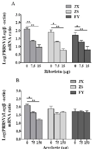
Susceptibility of PRRSV isolates to ribavirin and acyclovir: Ribavirin significantly decreased the RNA transcription levels of all three viral isolates at concentrations of 7.5 and 15 µg/mL in MARC-145 cells (Fig. 5A). By contrast, acyclovir did not show inhibition of RNA transcription of two viral isolates (ZS and FY) at concentrations of 75 and 150µg/mL (Fig. 5B). Treatment with 10µg/mL ribavirin could decrease the virus titer by
Fig. 5: Antiviral activity of ribavirin and acyclovir on PRRSV in MARC-145 cells shown as changes of viral RNA normalized to β-actin gene. Two-fold serial dilutions of ribavirin and acyclovir were prepared in DMEM and added to the MARC-145 cells for 1 h. The cells were infected with PRRSV at 100 TCID50. Viral RNA copies were determined
by real-time RT-PCR at 48h post-infection. Data represent means±SD of three experiments, each in triplicate (*P<0.05; **P<0.01).
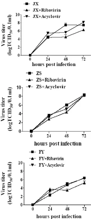
102.96 (JX), 101.2 (FY) and 101.65 (ZS) 48 hpi. Ribavirin did not seem to have lasting antiviral effect upon further treatment to 72 hpi, particularly for isolate ZS. Treatment with 150µg/mL acyclovir did not show apparent antiviral activity on isolates ZS and FY although it did show inhibition on isolate JX (Fig. 6).
DISCUSSION
Fig. 6: The antiviral activity of ribavirin and acyclovir on PRRSV shown as changes of viral titers at different time points. MARC-145 cells were pretreated with ribavirin (10 µg/mL) and acyclovir (150 µg/mL) for 1 h, and then were infected with PRRSV at 100 TCID50. Viral particles in the
culture supernatants were titrated at different time points. Data represent means±SD of three experiments, each in triplicate.
al., 2009). Some differences in growth characteristics were observed between the highly pathogenic and classically pathogenic isolates in vitro. However, all three isolates suppressed the production of IFN-β in response to poly (I:C) induction, similar to other PRRSVs (Kim et al., 2010; Li et al., 2010).
Anti-PRRSV drugs could be important at the early stages of an endemic, especially when specific PRRSV vaccines are unavailable or not effective. Ribavirin and acyclovir have antiviral effects on a variety of DNA and RNA viruses (Stranska et al., 2005; Tam et al., 2001).
However, their effects on PRRSV replication remain unknown. Here we show that ribavirin has potent antiviral activity against all three viruses at concentrations of 7.5 and 15µg/mL in MARC-145 cells, similar to findings in other Nidovirales (Kim and Lee, 2012). Acyclovir seems to be effective only against classically pathogenic PRRSV. Therefore, our work suggests that ribavirin could be resorted of as an antiviral drug when PRRSV is endemic without effective vaccines.
Conclusion: Of the three PRRSV isolates, two shared discontinuous deletion of 30 amino acids (aa) in NSP2 similar to the highly pathogenic type. All three isolates were highly or moderately susceptible to ribavirin, but only the classical pathogenic isolate is acyclovir-sensitive. Acknowledgement: This study is supported by a grant from Hangzhou Municipal Government (20102012A05) and Zhejiang Entry-Exit Inspection and Quarantine Bureau (ZK201109).
REFERENCES
An TQ, YJ Zhou, HJ Qiu, GZ Tong, YF Wang, JX Liu and JY Yang, 2005. Identification of a novel B cell epitope on the nucleocapsid protein of porcine reproductive and respiratory syndrome virus by phage display. Virus Genes,31: 81-87.
Collins JE, DA Benfield, WT Christianson, L Harris, JC Hennings, DP Shaw, SM Goyal, S McCullough, RB Morrison, HS Joo, D Gorcyca and D Chladek, 1992. Isolation of swine infertility and respiratory syndrome virus (isolate ATCC VR-2332) in North America and experimental reproduction of the disease in gnotobiotic pigs. J Vet Diagn Invest, 4: 117-126.
Gao Y, W Liu, H Gao, Qi X, Lin H, Wang X and R Shen, 2008. Effective inhibition of infectious bursal disease virus replication in vitro by DNA vector-based RNA interference. Antiviral Res, 79: 87-94. Geldhof MF, M Vanhee, W VanBreedam, J VanDoorsselaere, UU
Karniychuk and HJ Nauwynck, 2012. Comparison of the efficacy of autogenous inactivated Porcine Reproductive and Respiratory Syndrome Virus (PRRSV) vaccines with that of commercial vaccines against homologous and heterologous challenges. BMC Vet Res, 8: 182.
Godeny EK, L Chen, SN Kumar, SL Methven, EV Koonin and MA Brinton, 1993. Complete genomic sequence and phylogenetic analysis of the lactate dehydrogenase-elevating virus (LDV). Virology, 194: 585-596.
Han J, MS Rutherford and KS Faaberg, 2009. The porcine reproductive and respiratory syndrome virus nsp2 cysteine protease domain possesses both trans- and cis-cleavage activities. J Virol, 83: 9449-9463.
Hu HX, XL Li, ZF Zhang, JB Shuai, N Chen, GQ Liu and WH Fang, 2009. Porcine reproductive and respiratory syndrome viruses predominant in southeastern China from 2004 to 2007 were from a common source and underwent further divergence. Arch Virol, 154: 391-398.
Huang YW and XJ Meng, 2010. Novel strategies and approaches to develop the next generation of vaccines against porcine reproductive and respiratory syndrome virus (PRRSV). Virus Res, 154: 141-149.
Karuppannan AK, KX Wu, J Qiang, JJ Chu and J Kwang, 2012. Natural compounds inhibiting the replication of Porcine reproductive and respiratory syndrome virus. Antiviral Res, 94: 188-194.
Kim O, Y Sun, FW Lai, C Song and D Yoo, 2010. Modulation of type I interferon induction by porcine reproductive and respiratory syndrome virus and degradation of CREB-binding protein by non-structural protein 1 in MARC-145 and HeLa cells. Virology, 402: 315-326.
Kim Y and C Lee, 2012. Ribavirin efficiently suppresses porcine nidovirus replication. Virus Res, 171: 44-53.
Li Y, X Wang, K Bo, B Tang, B Yang, W Jiang and P Jiang, 2007. Emergence of a highly pathogenic porcine reproductive and respiratory syndrome virus in the Mid-Eastern region of China. Vet J, 174: 577-584.
Livak KJ and TD Schmittgen, 2001. Analysis of relative gene expression data using real-time quantitative PCR and the 2(-Delta Delta C(T)) Method. Methods, 25: 402-408.
Meng XJ, 2000. Heterogeneity of porcine reproductive and respiratory syndrome virus: implications for current vaccine efficacy and future vaccine development. Vet Microbiol, 74: 309-329.
Murtaugh MP and M Genzow, 2011. Immunological solutions for treatment and prevention of porcine reproductive and respiratory syndrome (PRRS). Vaccine, 29: 8192-8204.
Nelsen CJ, MP Murtaugh and KS Faaberg, 1999. Porcine reproductive and respiratory syndrome virus comparison: divergent evolution on two continents. J Virol, 73: 270-280.
Stranska R, R Schuurman, E Nienhuis, IW Goedegebuure, M Polman, JF Weel, PM Wertheim-Van Dillen, RJ Berkhout and AM van Loon, 2005. Survey of acyclovir-resistant herpes simplex virus in the Netherlands: prevalence and characterization. J Clin Virol, 32: 7-18. Tam RC, JY Lau and Z Hong, 2001. Mechanisms of action of ribavirin in
antiviral therapies. Antivir Chem Chemother, 12: 261-272. Tian KG, Xl Yu, TZ Zhao, YJ Feng, Z Cao, CB Wang, Y Hu, XZ Chen,
DM Hu, XS Tian, D Liu, S Zhang, XY Deng, YQ Ding, L Yang, YX Zhang, HX Xiao, MM Qiao, B Wang, LL Hou, XY Wang, XY Yang, LP Kang, M Sun, P Jin, SJ Wang, Y Kitamura, JH Yan and GF Gao, 2007. Emergence of fatal PRRSV variants: unparalleled outbreaks
of atypical PRRS in China and molecular dissection of the unique hallmark. PLoS One, 2: e526.
Verheije MH, MV Kroese, IF van der Linden, EA de Boer-Luijtze, PA van Rijn, JM Pol, JJ Meulenberg and PJ Steverink, 2003. Safety and protective efficacy of porcine reproductive and respiratory syndrome recombinant virus vaccines in young pigs. Vaccine, 21: 2556-2563.
Wensvoort G, C Terpstra, JM Pol, EA ter Laak, M Bloemraad, EP de Kluyver, C Kragten, L van Buiten, A den Besten, F Wagenaar, JM Broekhuijsen, PL Moonen, T Zetstra, EA de Boer, HJ Tibben, MF de Jong, P van’t Veld, GJ Greenland, JA Gennep, MTh Voets, JH Verheijden and J Braamskamp, 1991. Mystery swine disease in The Netherlands: the isolation of Lelystad virus. Vet Q, 13: 121-130. Witkowski JT, RK Robins, RW Sidwell and LN Simon, 1972. Design,
synthesis, and broad spectrum antiviral activity of 1-D-ribofuranosyl-1,2,4-triazole-3-carboxamide and related nucleosides. J Med Chem, 15: 1150-1154.
Wu CY, JT Jan, SH Ma, CJ Kuo, HF Juan, YS Cheng, HH Hsu, HC Huang, D Wu, A Brik, FS Liang, RS Liu, JM Fang, ST Chen, PH Liang and CH Wong, 2004. Small molecules targeting severe acute respiratory syndrome human coronavirus. Proc Natl Acad Sci, 101: 10012-10017.