2013
molecules
ISSN 1420-3049 www.mdpi.com/journal/molecules ArticleChemistry and Antiviral Activity of
Arrabidaea pulchra
(Bignoniaceae)
Geraldo Célio Brandão 1,2, Erna G. Kroon 3, Danielle E.R. Souza 1, José D. Souza Filho 4 and Alaíde Braga Oliveira 1,*
1
Faculdade de Farmácia, Departamento de Produtos Farmacêuticos, Universidade Federal de Minas Gerais, Av. Antônio Carlos, 6627, CEP 31.270-901, Belo Horizonte, MG, Brazil
2
Escola de Farmácia, Departamento de Farmácia, Universidade Federal de Ouro Preto, Rua Costa Sena, 171, CEP 35.400-000, Ouro Preto, MG, Brazil
3
Departamento de Microbiologia, ICB, Universidade Federal de Minas Gerais, Av. Antônio Carlos, 6627, CEP 31.270-901, Belo Horizonte, MG, Brazil 4
Departamento de Química, ICEX, Universidade Federal de Minas Gerais, Av. Antônio Carlos, 6627, CEP 31.270-901, Belo Horizonte, MG, Brazil
* Author to whom correspondence should be addressed; E-Mail: alaide.braga@pq.cnpq.br or alaidebraga@terra.com.br; Tel.: +55-31-3409-6972; Fax: +55-31-3441-5575.
Received: 26 April 2013; in revised form: 22 July 2013 / Accepted: 24 July 2013 / Published: 16 August 2013
Abstract: The aim of the present work was to carry out a bioguided isolation of antiviral chemical constituents from an ethanol extract of leaves from Arrabidaea pulchra (Cham.) Sandwith (EEAPL) that had shown in vitro activity in a previous screening using DNA and RNA viruses. The activity of EEPAL was evaluated against the DNA viruses Human herpesvirus 1 (HSV-1) and Vaccinia virus Western Reserve (VACV-WR) as well as against the RNA viruses Murine encephalomyocarditis virus (EMCV), and Dengue virus 2 (DENV-2) by the 3-(4,5-dimethylthiazol-2-yl)-2,5-diphenyltetrazolium bromide (MTT) colorimetric assay. Cytotoxicity was determined in LLCMK2 and Vero cells and the Selectivity Indexes (SI) were calculated. The most potent effect was observed against DENV-2 (EC50 46.8 ± 1.6 µg mL−1; SI 2.7). For HSV-1 and VACV-WR EC50 values > 200 µg mL−1 were determined, while no inhibition of the cytopathic effect was observed with EMCV. Bioguided fractionation of EEAPL by partition between immiscible solvents followed by chromatography over a Sephadex LH20 column afforded two arylpropanoid glycosides, verbascoside (AP 1) and caffeoylcalleryanin (AP 2), along with a terpenoid, ursolic acid (AP 3). AP 1 and AP 3 exhibited similar anti-DENV-2 profiles, with SI values
of 3.8 and 3.1, respectively, while AP 2 was the most effective anti-DENV-2 constituent, with a SI of 20.0. Our resultsshow that A. pulchra leaves ethanol extract (EEAPL) affords compounds with antiviral activity, mainly against DENV-2.
Keywords: antiviral activity; dengue virus; Arrabidaea pulchra; verbascoside; caffeoylcallerianin
1. Introduction
Viral infections represent a current problem in industrialized and developing countries, accounting for severe damages to human health and economic losses in livestock. Plants afford an extensive chemical diversity and represent rich and renewable sources of natural products with promising biological activities. Antivirals of ethnomedicinal origin are of great interest and have been widely explored [1]. Antiviral compounds can block or inhibit virus replication by interfering with virus attachment to cells, interfering with viral enzymes or suspending viral genome replication. To be effective, compounds working at each of these steps within the virus replication cycle must target the viral process, while avoiding similar ongoing cellular processes. Viruses of different families present different structures and replication schemes, and consequently offer different potential molecular targets [2].
Some viral diseases, such as dengue, which is considered the most important human arboviral disease, have a high impact in public health. It is estimated that 100 million cases occur every year around the World. The need for a safe and efficient approach either for treatment or prevention for virus infections has been a global priority, as neither an effective drug nor a vaccine for human use is available. However, investigations against dengue viruses have started in the previous decade and opportunities for the development of anti-dengue drugs have been pointed out by the WHO [3].
The genus Arrabidaea belongs to the tribe Bignonieae (family Bignoniaceae), a large clade of neotropical lianas occurring in Central America, Amazonia, the Atlantic forests of eastern Brazil, and the open dry forests and savannas of Argentina, Bolivia, Brazil, and Paraguay [4,5]. The Bignoniaceae family belongs to the order Lamiales, comprising eight tribes and 104 genera, with about 860 species which occur in the tropical regions, predominantely in the neotropics [6]. In Brazil, the species A. pulchra (Cham.) Sandwith occurs in the “cerrado” biome (savannas), in the states of Minas Gerais, São Paulo and Mato Grosso [7]. A recent publication reports the presence of dihydroxybenzaldehyde, p-coumaric acid, p-hydroxybenzoic acid, ursolic acid, and oleanolic acid in this species [8].
2. Results
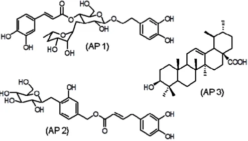
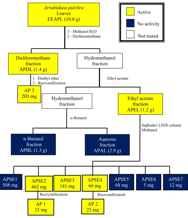
Preliminary fractionation of EEAPL by sequential extractions of its aqueous methanol solution with immiscible solvents (dichloromethane, ethyl acetate and n-butanol) led to four fractions: dichloromethane fraction (APDL), ethyl acetate fraction (APEL), n-butanol fraction (APBL) and aqueous fraction (APAL). APDL showed good activity against HSV-1 and DENV-2, with EC50 values of 18.6 ± 0.9 µg/mL and 15.4 ± 2.1 µg/mL, respectively. APEL was weakly active against HSV-1 (EC50 121.9 ± 9.8 µg/mL) and disclosed good activity against VACV (EC50 18.4 ± 1.9 µg/mL) and DENV-2 (EC50 12.2 ± 1.6 µg/mL), with SI values > 10 in these two last viruses. Chromatography of APEL over a Sephadex LH20 column led to the isolation of verbascoside (AP 1, 33 mg) and caffeoylcalleryanin (AP 2, 23 mg). APDL (1.4 g) was exhaustively extracted with diethyl ether affording ursolic acid (AP 3, 203 mg). Identification of the isolated compounds was based on spectrometric analyses (UV, IR, 1H- and 13C-NMR and MS) and comparison with literature data [11,12]. The chemical structures are shown in Figure 1 and antiviral activity in Table 1.
Figure 1. Chemical structures of verbascoside (AP 1), caffeoylcalleryanin (AP 2) and ursolic acid (AP 3).
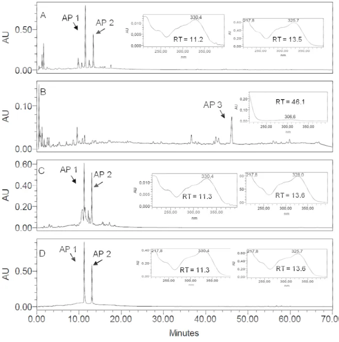
Figure 2. RP-HPLC-DAD fingerprints for the crude ethanol extract from Arrabidaea pulchra leaves (EEAPL) (A), APDL with AP 3 (RT = 46.1 min UV spectrum registered online detection 220 nm (B), APEF with UV spectra registered online for peaks corresponding to AP 1 (RT = 11.3 min) and AP 2 (RT = 13.6 min) (C) and for a mixture of the isolated arylpropanoids verbascoside (AP 1, RT 11.3 min) and caffeoylcallerianin (AP 2, RT 13.6 min) (D). Detection: 350 nm. Chromatographic conditions: see Experimental Section.
The ethanol extract from leaves of A. pulchra (EEAPL) did not show activity against EMCV, and revealed a low antiviral activity against HSV-1 (235.3 ± 9.7 μg/mL, SI > 2.21) and VACV-WR (245.2 ± 13.4 μg/mL, SI > 2.0), while good activity against DENV-2 was observed (EC50 46.8 ± 1.6 μg/mL, SI 2.7) (Table 1). The dichloromethane fraction (APDL) was inactive against HSV-1 and EMCV and showed good activity against VACV and DENV-2 (EC50 18.6 ± 0.9 μg/mL, SI 1.3 and
(EC50 18.4 ± 1.9 μg/mL, SI > 10.9) and DENV-2 (EC50 12.2 ± 1.6 μg/mL, SI > 16.3). Fractions APSE 2, 3 and 4 that were obtained from cromatography of APEL on a Sephadex LH20 column disclosed good activity against VAC-WR (EC50 15.9 ± 2.3 μg/mL, SI > 12.3 to EC50 18.1 ± 3.4 μg/mL, SI > 11.0) and against DENV-2 (EC50 8.8 ± 0.7 μg/mL, SI > 22.7 to EC50 11.9 ± 1.5 μg/mL, SI > 16.8).
Compounds AP 1, 2 and 3 were not active towards VAC-WR and EMCV, and AP 3 showed a low activity towards HSV-1. The three compounds disclosed good activity against DENV-2, although the SI values are low for AP 1 and AP 3, as a consequence of their toxicity to LLCMK2 cells. AP 2 was the most potent one, with SI > 10 (EC50 2.8 ± 0.4 μg/mL, SI 20.0, Table 1).
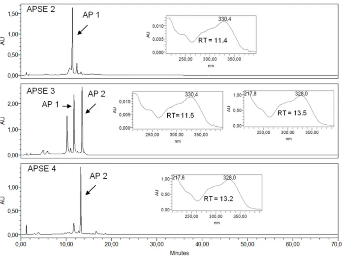
Figure 3. RP-HPLC-DAD profile of Sephadex LH 20 fractions APSE 2, APSE 3, APSE 4 with UV spectra registered online for peaks corresponding to AP 1 and to AP 2. RT: Retention Time. Detection: 350 nm. Chromatographic conditions: see Experimental Section.
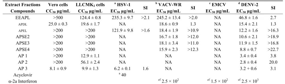
Table 1. Cytotoxicity (CC50, Vero and LLCMK2 cells), in vitro antiviral activity (EC50), selectivity index (SI) for ethanol extract from Arabidaea pulchra leaves (EEAPL), fractions APDL, APEL, APSE2-4 and compounds AP 1–3.
Extract Fractions Compounds
Vero cells CC50 µg/mL
LLCMK2 cells
CC50 µg/mL a
HSV-1 EC50 µg/mL
SI
b
VACV-WR EC50 µg/mL
SI
c
EMCV EC50 µg/mL
d
DENV-2 EC50 µg/mL
SI
EEAPL >500 124.4 ± 0.8 235.3 ± 9.7 >2.1 245.2 ± 13.4 >2.0 NA 46.8 ± 1.6 2.7
APDL 25.0 ± 0.3 19.6 ± 1.7 NA 18.6 ± 0.9 1.3 NA 15.4 ± 2.1 1.3
APEL >200 >200 121.9 ± 9.8 >1.6 18.4 ± 1.9 >10.9 NA 12.2 ± 1.6 >16.3
APSE2 >200 >200 NA 16.7 ± 1.8 >12.0 NA 10.6 ± 2.1 >18.9
APSE3 >200 >200 NA 18.1 ± 3.4 >11.0 NA 11.9 ± 1.5 >16.8
APSE4 >200 >200 NA 15.9 ± 2.3 >12.3 NA 8.8 ± 0.7 >22.7
AP 1 >200 12.9 ± 1.1 NA NA NA 3.4 ± 0.4 3.8
AP 2 >200 56.1 ± 2.4 NA NA NA 2.8 ± 0.4 20.0
AP 3 8.1 ± 0.9 9.9 ± 1.3 6.2 ± 0.1 1.6 NA NA 3.2 + 0.6 3.1
Acyclovir a40
α-2a Interferon ef 2.5 × 10² ef 1.5 × 10² ef 2.5 × 10³
SI, selective index; a viral titer TCID50/mL 2.5 × 10⁶ in 48 h; b viral titer TCID50/mL 1.0 × 10⁶ in 48 h; c viral titer TCID50/mL 1.0 × 10⁶ in 48 h; d viral titer TCID50/mL
1.0 × 10⁴ in 72 h; NA, no activity in the assayed concentrations; e 80 to 100% inhibition of cytopathic effect; f concentration in UI/mL; EEAPL, ethanol extract from
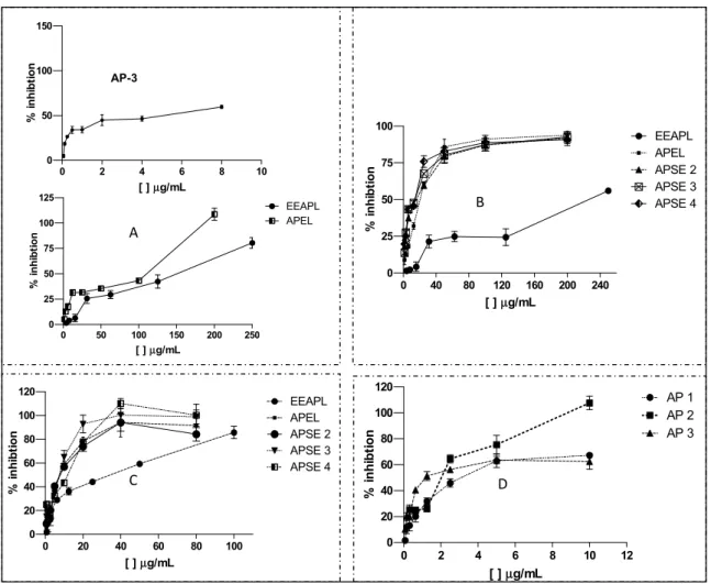
The strongest EEAPL antiviral effect was towards DENV-2 with 80% inhibition at 100 μg/mL. APEL and APSE2-4 caused approximately 100% inhibition at concentrations of 40 μg/mL. Finally, the highest potency of AP 2 against DENV-2 was demonstrated by the dose-response curves of Figure 4D, in which 100% of virus inhibition was observed at 10 μg/mL.
Figure 4. Dose-response curves for antiviral activity of ethanol extract, fractions, and constituents of Arrabidaea pulchra leaves. (A) EEAPL, APEL and AP 3 against HSV-1; (B) EEAPL, APEL, APSE 2–4 against VAC-WR; (C) EEAPL, APEL, APSE 2–4 against DENV-2; (D) AP 1, AP 2, AP 3 against DENV-2.
3. Discussion
According to our previously published results [10], the antiviral profile of the ethanol extract of the leaves of A. pulchra (EEAPL) is inactive towards EMCV, shows low activity against HSV-1 (235.3 ± 9.7 μg/mL, SI > 2.21) and VACV-WR (245.2 ± 13.4 μg/mL, SI > 2.0) and good activity towards DENV-2 (EC50 46.8 ± 1.6 μg/mL), although with a low SI (2.7, against LLCMK2 cells). (Table 1). Based on the EC50 value lower than 100 μg/mL as determined for EEAPL, the limit we established for a plant extract to be considered of interest for a bioguided fractionation, this extract was initially submitted to a liquid-liquid partition aiming to promote the separation of the EEPAL constituents according to their polarity/solubility. The dicloromethane fraction (APDL) inhibited the viral replication of VACV-WR and DENV-2, showing EC50 values of 18.6 ± 0.9 μg/mL (VACV-WR)
AP-3
0 40 80 120 160 200 240 0 25 50 75 100 EEAPL APEL APSE 2 APSE 3 APSE 4
[ ]µg/mL
% i n hi bt io n B
0 20 40 60 80 100 0 20 40 60 80 100 120 EEAPL APEL APSE 2 APSE 3 APSE 4
[ ]µg/mL
% i n hi bt ion
C D
0 2 4 6 8 10 12
0 20 40 60 80 100 120 AP 1 AP 2 AP 3
[ ]µg/mL
%
i
nhi
bt
ion
0 50 100 150 200 250
0 25 50 75 100 125 EEAPL APEL
[ ]µg/mL
% i nhi bt io n A
0 2 4 6 8 10
0 50 100 150
[ ]µg/mL
%
i
nhi
bt
and 15.4 ± 2.1 μg/mL (DENV-2), both of them with low SI (1.3). On the other hand, ursolic acid (AP 3), which was isolated from APDL, was inactive against VACV-WR, but has shown activity against HSV-1 and DENV-2 (EC50 6.2 ± 0.1 μg/mL and 3.2 + 0.6 μg/mL, respectively) although with low SI values (1.6 and 3.1, respectively), due to its cytotoxicity to Vero and LLCMk2 cells (8.1 ± 0.9 and 9.9 ± 1.3 μg/mL, respectively). APEL showed a better profile than APDL for both VACV and DENV-2, with good antiviral activity (EC50 < 20 μg/mL) and SI > 10 and, therefore, it was submitted to chromatography over a Sephadex LH20 column aiming at separation of its active constituents. Three fractions (APSE2-4) out of the seven fractions obtained have shown good activity against VACV-WR and DENV-2 with SI > 10, for both of these viruses, and particularly against DENV-2 (SI > 22.7). APSE 2 led to the isolation of verbascoside (AP 1) and caffeoylcallerianin (AP 2) from APSE4. AP 1 and AP 2 have inhibited the replication of DENV-2 with EC50 values of 3.4 μg/mL and 2.8 μg/mL, respectively, but AP 2 was less cytotoxic to LLCMk2 cells, thus resulting in a more favorable SI (20.0), while for AP 1 the SI value (3.8) is approximately five times lower. Indeed, the cytotoxicity of ursolic acid is recognized. It is reported to be involved in endoplasmic reticulum stress pathway [13] targeting apoptosis and its potent cancer-preventive activity and great cancer therapeutic potential have been pointed out [14]. None of the tested samples inhibited the multiplication of EMCV. EEAPL, fractions derived from it and isolated compounds have generally shown low cytotoxicity, except APDL and AP 1 with CC50 in the range of 19.6 μg/mL for LLCMK2 cells and 25.9 μg/mL for Vero cells. Concerning the effects against DENV-2, it is interesting to note that APEL, which contains similar contents of AP 1 and AP 2 (Figure 2), has shown an enhancement of the antiviral activity (SI > 16.3) in relation to the crude ethanol extract (EEAPL, SI 2.7) while APSE4 (SI > 22.7), containing predominantly AP-2 (Figure 3), was closer to the value for this compound (SI 20.0). Caffeoylcalleryanin (AP 2) was first isolated in 1968 from Pyrus calleryana (Rosaceae) [12], however this is the first report on its isolation from a Bignoniaceous species, as well as on its antiviral activity.
Verbascoside (AP 1, synonyms: acteoside, kusaginin) is a phenylethanoid glycoside isolated from various plant species that displays an interesting spectrum of biological activities [15]. In the genus Arrabidaea, verbascoside was isolated from A. harleyi [11] and its occurrence has been reported in some other genera of the family. It was also isolated from Markhamia lutea (Bignoniaceae) and has shown a potent antiviral effect against respiratory syncytial virus (RSV, EC50 0.80 µg/mL) [16]. Recently the activity against HSV-1 (EC50 58.0 µg/mL) and HSV-2 (EC50 8.9 µg/mL) was reported for this compound [17]. However, in the present work, no effect was observed for verbascoside against this virus. Our result is in agreement with a previous report in which this compound had not shown anti-herpes activity either [16] and these contradictory results might be related to different degrees of virulence of the strains used in the assays.
The mechanisms of antiviral action of this group of triterpenoids have been shown to involve inhibition of virus entry, of protease and of replication [1].
Verbascoside (AP 1) and caffeoylcalleryanin (AP 2) belong to the group of naturally occurring polyphenols that are known as phenylethanoid glycosides, supporting each one a moiety of glucose esterified by caffeic acid and bonded to an ethylcatechol unity by an ether linkage. The polyphenols often showed virucidal effects in several viral systems by attaching to the proteins and/or host cell surfaces, resulting in reduction or prevention of viral adsorption; inhibition of reverse transcriptase (RTase) of HIV-1 and RNA polymerase of influenza virus. The ortho-dihydroxy systems are probably responsible for polyphenols’ antiviral activity [1].
In light of our results, further investigations are needed to understand the mechanisms of the antiviral activity, especially of caffeoylcalleryanin, which shows the lowest toxicity in LLCMK2 cells of the tested compounds and was active only in DENV-2. Further assays are also needed to investigate virucidal activity and targets in the viral replication cycle.
4. Experimental
4.1. Plant Material
The leaves of A. pulchra were collected in Belo Horizonte, Minas Gerais, Brazil. The plants were taxonomically identified by Dr. J.A. Lombardi, Departamento de Botânica, Instituto de Biociências, UNESP, Rio Claro, Brazil. A voucher specimen was deposited at the BHCB/UFMG, Belo Horizonte, Minas Gerais, Brazil, under the number 21573.
4.2. Extraction and Isolation
Figure 5. Bioguided fractionation of the ethanol extract from Arrabidaea pulchra leaves.
4.3. Structural Determination
AP 1, AP 2 and AP 3 were identified on the basis of spectral analyses and comparison with literature data. 1H-NMR, 13C-NMR, NOESY, TOCSY, HSQC; HMBC spectra were registered in DMSO-d6 with TMS as internal standard and were recorded on a Bruker Advance DPX400
instrument. Chemical shifts are given as δ (ppm). LC-MS were obtained by electrospray ionization mass spectrometry (ESI-MS) in an Esquire 3000 Plus Bruker Daltonics equipment, Capillary: 4000 V, Nebulizer: 27 psi, Dry Gas: 7.0 L/min, Dry Temp: 320 °C, mass flux 100 uL/min, in the Central Analítica, Instituto de Química, Universidade de São Paulo, São Paulo, SP, Brazil.
1 - Methanol/H2O 2 - Dichloromethane
Hydromethanol fraction
Sephadex LH20 column Methanol
APSE1
508 mg 462 mg APSE2 141 mg APSE3
APSE4 66 mg
AP 1 33 mg
AP 2 23 mg
Recrystallization
Arrabidaea pulchra
Leaves EEAPL (10.0 g)
Hydromethanol fraction
Active
No activity
Ethyl acetate fraction APEL (1.2 g) Dichloromethane
fraction APDL (1.4 g)
AP 3 203 mg
Ethyl acetate
n-Butanol
n-Butanol fraction APBL (1.3 g)
Aqueous fraction APAL (2.9 g)
Recrystallization 1 –Diethyl ether 2 - Recrystallization
Not tested
APSE5 68 mg
APSE6 5 mg
4.4. Spectral Data
Verbascoside (AP 1). Light yellow powder. MP 145–148 °C, Lit. 149–151 °C [20]. UV (MeOH): λmax (log ε) 219.5 (4.6), 250 (sh), 290 (sh), 332.0 (4.6) nm. IR: υmax 3327, 1660, 1599, 1515, 1445, 1368, 1258, 1157, 1113, 1063, 1050, 853, 809, 784 cm−1. 1H-NMR (DMSO-d6): δ = 7.02 (1H, J = 2.0 Hz,
H-2′′′), 6.97 (1H, dd, J = 2.0 and 8.0 Hz; H-6′′′), 6.76 (1H, d, J = 8.0; H-5′′′), 6.64 (1H, dd, J = 8.0, H-5), 6.63 (1H, s, H-2), 6.49 (1H, dd, J = 2.0 and 8.0 Hz, H-6), 7.46 (1H, d, J = 16.0 Hz, H-7′′′), 6.19 (1H, d, J = 16.0 Hz; H-8′′′), 5.03 (1H, d, J = 8.0 Hz, H-1′), 4.35 (1H, d, J = 8.0 Hz, H-1′′). 13
C-NMR (DMSO-d6): δ = 129.1 (C1), 116.3 (C2), 145.0 (C3), 143.5 (C4), 115.4 (C5), 119.5 (C6),
35.5 (C7), 69.1 (C8), 101.2 (C1′), 75.0 (C2′), 79.5 (C3′), 69.6 (C4′), 72.1 (C5′), 61.2 (C6′), 102.3 (C1′′), 70.9 (C2′′), 70.7 (C3′′), 71.0 (C4′′), 69.2 (C5′′), 18.6 (C6′′), 125.5 (C1′′′), 114.7 (C2′′′), 145.5 (C3′′′), 148.4 (C4′′′), 115.8 (C5′′′), 121.3 (C6′′′), 145.0 (C7′′′), 113.6 (C8′′′), 165.7 (C9′′′). ESI-MS: found m/z 647.2 [M+Na]; calculated for C29H35O15Na m/z 647.
Caffeoylcalleryanin (AP 2). Light yellow powder. MP 167–171 °C. UV (MeOH): λmax (log ε) 217 (4.3), 250 (sh), 286.5 (4.1), 300 (sh), 329.5 (4.2) nm. IR: υmax 3270, 1660, 1599, 1510, 1443, 1353, 1253, 1175, 1150, 1035, 803 cm−1. 1H-NMR (DMSO-d6): δ = 7.07 (1H, d, J = 2.0 Hz; H-2′′), 7.02 (1H,
dd, J = 2.0 and 8.0 Hz; H-6′′), 6.78 (1H, d, J = 8.0; H-5′′), 7.12 (1H, dd, J = 8.0, H-5), 6.89 (1H, d, J = 2.0 Hz, H-2), 6.81 (1H, dd, J = 2.0 and 8.0 Hz, H-6), 7.52 (1H, d, J = 16.0 Hz, H-7′′′), 6.32 (1H, d, J = 16.0 Hz; H-8′′′), 4.71 (1H, d, J = 7.2 Hz, H-1′), 3.2–3,84 (H-2′, H-3′,H-4′, H-5′,H-6′a e 6′b, m). 13
C-NMR (DMSO-d6): δ = 131.0 (C1), 115.9 (C2), 145.1 (C3), 146.8 (C4), 116.7 (C5), 119.3 (C6),
65.1 (C7), 102.3 (C1′), 75.9 (C2′), 77.2 (C3′), 69.6 (C4′), 73.3 (C5′), 60.8 (C6′), 125.4 (C1′′), 114.8 (C2′′), 145.6 (C3′′), 148.6 (C4′′), 115.7 (C5′′), 121.4 (C6′′), 145.4 (C7′′), 113.7 (C8′′), 166.4 (C9′′). ESI-MS: found m/z 487.1 [M+Na]; calculated for C22H24O11Na m/z 487.0.
Ursolic acid (AP 3). Colourless crystals. MP 282–284 °C, Lit 285–287 °C [21]. IR: υmax 3392, 2973, 2852, 1687, 1620, 1456, 1387, 1259, 1089, 867, 798; 1H-NMR (DMSO-d6): 5.23 (bs, 1H, H-12), 3.0
(m, 1H, H-3), 2.11 (d, 1H, J11.2, H-18), 0.69; 0.71; 0.87; 0.91; 1.08; 1.29 (6s, 6xCH3). 13C-NMR (DMSO-d6): δ 38.1 (C-1), 26.9 (C-2), 76.7 (C-3), 32.6 (C-4), 54.7 (C-5), 17.9 (C-6), 36.4 (C-7), 39.5
(C-8), 46.9 (C-9), 38.3 (C-10), 22.8 (C-11), 124.5 (C-12), 138.1 (C-13), 41.6 (C-14), 27.4 (C-15), 23.7 (16), 46.7 (C-17), 52.3 (C-18), 38.4 (C-19), 38.4 (C-20), 30.1 (C-21), 36.2 (C-22), 28.2 (C-23), 15.1 (C-22), 28.2 (C-23), 15.1 (C-24), 16.0 (C-25), 16.8 (C-26), 23.2 (C-27), 178.5 (C-28), 16.9 (C-29), 21.0 (C-30). EI-MS m/z (%): 43 (100), 55 (56), 67 (26), 68 (36), 79 (23), 81 (30), 91 (23), 93 (23), 95 (26), 105 (24), 107 (20), 109 (16), 133 (44), 147 (17), 163 (7), 175 (9), 189 (15), 203 (31), 207 (26), 248 (76), 249 (13), 456 (M+) (2).
4.5. HPLC Analyses
dissolved by sonication in an ultrasound bath for 15 min, followed by centrifugation at 10,000 rpm for 10 min. The supernatant was filtered through a Millipore membrane (0.2 μm) and injected (10.0 μL) onto the equipment. Elution was carried out with a linear gradient of water (A) and acetonitrile (B) (from 5% to 95% of B in 60 min).
4.6. Cell Culture and Virus
Vero cells (ATCC CCL-81) and LLCMK2 cells were cultured in Dulbecco’s modified Eagle’s medium (DMEM, Cultilab, Campinas, SP, Brazil) at 37 °C, in 5% CO2 atmosphere, supplemented with 5% fetal bovine serum, 50 µg/mL gentamicin, 100 U/mL penicillin and 5 µg/mL amphotericin B. HSV-1 was obtained from the collection of Laboratório de Virus, UFMG, Belo Horizonte, Brazil. DENV-2, EMCV and VACV-WR were kindly donated by Dr. L. Figueiredo (USP, Ribeirão Preto, Brazil), Dr. I. Kerr (London Research Institute, London, UK) and Dr. C. Jungwirth (University of Würzburg, Würzburg, Germany), respectively. The viruses were titrated by TCID50 in Vero cells [22] and the titers were 2.5 × 10⁶; 1.0 × 10⁶; 1.0 × 10⁶ and 1.0 × 10⁴ TCID50/mL, respectively, for HSV-1, EMCV, VACV-WR and DENV-2 virus.
4.7. Cytotoxicity Assay
Vero and LLCMK2 cells were exposed to different concentrations of extracts/fractions/compounds for 48 and 72 h. After incubation, cell viability was assessed by the 3-(4,5-dimethylthiazol-2-yl)-2,5-diphenyltetrazolium bromide (MTT, Merck) assay at a concentration of 2 mg mL−1 in PBS [23]. Each sample was assayed in four replicates for concentrations ranging from 500 to 0.125 µg mL−1. The cytotoxicity of each sample was expressed as CC50, i.e. the concentration of sample that inhibited cell growth by 50%.
4.8. Antiviral Assays
The antiviral activity (EC50) was evaluated by the MTT assay [23]. Acyclovir (Calbiochem, Merck Brasil, São Paulo, SP, Brazil) and α-2a interferon (Bergamo Brasil, São Paulo, SP, Brazil) were used as positive controls. Experiments were carried out with eight different concentrations within the inhibitory range of the samples. The 50% inhibitor concentration of the viral effect (EC50) for each extract, fractions, and constituents were calculated from concentration-effect-curves after no linear regression analysis (Figure 5). The selective index (SI) is defined as CC50 over EC50.
5. Conclusions
Acknowledgements
This work was supported by funds from FAPEMIG–Fundação de Amparo à Pesquisa do Estado de Minas Gerais (Brazil) and CNPq (Brazil). CAPES (Brazil) and CNPq (Brazil) are also acknowledged for PhD (to GCB) and research fellowships (to ABO and EGK).
Conflicts of Interest
The authors declare no conflict of interest.
References
1. Chattopadhyay, D.; Naik, T.N. Antivirals of ethnomedicinal origin: Structure-activity relationship and scope. Mini Rev. Med. Chem. 2007, 7, 275–301.
2. Mukhtar, M.; Arshad, M.; Ahmad, M.; Pomerantz, R.J.; Wigdahl, B.; Parveen, Z. Antiviral potentials of medicinal plants. Virus Res. 2008, 131, 111–120.
3. Selisco, B.; Guillemot, J.C.; Alvarez, K.; Canard, B. Opportunities in the Development of Anti-Dengue Drugs. Available online: http://www.tropika.net/svc/review/ 061001-Dengue_Drugs (accessed on 22 July 2013).
4. Lohmann, L.G. Untangling the phylogeny of neotropical lianas (Bignonieae, Bignoniaceae). Am. J. Bot. 2006, 93, 304–318.
5. Gentry, A.H. A synopsis of Bignoniaceae ethnobotany and economic botany. Ann. Mo. Bot. Gard. 1992, 79, 53–64.
6. Fischer, E.; Theisen, I.; Lohmann, G.L. Bignoniaceae. In: The Families and Genera of Vascular Plants; Kubitsky, K., Ed.; Springer-Verlag: Heidelberg, Germany, 2004; Volume VII, pp. 9–38. 7. Scudeller, V.V.; Carvalho-Okano, R.M. A tribo Bignonieae Spreng (Bignoniaceae) no Parque
Estadual do Rio Doce (MG). Iheringia Série Botânica 1998, 51, 79–133.
8. Alvarenga, T.A.; Bêdo, T.R.O.; Braguine, C.G.; Gonçalves, U.O.; Magalhães, L.G.; Rodrigues, V.; Gimenez, V.M.M.; Groppo, M.; Silva, M.L.A.; Cunha, W.R.; Januário, A.H.; Pauletti, P.M. Evaluation of Cuspidaria pulchra and its isolated compounds against Schistosoma mansoni Adult Worms. Int. J. Biotechn. Wellness Ind. 2012, 1, 122–127.
9. Brandão, G.C.; Kroon, E.G.; Santos, J.R.; Stehmann, J.R.; Lombardi, J.A.; Oliveira, A.B. Antiviral activities of plants occurring in the state of Minas Gerais, Brazil. Part 2. Screening Bignoniaceae species. Braz. J. Pharmacogn. 2010, 20, 742–750.
10. Brandão, G.C.; Kroon, E.G.; Santos, J.R.; Stehmann, J.R.; Lombardi, J.A; Oliveira, A.B. Antiviral activity of Bignoniaceae species occurring in the State of Minas Gerais (Brazil): Part 1. Letters Appl. Microbiol. 2010, 51, 469–476.
11. Lima, C.S.A.; Amorim, E.L.C.; Sena, K.X.F.R.; Chiappeta, A.A.; Nunes, X.P.; Agra, M.F; Cunha, E.V.L.; Silva, M.S.; Barbosa-Filho, J.M. Antimicrobial activity of a mixture of two isomeric phenylpropanoid glycosides from Arrabidaea harleyi A.H. Gentry (Bignoniaceae). Braz. J. Pharm. Sci. 2003, 39, 77–81.
13. Zheng, Q.Y.; Li, P.P.; Jin, F.S.; Yao, C.; Zhang, G.H.; Zang, T.; Ai, X. Ursolic acid induces ER stress response to activate ASK1-JNK signaling and induce apoptosis in human bladder cancer T24 cells. Cell Signal. 2013, 25, 206–213.
14. Yang, H.; Dou, Q.P. Targeting Apoptosis Pathway with Natural Terpenoids: Implications for Treatment of Breast and Prostate Cancer. Curr. Drug Targets 2010, 11, 733–744.
15. Ghisalberti, E.L. Lantana camara L. Verbenaceae. Fitoterapia 2000, 71, 467–486.
16. Kernan, M.R.; Amarquaye, A.; Chen, J.L.; Chan, J.; Sesin, D.F.; Parkinson, N.; Ye, Z.; Barrett, M.; Bales, C.; Stoddart, C.A. Antiviral phenylpropanoid glycosides from the medicinal plant Markhamia lutea. J. Nat. Prod. 1998, 61, 564–570.
17. Martins, F.O.; Esteves, P.F.; Mendes, G.S.; Barbi, N.S.; Menezes, F.S.; Romanos, M.T. Verbascoside isolated from Lepechinia speciosa has inhibitory activity against HSV-1 and HSV-2 in vitro. Nat. Prod. Commun. 2009, 4, 1693–1696.
18. Lui, J. Pharmacology of oleanolic acid and ursolic acid. J. Ethnopharmacol. 1995, 49, 57–68. 19. Simões, L.R.; Maciel, G.M.; Brandão, G.C.; Kroon, E.G.; Castilho, R.O.; Oliveira, A.B. Antiviral
activity of Distictella elongata (Vahl) Urb. (Bignoniaceae), a potentially useful source of anti-dengue drugs from the state of Minas Gerais, Brazil. Lett. Appl. Microbiol. 2011, 23, 602–607. 20. Sarkhail, A.; Monsef-Esfehani, H.R.; Amin, G.; Surmaghi, M.H.S; Shafiee, A. Phytochemical
Study of Phlomis olivieri Benth. and Phlomis persica Boiss. DARU 2006, 14, 115–121.
21. Rodriguez, D.J.; Chulia, J.; Simões, C.M.O.; Amoros, M.; Mariotte, A.M.; Girre, L. Search for in vitro antiviral activity of a new isoflavonic glycoside from Ulex europaeus. Planta Med. 1990, 56, 59–62.
22. Betancur-Galvis, L.A.; Saez; Granados, J.H.; Salazar, A.; Ossa, J.E. Antitumor and antiviral activity of Colombian medicinal plants extracts. Mem. Inst. Oswaldo Cruz. 1999, 94, 531–535. 23. Twentyman, P.R.; Luscombe, M. A study of some variables in a tetrazolium dye (MTT) based
assay for cell growth and chemosensitivity. Br. J. Cancer. 1987, 56, 279–285.
24. Oliveira, A.B.; Kroon, E.G.; Brandão, G.C. Composições farmacêuticas antivirais contendo extrato, frações e/ou compostos isolados de Arrabidaea pulchra e uso. Brazilian Patent PI BR 014110003098 (INPI/MG), 27 November 2011.