RESEARCH ARTICLE
Molecular Characterization of
Enterotoxin-Producing
Escherichia coli
Collected in
2011
–
2012, Russia
Nikolay N. Kartsev1*, Nadezhda K. Fursova1, Dmitry M. Pachkunov2, Vasiliy A. Bannov1, Boris V. Eruslanov1, Edward A. Svetoch1, Ivan A. Dyatlov1
1Department of Molecular Microbiology, State Research Center for Applied Microbiology and Biotechnology, Obolensk, Russian Federation,2Department of Innovation Research, Volga State Technological University, Yoshkar-Ola, Russian Federation
Abstract
Enterotoxin-producingEscherichia coli(ETEC) are one of the main causative agents of diarrhea in children especially in developing countries and travel diarrhoea in adults. Pathogenic proper-ties of ETEC associated with their ability to produce a heat-stable (ST) and/or heat-labile (LT) enterotoxins, as well as adhesins providing bacterial adhesion to intestinal epithelial cells. This study presents the molecular characterization of the ETEC isolates collected from the Central and Far-Eastern regions of Russia in 2011–2012. It was shown that all ETEC under study (n=18) had the heat-labile enterotoxin-coding operonelt, and had no the genes of the heat-stable enterotoxin operonest. DNA sequencing revealed two types of nucleotide exchanges in theeltBgene coding subunit B of LT in isolates collected from Cherepovets city (Central region, Russia) and Vladivostok city (Far-East region, Russia). Only one ETEC strain carried genes cfaA,cfaB,cfaCandcfaDcoding adhesion factor CFA/I. Expression of LT in four ETEC isolates in the agglutination reaction was detected using a latex test-system. The isolates were assigned to serogroups O142 (n = 6), O6 (n = 4), O25 (n = 5), O26 (n = 2), and O115 (n = 1). Genotyping showed that they belonged to an earlier described sequence-type ST4 (n = 3) as well as to 11 novel sequence-types ST1043, ST1312, ST3697, ST3707, ST3708, ST3709, ST3710, ST3755, ST3756, ST3757 and ST4509. The ETEC isolates displayed different levels of antimi-crobial resistance. Eight isolates were resistant to only one drug, three isolates—to two drugs, one isolate—to three drugs, two isolates—to four antibacterials, and only one isolate to each of the five, six and ten antibacterials simultaneously. Genetic determinants of the resistance to beta-lactams and other classes of antibacterials on the ETEC genomes were identified. There areblaTEM(n = 10),blaCTX-M-15(n = 1), class 1 integron (n = 3) carrying resistance cassettes to
aminoglycosides and sulphonamidesdfrA17-aadA5anddfrA12-orfF-aadA2. One isolate ETE-C_Ef-6 was found to be a multidrug-resistant (MDR) pathogen that carried both the beta-lacta-mase gene and class 1 integron. These data suggest the circulation of ETEC in Russia. Further investigations are necessary to study the spread of the revealed ETEC sequence types (STs) and serotypes. Their role in the etiology of diarrhea should be also estimated.
PLOS ONE | DOI:10.1371/journal.pone.0123357 April 29, 2015 1 / 12
a11111
OPEN ACCESS
Citation:Kartsev NN, Fursova NK, Pachkunov DM, Bannov VA, Eruslanov BV, Svetoch EA, et al. (2015) Molecular Characterization of Enterotoxin-Producing
Escherichia coliCollected in 2011–2012, Russia.
PLoS ONE 10(4): e0123357. doi:10.1371/journal. pone.0123357
Academic Editor:Patrick Butaye, Ross University School of Veterinary Medicine, SAINT KITTS AND NEVIS
Received:December 19, 2014
Accepted:March 3, 2015
Published:April 29, 2015
Copyright:© 2015 Kartsev et al. This is an open access article distributed under the terms of the
Creative Commons Attribution License, which permits unrestricted use, distribution, and reproduction in any medium, provided the original author and source are credited.
Data Availability Statement:All relevant data are within the paper and its Supporting Information files.
Funding:Grant 037, Rospotrebnadzor, Grant 039, Rospotrebnadzor.
Introduction
Escherichia colibacteria causing human intestinal infections are subdivided into six pathotypes, namely enterotoxigenic (ETEC), enteropathogenic (EPEC), enteroinvasive (EIEC), enterohae-morrhagic (EHEC), enteroaggregative (EAgEC) and diffusely-aggregative (DAEC) [1].
ETEC are the enteric pathogens that may cause cholera-like diarrhea in animals and hu-mans. Of all pathogenicE.colibacteria ETEC are the most common causative agents of human diarrhea worldwide. More than 650 million cases of ETEC-infections are registered annually in 2000s among which 800 thousands cases are fatal [2]. It is estimated that each year on the peri-od from 2009 to 2012 nearly 600,000 children less than five years of age die from severe dehy-drating diarrhea, mostly in the developing world [3]. Among the many causes of diarrheal disease, enterotoxigenicE.coliandShigellaare the two most important bacterial pathogens [4]. ETEC can also cause so called traveler’s diarrhea, the most frequent illness reported and related to people who traveled from an industrialized country to a developing country, including intes-tinal infections in the military contingent of the United Nations. The incidence of diarrhea among visitors of the tropics and subtropics varies from 10% to 60%. A high percentage of inci-dences are registered in Latin America, Africa and the Indian subcontinent [5]. The seasonal prevalence of ETEC-associated diarrhea has been established. The morbidity rate increases by 7% per each degree of rise in ambient temperature that is accompanied by the growth and spread of bacteria contaminating food and water [6].
ETEC strains isolated from humans belong to different O-groups, but more often (60–70% strains collected worldwide)—to O6, O8, O25, O78, O128, and O153 groups. The remaining 30–40% constitutes a great number of other serogroups [13]. ETEC isolates of O148 serogroup are also isolated in Russia [14]. PCR-serotyping ofE.coliidentifies the genes coding enzymes involved into the O-antigen synthesis. These determinants locate in the O-cluster formerly known asrfb-cluster codingE.colihousekeeping genes [15]. Genetic structures of microbial populations and a contribution of the specific clone to the development of the epidemiological situation are studied by various genotyping methods including ribotyping, Pulsed Field Gel Electrophoresis (PFGE), Random Amplified Polymorphic DNA (RAPD), and Multilocus Se-quence Typing (MLST). The MLST analysis of bacterial isolates collected during an outbreak allows one to follow up their relationships (local epidemiology) and to establish their potential links with similar isolates from other geographic regions (global epidemiology). MLST algo-rithm developed for pathogenicE.coliis based on the polymorphism of seven chromosomal housekeeping genes:adk(adenylate kinase),fumC(fumarate hydratase),gyrB(DNA gyrase), icd(isocitrate/isopropylmalate dehydrogenase),mdh(malate dehydrogenase),purA (adenylo-succinate dehydrogenase),recA(ATP/GTP binding motif) (Mark Achtman Database,http:// mlst.warwick.ac.uk/mlst/dbs/Ecoli). The genetic diversity of ETEC strains was explained by the ability ofE.colito capture virulence plasmids at rapid evolutionary processes and great struc-tural variety of their chromosome [16]. The analysis of many strains from various sources col-lected worldwide confirmed the intensive exchange of enterotoxin and adhesin genes among ETEC of different genetic lineages [17].
The objective of this study is to characterize ETEC strains isolated during the food-borne outbreak in Cherepovets in August, 2011, as well as those collected during a sporadic case of food-borne infection in Vladivostok in September, 2012. ETEC phenotypes responsible for an-timicrobial susceptibility and LT-production were determined. Genetic determinants coding enterotoxins and adhesins, and antibacterial resistance genes in their genomes were detected by PCR. O-groups and sequence types (ST) of the strains were identified.
Material and Methods
Ethics Statement
Our laboratory is a reference center for the study of food-borne infections agents, and we do not have direct contact with patients. Materials for research were obtained from the regional centers of Hygiene and Epidemiology. The study was not reviewed and approved by an institu-tional review board (ethics committee) before the study began because we studiedE.coli strains, isolated from the fecal samples without specifying the names of patients, the race/eth-nicity, age, religion, sex/gender, sexual orientation, or other socially constructed groupings. Our task was to identify the agent of food-borne outbreak and identify the source in food. However, in accordance with the rules of the Russian Federation, every patient entering the hospital signed informed consent to medical procedures and diagnostic tests.
Microbiology
Bacterial isolates and strains. LT-positive ETEC isolates were collected from the sick peo-ple with moderate gastroenteritis during an outbreak of food-borne infection in Cherepovets (Central Region of Russia) in August, 2011 (n = 12) and from a patient with moderate food-borne infection in Vladivostok (Far Eastern region of Russia) in September, 2012 (n = 6).
Bacterial isolation and growing. PathogenicE.coliwere isolated from the fecal and food samples using nutrient enrichment mediums «Nutrient medium No. 11 GRM», «RVS-broth», «Nutrient medium No. GRM», «SDS-broth» (SRCAMB, Obolensk, Russia) followed by
ETEC in Russia 2011–2012
growing on the selective agar mediums «Nutrient medium No. 1 GRM», «E.coliO157:H7 Sor-bitol-agar », «XLD-agar», «Iron-glucose-lactose agar with urea», «Endo-agar GRM» (SRCAMB, Obolensk, Russia). IdentifiedE.colistrains were grown on agar nutrient media «Muller-Hinton» (Himedia, India), «Luria Bertani broth» (Difco, USA), and «Nutrient medium No. 1 GRM» (SRCAMB, Obolensk, Russia). Bacterial isolates were stored in 10% glycerol at minus 70 degrees on Celsius.
LT induction. Mundell nutrient medium [18] was inoculated by 109CFU/mlE.coli bacte-rial suspension in proportion 10:1, than incubated at aeration (120 rpm/min) at 37 degrees on Celsius for 24 h. After that 25μl of polymixin B (10000 U/ml) was added into 1 ml bacterial culture and incubated without aeration at 37 degrees on Celsius for 4 h. Bacterial cells were re-moved by centrifugation at 900 g for 20 min. Super was filtrated using «Ultrafree-MC Micro-centrifuge Filters» (Sigma-Aldrich, CIIIA) with pores 0,22μm. Obtained filtrate was the object for LT detection using latex-agglutination assay (SRCAMB, Obolensk, Russia).
Latex agglutination. Latex agglutination was done by mixing 20μl of the bacterial filtrate and 20μl of the latex suspension for LT detection. Pure cholera toxin (Oxoid, UK) in concen-tration of 2 mg/ml was used as positive control. Bacterial filtrate of the non-LT-producing E.colistrain HB101 was used as negative control.
Susceptibility to antibacterials. Susceptibility to antibacterials (ABs) was measured by disk-diffusion method and microdilution method in broth according to EUCAST guide-lines (http://www.eucast.org/clinical_breakpoints/).E.colistrain ATCC 25922 was used as control.
Molecular Genetics Methods
Detection of pathogenicE.coliin clinical samples by real-time PCR. Preliminary detec-tion of ETEC, EPEC, EHEC, EAgEC and EIEC in clinical specimens, samples of food and enrichment cultures of pathogenic ofE.coliwas done using commercial assay“AmpliSens Escherichioses-FRT”(InterLabService, Russia). The assay is based on the amplification of path-ogen genome specific region using specificE.coliprimers and oligonucleotide probes. The amplified product is detected in real-time PCR (RT-PCR). The kit is a qualitative test that con-tains the internal control (http://www.interlabservice.ru/en/catalog/index.php?sid=1104&id= 8415).
Detection of the resistance genetic determinants. Beta-lactamase TEM- and
CTX-M-type genes as well as class 1 and 2 integron were detected by PCR with specific primers [19]. Bacterial strain serotyping. ETEC serotyping was done both by i) agglutination with se-rums of the kits“OK-polyvalent Escherichia serums for agglutination» and «O-serogroup Escherichia serums for agglutination”(Biomed, Russia), and ii) PCR-serotyping using specific oligonucleotide primers [20–22] in 25μl reaction mix consisting of 2.5μl 10×Taq-buffer with (NH4)2SO4and 20 mM MgCl2, 2.5μl of 2.5 mM solution of each dNTPs, 0.25μl of BSA, 12.5 pM each primer, 0.7 U of recombinantTaq-polymerase, and 5μl of the bacterial lysate at the program consisting of initial denaturation for 5 min at 95°C; then 30 cycles of denaturation for 30 sec at 95°C, annealing for 30 sec at 58°C, and elongation for 40 sec at 72°C; final elongation for 5 min at 72°C.
cycles of denaturation at 45 sec for 95°C, annealing for 45 sec at appropriate Ta°C (Table 1) and elongation for 45 sec at 72°C; final elongation for 5 min at 72°C.
Plasmid DNA extraction. DNA extraction was done by alkaline method [23]. Plasmid profiles were analyzed by electrophoresis at 0.8% agarose gel.
Strains genotyping. Strains genotyping was done by RAPD-PCR using «random» primer
OPA11 [24]. DNA amplification was carried in 25μl of the reaction mixture contained 2.5μl of 10×Taq-buffer with (NH4)2SO4and 20 mM MgCl2, 2.5μl of 2.5 mM solution of each dNTPs, 20 pM of each primer, 0.7 U of the recombinantTaq-polymerase, and 5μl of cell lysate at the program involving initial denaturation for 3 min at 95°C; 5 cycles of denaturation for 1 min at 94°C, annealing for 1 min at 35°C, and elongation for 1 min at 72°C; then 40 cycles in-cluding denaturation for 30 sec at 94°C, annealing for 30 sec at 35°C, and elongation for 2 min at 72°C, then final elongation for 10 min at 72°C.
Multi Locus Sequence Typing. MLST of the ETEC strains was done using sequencing of
seven house-keeping genes, namelyadk(adenylate kinase),fumC(fumarate hydratase),gyrB (DNA gyrase),icd(isocitrate/isopropylmalate dehydrogenase),mdh(malate dehydrogenase), purA(adenyl-succinate dehydrogenase), andrecA(ATP/GTP-binding motif) according the approach presented by web-site of the MLST.UCC Mark Achtman database (http://mlst. warwick.ac.uk/mlst/dbs/Ecoli/). DNA sequence analysis was done by Vector NTI Advance 11.5 (Invitrogen, USA), BLAST (http://blast.ncbi.nlm.nih.gov/Blast.cgi?PROGRAM=
blastn&PAGE_TYPE=BlastSearch&LINK_LOC=blasthome), and Chromas Version 1.5 (Tech-nelysium Ply Ltd, Australia).
Phylogenetic analysis. Dendrogram showing the genetic relationship of the ETEC
strains under study was constructed based on MLST sequences by Mega6 software. Phyloge-netic reconstruction was performed using «neighbor-joining» method based on a two-parameter model nucleotide changes, branches were generated by bootstrapping with 1000 replications [25].
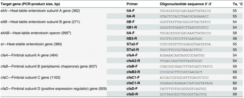
Table 1. Oligonucleotide primers for enterotoxin and adhesin genes detection.
Target gene (PCR-product size, bp) Primer Oligonucleotide sequence 5’-3’ Ta, °C
eltA—Heat-labile enterotoxin subunit A gene (362) ltA-F TGCACATGGCGACAAATTATACCG 55
ltA-R GTACTCCACCTAACGCAGAAACC 55
eltB—Heat-labile enterotoxin subunit B gene (271) ltB-F GGGTTATTTACGGCGTTACTATCC 54
ltB1-R GGGGTGTGAATCTTAATGTGTCC 54
eltAB—Heat-labile enterotoxin operon (995a) ltA-F TGCACATGGCGACAAATTATACCG 58
ltB3-R TCCTTCCTCCTTTCAATGGC 58
st—Heat-stable enterotoxin gene (380) STa2-F CGTCGTGTTTCGGAGGTAATATGA 55
STa2-R TGCCTTCCGCTAACACTTCC 55
cfaA—Fimbrial subunit A gene (464) cfaA-F AGAAAACAATAGGCGCAATGG 54
cfaA2-R TTGACCAGCTGTTAGTGCGC 54
cfaB—Fimbrial subunit B (periplasmic chaperone) gene (637) cfaB-F CGACGGCGAACTTTATGATCTATCC 59
cfaB2-R CCCGCGCTTCCATCAACACT 59
cfaC—Fimbrial subunit C gene (1163) cfaC1-F ACCACCGTGGCATTTGAGTCTCC 60
cfaC1-R GGGAAGCAGAAACCATCAGTATAGG 60
cfaD—Fimbrial subunit D (positive expression regulator) gene (929) cfaD-F TATTTTGTGCGGTGGTCAGTGC 59
cfaD-R GCCTAGCATCTGCGGTTACTCG 59
Note: a
product size varies depending oneltBgene variability
doi:10.1371/journal.pone.0123357.t001
ETEC in Russia 2011–2012
Results and Discussion
Heat-labile enterotoxin-producingE.colistrains collected during an outbreak of food-borne infection in Cherepovets (Central Region, Russia) in August, 2011 (n = 12) and from a patient with moderate food-borne infection in Vladivostok (Far Eastern region, Russia) in September, 2012 (n = 6) are characterized in this study.
The outbreak in Cherepovets involved workers who had taken their meal from the factory canteen. Mild disease developed in 97 infected persons, while moderate disease (abdominal pain, multiple liquid stools, vomiting, and temperature of 37°C to 38°C) was diagnosed in 10 patients. Totally, 76 clinical samples comprising 31 fecal samples from sick persons, 19 fecal samples from healthy canteen personnel, and 26 samples of foodstuffs (vegetables, greens and dairy products) have been analyzed. The preliminary analysis of the clinical materials and foodstuffs by the RT-PCR revealed the presence of DNAs of pathogenicE.colibacteria, namely ETEC (n = 23), EPEC (n = 18), EAgEC (n = 5), and EIEC (n = 1) in the samples. Totally 70 E.coliisolates were collected. All of them were tested on belonging to the listed aboveE.coli pathogenic groups including presence of heat labile toxineltand heat stable toxinestgenes. As a result 11 ETEC strains were isolated from nine patients’clinical material, and one ETEC strain was isolated from a sample of sour cream (Table 2).
A sporadic case of the moderate food-borne infection was registered in Vladivostok in Sep-tember, 2012. A patient complained of an abdominal pain, diarrhea, vomiting, and fever. ETEC DNA was identified in clinical specimens (feces) by means of the RT-PCR test system "AmpliSens Escherihioses-FRT». The presence of heat labile enterotoxin geneseltAandeltBin 6 of 10E.coliisolates (ETEC_Ef-2, ETEC_Ef-4, ETEC_Ef-5, ETEC_Ef-6, ETEC_Ef-7 and ETE-C_Ef-9) was confirmed by the conventional PCR (Table 2).
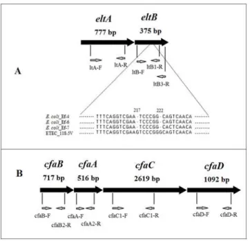
To determine genetic structure of main ETEC pathogenicity factors, a set of specific primers including those to detect A and B subunits of heat-labile enterotoxin geneseltAandeltB, com-plete operonelt, as well as A subunit of heat-stable enterotoxin geneestA, and subunits of the adhesion factor CFA/I (cfaA,cfaB,cfaCandcfaD) were designed (Table 1).
It was shown that all ETEC isolates collected from the food-borne outbreak and the sporadic case in Cherepovets and Vladivostok respectively, had the complete length copy of theelt oper-on (Fig 1), and did not carry the heat-stable enterotoxinestAgene. Sequence of PCR products ofeltAandeltBgenes amplified using primers LT-A(For) and LT-B3(Rev) has shown differ-ences between nucleotide sequdiffer-ences of theeltoperon in the strain ETEC_118-5V (GenBank: JX504011) (clinical specimens; Cherepovets, 2011) and those of theeltoperon in the ETE-C_Ef-4 (GenBank: KF733766), ETEC_Ef-6 (GenBank: KF733765) and ETEC_Ef-7 (GenBank: KF733767) strains (Vladivostok, 2012). In the subunit B gene there were found differences in nucleotide positions 217 and 222 (Fig 1), reflecting the known fact of this gene heterogeneity.
From one to six molecular weight-varying plasmids were found in ETEC strains under study. However, theeltoperon in these strains is likely to locate not on a plasmid as described in some studies [2], but in a chromosome because it was not transmitted via conjugation. This observation agrees with data from retrospective molecular genetic analyses ofeltoperons of ETEC strains collected from numerous sources worldwide. It has been shown that theeltlocus may locate inside Lambda prophages [26].
Heat-labile enterotoxin gene expression in ETEC_27–1, ETEC_73–7, ETEC_Ef-4 and ETE-C_Ef-6 strains was confirmed in latex agglutination tests. The LT production revealed in the strains was some lower compared to the control (purified cholera toxin, 2 g/L). The LT was not identified in other ETEC strains that indicate a low level of expression.
Table 2. Characteristics of ETEC isolates collected in 2011–2012.
ETEC Isolate
City Patient Isolation source
Accession in “SCPM-Obolensk”
O-group adk fumC icd purA gyrB recA mdh ST Clonal complex
Antibacterial resistance
Resistance genes
16-8V Ch 16 feces B-7205 O25 66 11 223 8 4 2 8 3755 - AMI CML
-22-2V Ch 22 feces B-7203 O6 6 5 8 8 4 2 8 4 10 CML DOC
- 24-10V
Ch 24 feces B-7204 O25 6 11 8 78 4 2 8 1312 - CML
- 26-15V
Ch 26 feces B-7196 O6 127 24 299 8 4 2 8 3756 - AMP blaTEM
27-1V Ch 27 feces B-7207 O25 66 11 8 78 4 2 298 3697 - DOC
-73-7V Ch 73 feces B-7199 O25 232 459 39 27 4 2 8 3708 - AMP NAL
TRM
- 73-10V
Ch 73 feces B-7206 O26 56 11 299 78 4 2 8 3709 - -
-85-4V Ch 85 feces B7202 O26 332 11 299 8 331 2 8 3757 - AMI AMP
CML DOC NAL TRM
blaTEM
85-17V
Ch 85 feces B-7197 O115a 232 37 25 5 29 73 4 4509 - AMI CML int1-dfrA17-aadA5
112-2V
Ch - sour
cream
B-7201 O6 6 5 8 8 4 2 8 4 10 AMI AMP
DOC NAL
blaTEM
118-5V
Ch 118 feces B-7198 O6 232 68 223 8 347 6 8 3710 - AMP blaTEM
121-3V
Ch 121 feces B-7200 O25 10 11 8 78 4 2 8 1043 - AMP CML
CTX DOC
blaTEM
Ef-2 V Ef feces B-7684 O142 ND ND ND ND ND ND ND ND ND AMP blaTEM
Ef-4 V Ef feces B-7685 O142 ND ND ND ND ND ND ND ND ND AMP blaTEM
Ef-5 V Ef feces B-7686 O142 ND ND ND ND ND ND ND ND ND AMP blaTEM
Ef-6 V Ef feces B-7687 O142 6 19 16 5 15 44 9 3707 - AMI AMP CAZ
CIP CTA CTX DOC FEP GEN TRM
blaTEM,blaCTX-M-15 int1-dfrA12-orfF-aadA2
Ef-7 V Ef feces B-7688 O142 6 5 8 8 4 2 8 4 10 AMI AMP
DOC GEN TRM
blaTEMint1
Ef-9 V Ef feces B-7689 O142 ND ND ND ND ND ND ND ND ND AMP blaTEM
Note: Ch—Cherepovets; V—Vladivostok;“SCPM-Obolensk”—The Strain Collection of Pathogenic Microorganisms of the State Research Center for Applied Microbiology and Biotechnology; AMI—amikacin; AMP—ampicillin; CAZ—ceftazidime; CIP—ciprofloxacin; CML—chloramphenicol; CTA—ceftriaxone; CTX—cefotaxime; DOC—doxycycline; NAL—
nalidixic acid; TRM—trimethoprim; FEP—cefepime; GEN—gentamicin; ND—not detected; «-»—no adata obtained by serotyping without confirmation by PCR
isolated from the patient in Cherepovets in 2011. Nucleotide sequences ofcfaA,cfaB,cfaC, and cfaDgenes were submitted to the GenBank database (JX504012, JX504013, JX504014, and JX504015, correspondingly). The discovery of the adhesion genes in only one ETEC isolate can be explained by the genetic heterogeneity of adhesion and colonization determinants. It is also possible that the only set of specific PCR primers designed for a specific CFA-type fails to de-tect these patterns.
Isolates obtained in Cherepovets were attributed toО6 (n = 3),О25 (n = 5) andО26 (n = 2)
groups by serotyping using poly- and monovalent agglutinating sera as well as by serotyping using PCR with specific primers for the flippasewzxgene and the O-antigen polymerasewzy gene. Isolates obtained in Vladivostok were attributed toО142 group (Table 2).E.coliof O6,
O25, O115 and O142 groups identified in the research are typical representatives of ETEC strains being also isolated in some other countries, e.g. in China, Japan, and Bangladesh [27–
29]. Before the research there were reported about identification of ETEC ofО148 serogroup
in Russia [14]. Of special interest is identification of the ETEC strains belonging to O26 group that is more common for Shiga toxin-producingE.coli(STEC). Such ETEC strains were isolat-ed not so often in the world, one example is isolation of O26 ETEC from bulls Mithun in India were reported in 2009 [30].
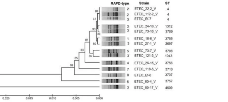
The MLST analysis of ETEC isolates under study revealed 12 different sequence-types. It was shown that two strains from Cherepovets (ETEC_22-2V and ETEC_112-2V) and one strain from Vladivostok (ETEC_Ef 7) belonged to the previously known sequence-type ST4 that had been presented in the MLST database by the ETEC strain (ETEC 117/86) isolated in Germany in 1986. It should be noted that the latter belongs toО6 serogroup as do our strains
ETEC_22-2V and ETEC_112-2V isolated from two patients and sour cream sample during the outbreak in Cherepovets in 2011. It is noteworthy that the Clonal complex ST10 which ST4 se-quence-type belongs to is quite typical for ETEC isolates worldwide [7]. The fact that ETEC strains of the sequence-type ST4 were isolated not only from human (ETEC_22-2V) but also Fig 1. Primer arrangement on the genes. A—theeltAand theeltBgenes; alignment of theeltBgene nucleotides from fourE.coliisolates.B—CFA/I operon consisting of thecfaA,cfaB,cfaCandcfaCgenes. Nucleotide numbers indicate their position from the start of the open reading frame.
from sour cream (ETEC_112-2V) allows one to consider sour cream as a potential source of the outbreak in Cherepovets. This observation agrees with earlier reports stating milk and dairy products as a potential reservoir for ETEC-associated infections. In addition to the sequence-type ST4, 11 novel sequence-types (ST1043, ST1312, ST3697, ST3707, ST3708, ST3709, ST3710, ST3755, ST3756, ST3757, and ST4509) were described in our research. They differ from each other and from the ST4 by sets of "housekeeping gene" alleles. The most re-mote from the ST4 is ST4509 of the strain ETEC_85-17V. It differs by all seven gene alleles. Other STs differ from the ST4 by two to six gene alleles. Theadk,fumC,icdandpurAgenes were the most variable, while thegyrB,recAandmdhgenes were more conservative (Table 2). It is interesting that some isolates that differed in their sequence-types, serogroups and resis-tance genes were collected from one and the same patient, for example, ETEC_85-4V and ETEC_85-17V strains, and ETEC_73-7V and ETEC_73-10V strains were isolated from the pa-tient 85 and papa-tient 73 respectively. The analysis of the phylogenic tree structure and RAPD data of the ETEC strains described in our study allow one to combine the ST4, ST1312, ST3709, ST3755, and ST3697 that are genetically related into one cluster, the ST3708 and ST1043 into other cluster, while remaining STs were unclustered (Fig 2).
It was shown that the ETEC strains had very different spectra of antibacterial susceptibility to antibiotics of several functional classes. Eight isolates were resistant to one drug, three isolates—to two drugs, one isolate—to three drugs, two isolates—to four drugs, one isolate—to five drugs, one isolate—to six drugs, and one isolate—to ten drugs (Table 2). The genetic deter-minants of the beta-lactam resistance were detected in six ETEC strains. Namely they were beta-lactamase of TEM-type gene (ETEC_26-15V, ETEC_85-4V, ETEC_112-2V, ETEC_118-5V, ETEC_121-3V, and ETEC_Ef-6 strains) and the epidemiologically significantblaCTX-M-15 gene (ETEC_Ef-6 strain). Thus, it is shown that theblaCTX-M-15gene is present inE.coli bacte-ria of the novel ST3707 sequence-type. It should be noted that CTX-M is the main type of ex-tended spectrum beta-lactamases (ESBLs) inEnterobacteriaceae, while they are less common in diarrheagenicE.coli(<3.2% of isolates identified). The spread of CTX-M-producing ETEC
in the future may affect and hamper an antibiotic therapy of diarrhea caused by these patho-gens [31].
Genetic determinants conferring resistance to antibacterials of some other drug classes (aminoglycosides and sulfonamides) were revealed into the class 1 integron In54 carrying the dfrA17-aadA5(GenBank KM236803) genetic cassette and class 1 integron In27 carrying the dfrA12-orfF-aadA2(GenBank KM236804) genetic cassette in two isolates ETEC_85-17V and Fig 2. MLST phylogenetic tree of the 14 ETEC strains as reconstructed from the sequences of seven
“housekeeping”genes.Phylogenetic tree constructed after assembly and alignment of MLST DNA sequences using the Mega 6 program. MLST and RAPD experiments are described in Materials and Method.
doi:10.1371/journal.pone.0123357.g002
ETEC in Russia 2011–2012
ETEC_Ef-6 ccorrespondingly, and the class 1 integron carrying unidentified genetic cassette in ETEC_Ef-7 isolate (Table 2). According to reported data, integron structures are widespread in ETEC genomes, class 1 and class 2 integrons have been detected in 35% and 18% of the strains, respectively [32]. Of all our LT-producingE.colistrains, there was a single strain ETEC_Ef-6 that can be considered as a multidrug-resistant (MDR) bacterial pathogen carrying concurrent-ly beta-lactam resistance genes and resistance cassettes to some other classes of antibacterials (aminoglycosides, sulfonamides). Interestingly, the other five ETEC isolates collected from the same patient Ef varied notably by the phenotype of their antimicrobial susceptibility and genet-ic markers (Table 2). The obtained results are consistent with earlier publications that there is no direct link between a sequence-type of ETEC and a serogroup they belong to, and there is no connection between the presence of pathogenic factors (enterotoxins and adhesion factors) and resistance genes in bacterial genomes [16].
Conclusion
In this study we characterized the molecular biological properties of LT-producing ETEC strains which caused food-borne diseases in two large geographically remote cities of Russia. Collected strains were attributed to eleven novel sequence-types and to ST4 (ST10 Complex) which commonly spread among ETEC bacteria. The fact that ETEC strains belonging to ST4 and O6 serogroup were isolated from a patient and from sour cream allowed one to consider sour cream as a potential source of the food-borne outbreak. A correlation between ETEC sequence-types and the serogroups they belong to, as well as between the presence of pathoge-nicity genes and antimicrobial resistance was not found. The emergence of multi-drug resistant ETEC is of special concern and may pose serious problems to public health within the next few years. In conclusion, it should be emphasized that genetic heterogeneity of ETEC strains identi-fied in the study can be explained by a high degree ofE.coligenetic plasticity and the possibility of horizontal transfer of virulence factors and antibacterial resistance genes in populations of bacteria colonizing humans.
Supporting Information
S1 Fig. LT-detection using the latex agglutination assay. (PDF)
Acknowledgments
This research was carried out under Russian R&D Projects #037, #039, Rospotrebnadzor. DNA sequencing was performed in SYNTOL MolBiol center (Moscow, Russia).
Author Contributions
Conceived and designed the experiments: NNK NKF EAS IAD. Performed the experiments: NNK DMP VAB BVE. Analyzed the data: NNK NKF EAS IAD. Contributed reagents/ materials/analysis tools: NKF BVE EAS IAD. Wrote the paper: NNK NKF EAS. DNA sequenc-ing: NNK NKF.
References
1. Clarke SC. DiarrhoeagenicEscherichia coli—an emerging problem? Diagn Microbiol Infect Dis. 2001; 41(3): 93–98. PMID:11750160
3. United Nations Children’s Fund. Committing to child survival: promise renewed progress report 2013. UNICEF; 2013. Available:http://www.unicef.org/lac/Committing_to_Child_Survival_APR_9_Sept_ 2013.pdf.
4. Walker RI. An assessment of enterotoxigenicEscherichia coliandShigellavaccine candidates for in-fants and children. Vaccine. 2015; 33(8): 954–965. doi:10.1016/j.vaccine.2014.11.049PMID: 25482842
5. DuPont HL. Systematic review: the epidemiology and clinical features of travellers' diarrhoea. Aliment Pharmacol Ther. 2009; 30: 187–196. doi:10.1111/j.1365-2036.2009.04028.xPMID:19392866
6. Paredes-Paredes M, Okhuysen PC, Flores J, Mohamed JA, Padda RS, Gonzalez-Estrada A, et al. Seasonality of Diarrheagenic Escherichia coli pathotypes in U.S. Students Acquiring Diarrhea in Me-xico. J Travel Med. 2011; 18(2): 121–125. doi:10.1111/j.1708-8305.2010.00488.xPMID:21366796
7. Jiang Z-D, Butzler J-P. The bacterial pathogens. In: Travelers’diarrhea. 2nd ed. BC Decker Inc.; 2008. pp. 6–17.
8. Shahrokhi N, Bouzari S, Jafari A. Comparison of virulence markers and antibiotic resistance in entero-toxigenicEscherichia coliisolated ten years apart in Tehran. J Infect Dev Ctries. 2011; 5(4): 248–254. PMID:21537065
9. Froehlich B, Parkhill J, Sanders M, Quail MA, Scott JR. The pCoo plasmid of enterotoxigenic Escheri-chia coliis a mosaic cointegrate. J Bacteriol. 2005; 187: 6509–6516. PMID:16159784
10. Belaia YA, Belyi YF, Petruchin VG, Ruzhanskaia TV, Cheklakova LA. [Escherichia coliheat-labile en-terotoxin in biosubstrates of intestinal infections patients]. Modern Sci Tech. 2005; 10: 35. Russian. Available:http://elibrary.ru/item.asp?id=11523862
11. Nada RA, Shaheen HI, Khalil SB, Mansour A, El-Sayed N, Touni I, et al. Discovery and phylogenetic analysis of novel members of class b enterotoxigenicEscherichia coliadhesive fimbriae. J Clin Micro-biol. 2011; 49(4): 1403–1410. doi:10.1128/JCM.02006-10PMID:21289147
12. Turner SM, Scott-Tucker A, Cooper LM, Henderson IR. Weapons of mass destruction: virulence factors of the global killer enterotoxigenicEscherichia coli. FEMS Microbiol Lett. 2006; 263: 10–20. PMID: 16958845
13. Wolf MK. Occurrence, distribution, and associations of O and H serogroups, colonization factor anti-gens, and toxins of enterotoxigenic Escherichia coli. Clin Microbiol Rev. 1997; 10: 569–584. PMID: 9336662
14. Makarova MA, Kaftyreva LA, Egorova SA. [OpportunicticEnterobacteriacaeas the cause of the acute diarrhea and gut disbiosis]. Infect Immun. 2011; 1(2): 181–184. Russian. Available:http://elibrary.ru/ item.asp?id=16382523
15. DebRoy C, Fratamico PM, Roberts E, Davis MA, Liu Y. Development of PCR Assays Targeting Genes in O-Antigen Gene Clusters for Detection and Identification ofEscherichia coliO45 and O55 Ser-ogroups. Appl Environment Microbiol. 2005; 71(8): 4919–4924. PMID:16085897
16. Rodas C, Klena JD, Nicklasson M, Iniguez V, Sjöling A. Clonal relatedness of enterotoxigenic Escheri-chia coli(ETEC) strains expressing LT and CS17 isolated from children with diarrhoea in La Paz, Bo-livia. PLoS One. 2011; 6(11): e18313. doi:10.1371/journal.pone.0018313PMID:22140423
17. Steinsland H, Lacher DW, Sommerfelt H, Whittam TS. Ancestral lineages of human enterotoxigenic
Escherichia coli. J Clin Microbiol. 2010; 48(8): 2916–2924. doi:10.1128/JCM.02432-09PMID: 20534806
18. Mundell DH, Anselmo CR, Wishnow RM. Factors influencing heat-labileEscherichia colienterotoxin activity. Infect Immun. 1976; 14: 383–388. PMID:9363
19. Priamchuk SD, Fursova NK, Abaev IV, Kovalev IuN, Shishkova NA, Pecherskikh EI, et al. [Genetic de-terminants of antibacterial resistance among nosocomial Escherichia coli, Klebsiella spp., and Entero-bacter spp. isolates collected in Russia within 2003–2007] Antibiot Khimioter. 2010; 55(9–10): 3–10. Russian. PMID:21140564
20. Li D, Liu B, Chen M, Guo D, Guo X, Liu F, et al. A multiplex PCR method to detect 14 Escherichia coli serogroups associated with urinary tract infections. J Microbiol Meth. 2010; 82: 71–77. doi:10.1016/j. mimet.2010.04.008
21. Paddock Z, Shi X, Bai J, Nagaraja TG. Applicability of a multiplex PCR to detect O26, O45, O103, O111, O121, O145, and O157 serogroups ofEscherichia coliin cattle feces. Vet Microbiol. 2012; 156: 381–388. doi:10.1016/j.vetmic.2011.11.017PMID:22177888
22. Wang Q, Ruan X, Wei D, Hu Z, Wu L, Yu T, et al. Development of a serogroup-specific multiplex PCR assay to detect a set ofEscherichia coliserogroups based on the identification of their O-antigen gen-eclusters. Mol Cell Prob. 2010; 286: e290. doi:10.1016/j.mcp.2010.06.002
23. Maniatis T, Sambrook J, Fritsch EF, ed. Molecular Cloning: A Laboratory Manual. Cold Spring Harbor Laboratory Press:1982.
ETEC in Russia 2011–2012
24. Zimmer M, Barnhart H, Idris U, Lee MD. Detection ofCampylobacter jejunistrains in the water lines of a commercial broiler house and their relationship to the strains that colonized the chickens. Avian Dis. 2003; 47(1): 101–107. PMID:12713164
25. Tamura K, Stecher G, Peterson D, Filipski A, Kumar S. MEGA6: Molecular Evolutionary Genetics Anal-ysis Version 6.0. Molecular Biology and Evolution. 2013; 30: 2725–2729. doi:10.1093/molbev/mst197 PMID:24132122
26. Jobling MG, Holmes RK. Type II heat-labile enterotoxins from 50 diverseEscherichia coliisolates be-long almost exclusively to the LT-IIc family and may be prophage encoded. PLoS One. 2012; 7(1): e29898. doi:10.1371/journal.pone.0029898PMID:22242186
27. Del Canto F, Valenzuela P, Cantero L, Bronstein J, Blanco JE, Blanco J, et al. Distribution of classical and nonclassical virulence genes in enterotoxigenicEscherichia coliisolates from Chilean children and tRNA gene screening for putative insertion sites for genomic islands. J Clin Microbiol. 2011; 49(9): 3198–3203. doi:10.1128/JCM.02473-10PMID:21775541
28. Konishi N, Obata H, Monma C, Nakama A, Kai A, Tsuji T. Bacteriological and epidemiological charac-teristics of enterotoxigenicEscherichia coliisolated in Tokyo, Japan, between 1966 and 2009. J Clin Microbiol. 2011; 49(9): 3348–3351. doi:10.1128/JCM.02576-10PMID:21752981
29. Ansaruzzaman M, Bhuiyan NA, Begum YA, Kühn I, Nair GB, Sack DA, et al. Characterization of entero-toxigenicEscherichia colifrom diarrhoeal patients in Bangladesh using phenotyping and genetic profil-ing. J Med Microbiol. 2007; 56(Pt 2): 217–222. PMID:17244803
30. Rajkhowa S, Hussain I, Rajkhowa C. Detection of heat-stable and heat-labile enterotoxin genes of
Escherichia coliin diarrhoeic faecal samples of mithun (Bos frontalis) calves by polymerase chain reac-tion. J Appl Microbiol. 2009; 106(2): 455–458. doi:10.1111/j.1365-2672.2008.04013.xPMID: 19200312
31. Kim JS, Kim J, Kim SJ, Jeon SE, Oh KH, Cho SH, et al. Characterization of CTX-M-type extended-spectrum beta-lactamase-producing diarrheagenicEscherichia coliisolates in the Republic of Korea during 2008–2011. J Microbiol Biotechnol. 2014; 24(3): 421–426. PMID:24509253