Apamin Boosting of Synaptic Potentials in
Ca
V
2.3 R-Type Ca
2+
Channel Null Mice
Kang Wang1, Melissa H. Kelley1, Wendy W. Wu2¤, John P. Adelman1, James Maylie2*
1Vollum Institute, Oregon Health & Science University, Portland, Oregon 97239, United States of America,
2Department of Obstetrics and Gynecology, Oregon Health & Science University, Portland, Oregon 97239, United States of America
¤ Current address: FDA, White Oak Campus, Silver Spring, MD 20993, United States of America *[email protected]
Abstract
SK2- and KV4.2-containing K+channels modulate evoked synaptic potentials in CA1
pyra-midal neurons. Each is coupled to a distinct Ca2+source that provides Ca2+-dependent
feedback regulation to limit AMPA receptor (AMPAR)- and NMDA receptor (NMDAR)-medi-ated postsynaptic depolarization. SK2-containing channels are activ(NMDAR)-medi-ated by Ca2+entry
through NMDARs, whereas KV4.2-containing channel availability is increased by Ca2+
entry through SNX-482 (SNX) sensitive CaV2.3 R-type Ca2+channels. Recent studies have
challenged the functional coupling between NMDARs and SK2-containing channels, sug-gesting that synaptic SK2-containing channels are instead activated by Ca2+entry through R-type Ca2+channels. Furthermore, SNX has been implicated to have off target affects, which would challenge the proposed coupling between R-type Ca2+channels and KV
4.2-containing K+channels. To reconcile these conflicting results, we evaluated the effect of SK channel blocker apamin and R-type Ca2+channel blocker SNX on evoked excitatory post-synaptic potentials (EPSPs) in CA1 pyramidal neurons from CaV2.3 null mice. The results
show that in the absence of CaV2.3 channels, apamin application still boosted EPSPs. The
boosting effect of CaV2.3 channel blockers on EPSPs observed in neurons from wild type
mice was not observed in neurons from CaV2.3 null mice. These data are consistent with a
model in which SK2-containing channels are functionally coupled to NMDARs and KV
4.2-containing channels to CaV2.3 channels to provide negative feedback regulation of EPSPs
in the spines of CA1 pyramidal neurons.
Introduction
On hippocampal CA1 pyramidal neurons, dendritic spines are specialized membrane compart-ments that protrude from the dendrites and house proteins that mediate and shape excitatory
postsynaptic responses[1]. Even within the small spine volume (~0.05 fL)[2], synaptic proteins
are organized into discrete, functional domains. The postsynaptic density (PSD) is an electron-dense structure that contains ionotropic glutamate receptors, AMPARs and NMDARs that mediate excitatory postsynaptic responses. SK2-containing channels are also localized in the OPEN ACCESS
Citation:Wang K, Kelley MH, Wu WW, Adelman JP, Maylie J (2015) Apamin Boosting of Synaptic Potentials in CaV2.3 R-Type Ca2+Channel Null Mice.
PLoS ONE 10(9): e0139332. doi:10.1371/journal. pone.0139332
Editor:Vadim E. Degtyar, University of California, Berkeley, UNITED STATES
Received:May 26, 2015
Accepted:September 11, 2015
Published:September 29, 2015
Copyright:This is an open access article, free of all copyright, and may be freely reproduced, distributed, transmitted, modified, built upon, or otherwise used by anyone for any lawful purpose. The work is made available under theCreative Commons CC0public domain dedication.
Data Availability Statement:All relevant data are within the paper.
Funding:Funding provided by National Institute of Health, 5R01NS038880-15, JPA; National Institute of Health, 5R01MH093599-03, JPA.
PSD[3]. These channels are activated by synaptically evoked Ca2+influx through NMDARs, and their repolarizing conductance reduces glutamate-evoked excitatory postsynaptic
responses and Ca2+transients within the spine head. Thus, blocking synaptic SK2-containing
channels with apamin increased EPSPs and the associated spine Ca2+transients, while blocking
NMDARs occludes the effects of apamin[4,5]. Several classes of ion channels and receptors
reside in the extrasynaptic domain of the spine head. Among them are KV4.2-containing K+
channels and CaV2.3 R-type Ca2+channels[6]. Previous experiments using glutamate uncaging
onto individual spines or direct afferent stimulation have reached different conclusions about
the role of R-type Ca2+channels in regulating EPSPs in CA1 pyramidal neurons. While both
sets of experiments showed that blocking R-type Ca2+channels with SNX boosted EPSPs, the
effects of SNX and apamin were mutually exclusive when spines were stimulated by glutamate
uncaging, suggesting that SK2-containing channels are gated by Ca2+influx through R-type
Ca2+channels[5]. In contrast, the boosting effects of SNX and apamin were additive when
direct afferent stimulation was applied[7]. Subsequent work showed that the boosting effect of
SNX on EPSPs induced by glutamate uncaging was lost in CaV2.3 null mice[8]. However, a
recent report showed that in addition to blocking R-type Ca2+channels, SNX blocks A-type K+
currents in dissociated dopamine neurons from substantia nigra pars compacta and cloned
KV4.3 channels were much more sensitive to SNX compared to KV4.2 channels[9].
Further-more, in cerebellar stellate cells where T-type Ca2+channels couple to A-type K+currents SNX
had no effect on A-type channel availability, nor in tsA-201 cells co-expressing R-type
(CaV2.3) channels with KV4.2[10]. Therefore, we used synaptic stimulations to evoke EPSPs
from CA1 pyramidal neurons in slices from CaV2.3 R-type null mice to determine whether in the absence of CaV2.3 channels, apamin and SNX still boosted EPSPs.
Materials and Methods
Animal Handling and Slice Preparation
All procedures were approved in accordance with the guidelines of the Institutional Animal Care and Use Committee (IACUC) of the Oregon Health & Science University (IACUC:
IS00002421). Hippocampal slices were prepared from 4–6 week-old CaV2.3 null (CaV2.3-/-,
C57BL/6J background) and wild type mice (C57BL/6J background). Mice were anesthetized by isofluorane, rapidly decapitated, and brains removed and placed into ice-cold sucrose-aCSF of
the following composition (equilibrated with 95%O2/5%CO2) [7]. Transverse hippocampal
slices (300μm) were cut with a Leica VT1200S and transferred into a holding chamber
con-taining regular aCSF (in mM: 125 NaCl, 2.5 KCl, 21.5 NaHCO3, 1.25 NaH2PO4, 2.0 CaCl2, 1.0
MgCl2, 12 glucose) and equilibrated with 95%O2/5%CO2. Slices were incubated at 35°C for 30–
45 min and then recovered at room temperature (22–24°C) for1 hr before recordings were
performed.
Electrophysiology
CA1 pyramidal cells were visualized with infrared–differential interference contrast optics
(Zeiss Axioskop 2FS, Zeiss Axio Examiner or Leica DM LFS). Whole-cell patch-clamp record-ings were obtained from CA1 pyramidal cells using an Axopatch 1D (Molecular Devices, Sun-nyvale, CA) interfaced to an ITC-16 analog-to-digital converter (Heka Instruments, Bellmore, NY), EPC 10 (Heka Instruments, Bellmore, NY) patch clamp amplifier or Multiclamp 700B interfaced to a Digidata 1440A (Molecular Devices, Sunnyvale, CA). Data were transferred to a computer using Patchmaster software (Heka Instruments, Bellmore, NY) or pClamp10
soft-ware (Molecular Devices, Sunnyvale, CA). Patch pipettes (open pipette resistance, 2.5–3.5 MO)
133 K-gluconate, 4 KCl, 4 NaCl, 2 MgCl2, 10 HEPES, 4 MgATP, 0.3 Na3GTP, 10 K-phospho-creatine (pH 7.3). EPSPs were recorded in whole-cell current-clamp mode and voltages were not corrected for a junction potential of -13 mV. All recordings used cells with a resting mem-brane potential less than -50 mV and a stable input resistance that did not change by more than 20%. Cells were biased to -65 mV and the input resistance was determined from a 25-pA hyperpolarizing current injection pulse given 500 ms after each synaptically evoked EPSP. There was not obvious difference in resting membrane properties in CA1 neurons from
CaV2.3-/-compared to WT (average input resistance = 188.5 ± 6.3 MO(n = 55) in CaV2.3
-/-and 201.4 ± 8.6 (n = 33) in WT; average bias current = -73.3 ± 7.1 pA (n = 55) in CaV2.3-/-and
-76.5 ± 7.5 pA (n = 33) for WT).
Synaptic stimulation
CA3 axons in the stratum radiatum were stimulated using capillary glass pipettes filled with
aCSF, with a tip diameter of ~5μm, connected to an Iso–Flex (A.M.P.I., Israel) or Digitimer
DS3 (Automate Scientific, Berkeley, Ca) stimulus isolation unit. Stimulation electrodes were
placed at ~100μm from the soma and ~20μm adjacent to the dendrite of the recorded cell.
GABAergic blockers SR95531 (2μM) and CGP55845 (1μM) were present throughout the
recordings to block GABAAand GABABreceptors, respectively. To prevent recurrent
excita-tion in the CA3 region in the presence of GABAergic blockers, the CA3 region was cut away
before recording. Subthreshold EPSPs were elicited by 100-μs current injections (20–30μA)
that were approximately one-third of the stimulus required for evoking an action potential. No
obvious difference in stimulation amplitude was observed between slices from CaV2.3-/-and
WT.
Data analysis
Data were analyzed using Igor Pro (WaveMetrics, Lake Oswego, OR). Data are expressed as mean ± s.e.m. Paired t-tests or Wilcoxon-Mann-Whitney 2-sample rank test was used to
deter-mine significance; P<0.05 was considered significant.
Pharmacology
Apamin was purchased from Calbiochem; D-AP5, SR95531, and CGP55845 from Tocris Cookson; and SNX-482 from Peptide Institute.
Results
To determine whether blocking SK2-containing channels in spines lacking R-type CaV2.3 Ca2+
channels boosts EPSPs, synaptic stimulations were delivered to the Shaffer collateral axons in the stratum radiatum in freshly prepared hippocampal slices from CaV2.3 null mice, and EPSPs were measured from individual CA1 pyramidal neurons. After establishing a stable
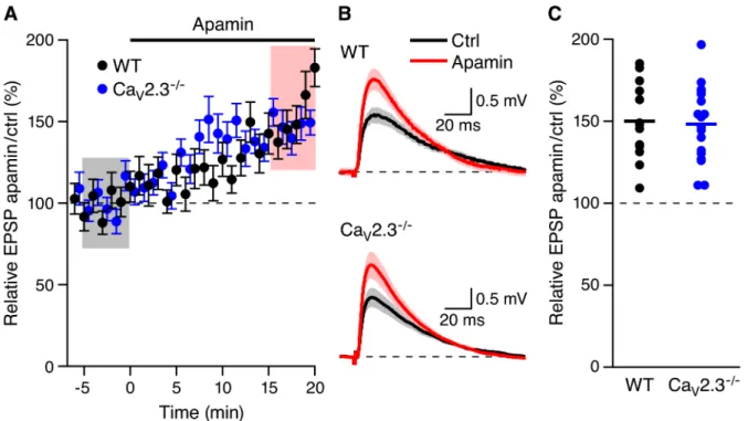
baseline, apamin (100 nM) was added to the bath solution. As shown inFig 1A and 1B, apamin
application boosted EPSPs (148.2 ± 5.4%, n = 18, P<0.001). The increase in EPSP amplitude
by apamin in mice lacking R-type Ca2+channels was not different (p = 0.82) than the boosting
effect of apamin in WT mice (158.2 ± 7.3%, n = 13, p<0.001) (Fig 1C). These results indicate
that Ca2+influx through CaV2.3 channels is not necessary to activate synaptic SK2-containing
channels.
In contrast, SNX (300 nM) that increased EPSPs in WT mice (166.2.0 ± 8.5%, n = 11,
p<0.001) did not affect EPSP amplitudes in mice lacking CaV2.3 Ca2+channels (103.1 ±
Fig 1. Apamin boosts EPSPs in CaV2.3-/-mice.(A) Time course of the normalized EPSP amplitude (mean±s.e.m.) for baseline in control aCSF (Ctrl) and
during wash-in of apamin (100 nM) as indicated above (n = 18) in CaV2.3-/-(blue symbols) and WT (black symbols) mice. (B) Average of 15 EPSPs taken
from indicated shaded time points in aCSF (black) and 16–20 min after application of apamin (red); shaded areas are mean±s.e.m. (C) Scatter plot of
relative ESPS peak compared to baseline (Ctrl) from the individual slices in panel A for CaV2.3-/-(blue symbols) and WT (black symbols). Horizontal bar
reflects mean response.
doi:10.1371/journal.pone.0139332.g001
Fig 2. Boosting of EPSPs by SNX requires CaV2.3 R-type Ca2+channels.(A) Time course of the normalized EPSP amplitude (mean±s.e.m.) for
baseline in control aCSF (Ctrl) and during wash-in of SNX (300 nM) as indicated above in CaV2.3-/-(blue symbols) and WT (black symbols) mice. (B)
Average of 15 EPSPs taken from indicated shaded time points in aCSF (black) and 19–23 min after application of SNX (red); shaded areas are mean±s.e.
m. (C) Scatter plot of relative ESPS peak compared to baseline (Ctrl) from the individual slices in panel A for CaV2.3-/-(blue symbols) and WT (black
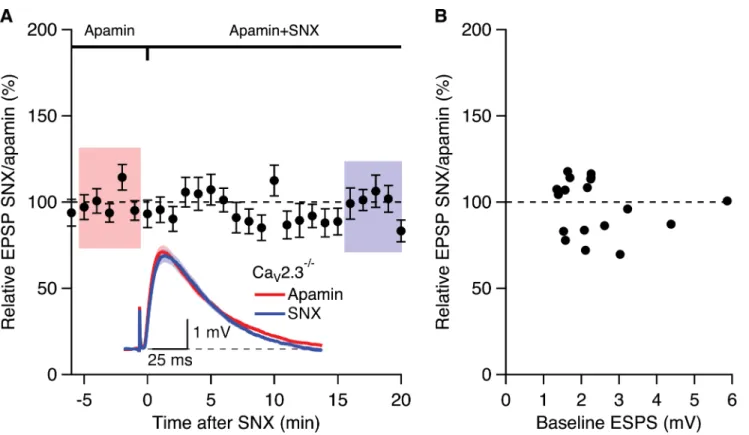
in slices from WT mice (157.0 ± 7.8%, n = 21, p<0.0001; seeFig 1in Wang et al., 2014 [7])
did not affect EPSP amplitudes in slices from mice lacking CaV2.3 R-type Ca2+channels
pre-treated with apamin for 20–30 min (98.9 ± 3.8%; n = 19) (Fig 3A). Previously we showed that,
in WT slices the apamin- and SNX-induced increase of EPSP were independent of initial EPSP
size[7,11].Fig 3Bshows that the relative EPSP of SNX/apamin was independent of initial EPSP
size; Fisher’s r to z analysis of the EPSP increase by SNX in the presence of apamin compared
to the initial EPSP size in apamin yielded no correlation. These results suggests that the SNX
boosting of EPSPs in CA1 neurons require CaV2.3 R-type Ca2+channels
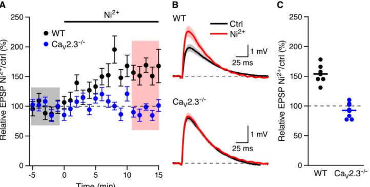
SNX may have off target effects as recently reported[12]. Low concentrations of Ni2+
(100μM) have also been used to block low voltage activated T-type and R-type Ca2+channels
[13–16]. As shown inFig 4Ni2+(100μM) increased EPSPs in WT mice (153.8 ± 5.9%, n = 7,
P<0.01) but not in CaV2.3-/-mice (92.4 ± 4.6%, n = 8,Fig 2). The sensitivity of CaV2.3 Ca2+
channels to 100μM Ni2+is greatest at voltages less than -10 mV (>85% block)[17] a the
volt-age range that is likely not surpassed in dendritic spines during synaptic input[18]. Therefore,
these results indicate that CaV2.3 R-type Ca2+channels are necessary for the boosting of EPSPs
by SNX and Ni2+in CA1 pyramidal neurons.
Discussion
These results show that in the absence of CaV2.3 R-type Ca2+channels, blocking
SK2-contain-ing channels with apamin boosts synaptic responses. Consistently, in these mice, we also found
that SNX or Ni2+provides no additional increase to EPSPs. These findings are consistent with
Fig 3. Boosting of EPSPs by SNX in the presence of apamin requires CaV2.3 R-type Ca2+channels.(A) Time course of the normalized EPSP
amplitude (mean±s.e.m.) for baseline in apamin (100 nM) and during wash-in of SNX (300 nM) in the presence of apamin (n = 19). Inset shows the average of 15 EPSPs taken from indicated shaded time points in apamin and 15–20 min after co-application of SNX; shaded areas are mean±s.e.m. (B) Plot of
relative EPSPs after SNX application in the presence of apamin versus the basline EPSP in apamin alone from all cells (n = 19).
synaptically evoked Ca2+entry through NMDARs gating the synaptic SK2-containing
chan-nels[4]. They also support previous conclusions that Ca2+influx through R-type Ca2+channels
binds to KChIPs to increase availability of KV4.2-containing A-type K+channels, and blocking
R-type Ca2+channels with SNX or Ni2+boosted synaptic potentials by decreasing availability
of the repolarizing A-type K+current[7]. Consistent with this, KV4.2 and CaV2.3 proteins have
been localized to the extrasynaptic region in CA1 spines[6,19]. This implies distinct Ca2+
sig-naling domains within the spine head, one coupling Ca2+influx through NMDARs to activate
SK2-containing channels and another coupling Ca2+influx through R-type Ca2+channels that
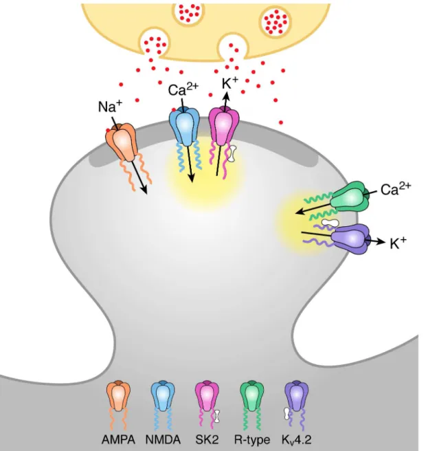
activates KV4.2-contianing channels via KChIPs (Fig 5).
A recent report revealed that in addition to blocking R-type Ca2+channels, SNX also blocks
KV4-containing channels[9]. The lack of effect of SNX and Ni2+in CA1 pyramidal neurons of
CaV2.3 mice, and the previous findings using CaV2.3 R-type Ca2+channel null mice[8],
sup-port the conclusion that R-type channels are necessary for the effects SNX and Ni2+in CA1
pyramidal neurons. However, the present results do not address whether SNX also blocks
KV4.2-containing K+channels, as they may not be available to participate in synaptic responses
in the absence of Ca2+influx through R-type Ca2+channels in CaV2.3 null mice. Importantly,
the lack of effect of Ni2+and SNX in CaV2.3 null mice supports the model that Ca2+influx
through R-type channels in CA1 provides the Ca2+source to modulate KV4.2-containing K+
channel availability via associated KChIPs[7]. This is in contrast to cerebellar granule and
stel-late cells in which T-type Ca2+channels couple to A-type K+currents and SNX had no effect
A-type channel availability[10,20].
Based upon results obtained using glutamate uncaging onto single dendritic spines, a model has been proposed in which the NMDAR dependence of synaptic boosting by apamin was not
Fig 4. Boosting of EPSPs by Ni2+requires Ca
V2.3 R-type Ca2+channels.(A) Time course of the normalized EPSP amplitude (mean±s.e.m.) for baseline
in control aCSF (Ctrl) and during wash-in of 100μM Ni2+in WT (black symbols, n = 7) and Ca
V2.3-/-mice (blue symbols, 8). (B) Average of 15 EPSPs taken
from indicated shaded time points in aCSF (black) and 16–20 min after application of Ni2+(red); shaded areas are mean±s.e.m. (C) Scatter plot of relative
ESPS peak compared to baseline (Ctrl) from the individual slices in panel A for CaV2.3-/-(blue symbols) and WT (black symbols). Horizontal bar reflects
mean response.
directly due to Ca2+influx through NMDARs activating SK2-containing channels. Rather that NMDAR activation provided a necessary component of depolarization that activated R-type
Ca2+channels, and they provided the Ca2+to fuel SK2-containing channel activation[5]. The
present results used synaptic stimulations and suggest alternate conclusions. It should be noted that there are several distinctions between these studies that may be very significant. First,
Kv4.2 and SK2 expression change with age[21,22] and the ages of the animals employed are
different, P 16–18 for the uncaging studies while we used 4–8 week old mice. Second, we
Fig 5. Model of activation of SK2 and KV4.2 containing channels by distinct Ca2+microdomains during synaptic stimulation.Schaffer collateral
stimulation releases glutamate (red particles) from the presynaptic terminal (ivory). Glutamate binding to AMPA and NMDA receptors in the postsynaptic density (PSD; dark grey) of the spine head depolarizes the spine membrane potential and releases voltage-dependent Mg2+block from NMDA receptors
allowing for Ca2+influx during the EPSP. This Ca2+activates closely associated SK2 channels via binding to calmodulin (barbell structure) bound to
C-terminus of SK2 subunits. Spine depolarization also activates R-type Ca2+channels located extrasynaptically that are close to K
V4.2-containing K+channels.
Ca2+entering through R-type channels binds to KChIPs (peanut structure) associated with K
V4.2 channels shifting the voltage-dependence of availability to
more negative potentials and allowing for KV4.2 activation during an EPSP. The yellow clouds represent the microdomain for each Ca2+source.
cannot be precisely sure of the location of the spines that are stimulated while the uncaging
studies used spines on first oblique branches within 100μm of the soma. Third, we do not
know the nature of the stimulated spines, and the uncaging studies chose mushroom type spines. Given these differences, and while we cannot rule out the possibility that in the absence
of R-type Ca2+channels, the Ca2+signaling domain architecture in the spine head is
compro-mised, the present results are more consistent with Ca2+influx through NMDARs fueling
SK2-containing channel activation, a model supported by immuno-electron microscopy that
showed close anatomical localization of SK2 and NMDAR within the PSD[3].
Acknowledgments
We thank Dr. Miller for the generous gift of CaV2.3 null mice.
Author Contributions
Conceived and designed the experiments: JPA JM. Performed the experiments: KW MHK WWW. Analyzed the data: JM. Wrote the paper: JPA JM.
References
1. Nimchinsky EA, Sabatini BL, Svoboda K. Structure and function of dendritic spines. Annu Rev Physiol. 2002; 64: 313–353. doi:10.1146/annurev.physiol.64.081501.160008PMID:11826272
2. Dumitriu D, Rodriguez A, Morrison JH. High-throughput, detailed, cell-specific neuroanatomy of den-dritic spines using microinjection and confocal microscopy. Nat Protoc. 2011; 6: 1391–1411. doi:10.
1038/nprot.2011.389PMID:21886104
3. Lin MT, Luján R, Watanabe M, Adelman JP, Maylie J. SK2 channel plasticity contributes to LTP at Schaffer collateral-CA1 synapses. Nat Neurosci. 2008; 11: 170–177. doi:10.1038/nn2041PMID:
18204442
4. Ngo-Anh TJ, Bloodgood BL, Lin M, Sabatini BL, Maylie J, Adelman JP. SK channels and NMDA recep-tors form a Ca2+-mediated feedback loop in dendritic spines. Nat Neurosci. 2005; 8: 642–649. doi:10.
1038/nn1449PMID:15852011
5. Bloodgood BL, Sabatini BL. Nonlinear regulation of unitary synaptic signals by CaV(2.3) voltage-sensi-tive calcium channels located in dendritic spines. Neuron. 2007; 53: 249–260. doi:10.1016/j.neuron.
2006.12.017PMID:17224406
6. Parajuli LK, Nakajima C, Kulik A, Matsui K, Schneider T, Shigemoto R, et al. Quantitative Regional and Ultrastructural Localization of the Cav2.3 Subunit of R-type Calcium Channel in Mouse Brain. J Neu-rosci. 2012; 32: 13555–13567. doi:10.1523/JNEUROSCI.1142-12.2012PMID:23015445
7. Wang K, Lin MT, Adelman JP, Maylie J. Distinct Ca2+sources in dendritic spines of hippocampal CA1
neurons couple to SK and KV4 channels. Neuron. Elsevier; 2014; 81: 379–387. doi:10.1016/j.neuron.
2013.11.004PMID:24462100
8. Giessel AJ, Sabatini BL. Boosting of synaptic potentials and spine Ca transients by the peptide toxin SNX-482 requires alpha-1E-encoded voltage-gated Ca channels. PLoS ONE. 2011; 6: e20939. doi: 10.1371/journal.pone.0020939PMID:21695265
9. Kimm T, Bean BP. Inhibition of A-Type Potassium Current by the Peptide Toxin SNX-482. Journal of Neuroscience. 2014; 34: 9182–9189. doi:10.1523/JNEUROSCI.0339-14.2014PMID:25009251 10. Heath NC, Rizwan AP, Engbers JDT, Anderson D, Zamponi GW, Turner RW. The Expression Pattern
of a Cav3-Kv4 Complex Differentially Regulates Spike Output in Cerebellar Granule Cells. Journal of Neuroscience. 2014; 34: 8800–8812. doi:10.1523/JNEUROSCI.0981-14.2014PMID:24966380 11. Lin MT, Luján R, Watanabe M, Frerking M, Maylie J, Adelman JP. Coupled activity-dependent
traffick-ing of synaptic SK2 channels and AMPA receptors. Journal of Neuroscience. 2010; 30: 11726–11734.
doi:10.1523/JNEUROSCI.1411-10.2010PMID:20810893
12. Bourinet E, Bourinet E, Stotz SC, Stotz SC, Spaetgens RL, Spaetgens RL, et al. Interaction of SNX482 with domains III and IV inhibits activation gating of alpha(1E) (Ca(V)2.3) calcium channels. Biophys J. Elsevier; 2001; 81: 79–88. doi:10.1016/S0006-3495(01)75681-0
13. Tsien RW, Ellinor PT, Horne WA. Molecular diversity of voltage-dependent Ca2+ channels. Trends Pharmacol Sci. 1991; 12: 349–354. Available:http://eutils.ncbi.nlm.nih.gov/entrez/eutils/elink.fcgi?
14. Zhang JF, Randall AD, Ellinor PT, Horne WA, Sather WA, Tanabe T, et al. Distinctive pharmacology and kinetics of cloned neuronal Ca2+ channels and their possible counterparts in mammalian CNS neurons. Neuropharmacology. 1993; 32: 1075–1088. PMID:8107963
15. Lee JH, Gomora JC, Cribbs LL, Perez-Reyes E. Nickel block of three cloned T-type calcium channels: low concentrations selectively block alpha1H. Biophys J. 1999; 77: 3034–3042. doi:
10.1016/S0006-3495(99)77134-1PMID:10585925
16. Sochivko D, Pereverzev A, Smyth N, Gissel C, Schneider T, Beck H. The Ca(V)2.3 Ca(2+) channel subunit contributes to R-type Ca(2+) currents in murine hippocampal and neocortical neurones. J Phy-siol (Lond). 2002; 542: 699–710.
17. Zamponi GW, Bourinet E, Snutch TP. Nickel block of a family of neuronal calcium channels: subtype-and subunit-dependent action at multiple sites. J Membr Biol. 1996; 151: 77–90. PMID:8661496 18. Palmer LM, Stuart GJ. Membrane potential changes in dendritic spines during action potentials and
synaptic input. J Neurosci. 2009; 29: 6897–6903. doi:10.1523/JNEUROSCI.5847-08.2009PMID:
19474316
19. Kim J, Jung S-C, Clemens AM, Petralia RS, Hoffman DA. Regulation of dendritic excitability by activity-dependent trafficking of the A-type K+ channel subunit Kv4.2 in hippocampal neurons. Neuron. 2007; 54: 933–947. doi:10.1016/j.neuron.2007.05.026PMID:17582333
20. Anderson D, Mehaffey WH, Iftinca M, Rehak R, Engbers JDT, Hameed S, et al. Regulation of neuronal activity by Cav3-Kv4 channel signaling complexes. Nat Neurosci. 2010; 13: 333–337. doi:10.1038/nn.
2493PMID:20154682
21. Maletic-Savatic M, Lenn NJ, Trimmer JS. Differential spatiotemporal expression of K+ channel polypep-tides in rat hippocampal neurons developing in situ and in vitro. J Neurosci. 1995; 15: 3840–3851.
PMID:7751950