Improvement of ENU Mutagenesis Efficiency
Using Serial Injection and Mismatch Repair
Deficiency Mice
Jabier Gallego-Llamas1,2☯, Andrew E. Timms1☯, Rose Pitstick3, Janet Peters3, George A. Carlson3, David R. Beier1,2*
1Center for Developmental Biology and Regenerative Medicine, Seattle Children’s Research Institute, Seattle, WA, United States of America,2Department of Pediatrics, University of Washington School of Medicine, Seattle, WA, United States of America,3McLaughlin Research Institute, Great Falls, MT, United States of America
☯These authors contributed equally to this work.
Abstract
ENU mutagenesis is a powerful method for generating novel lines of mice that are informa-tive with respect to both fundamental biological processes and human disease. Rapid developments in genomic technology have made the task of identifying causal mutations by positional cloning remarkably efficient. One limitation of this approach remains the mutation frequency achievable using standard treatment protocols, which currently generate approxi-mately 1–2 sequence changes per megabase when optimized. In this study we used two strategies to attempt to increase the number of mutations induced by ENU treatment. One approach employed mice carrying a mutation in the DNA repair enzymeMsh6. The second strategy involved injection of ENU to successive generations of mice. To evaluate the num-ber of ENU-induced mutations, single mice or pooled samples were analyzed using whole exome sequencing. The results showed that there is considerable variability in the induced mutation frequency using these approaches, but an overall increase in ENU-induced vari-ants from one generation to another was observed. The analysis of the mice deficient for
Msh6also showed an increase in the ENU-induced variants compared to the wild-type
ENU-treated mice. However, in both cases the increase in ENU-induced mutation fre-quency was modest.
Introduction
Forward genetic screens usingN-ethyl-N-nitrosurea(ENU) as a chemical mutagen have
revealed a wide spectrum of biological and disease processes [1]. ENU causes DNA damage by transferring a methyl or ethyl group to the oxygen and nitrogen atoms of nucleotide bases (reviewed in [2] and [3]). The resulting base adducts tend to mispair during semi-conservative replication. If this is not corrected, the following round of replication will convert the mismatch to a point mutation. This method creates random mutations throughout the genome, which a11111
OPEN ACCESS
Citation:Gallego-Llamas J, Timms AE, Pitstick R, Peters J, Carlson GA, Beier DR (2016) Improvement of ENU Mutagenesis Efficiency Using Serial Injection and Mismatch Repair Deficiency Mice. PLoS ONE 11 (7): e0159377. doi:10.1371/journal.pone.0159377
Editor:Javier Marcelo Di Noia, Institut de Recherches Cliniques de Montréal (IRCM), CANADA
Received:May 11, 2016
Accepted:July 3, 2016
Published:July 21, 2016
Copyright:© 2016 Gallego-Llamas et al. This is an open access article distributed under the terms of the
Creative Commons Attribution License, which permits unrestricted use, distribution, and reproduction in any medium, provided the original author and source are credited.
Data Availability Statement:Sequence data have been deposited in the SRA repository with the accession number SRP074832.
Funding:This work was supported by NIH grants R01NS41997, R01MH081187 and R01HD36404. The funders had no role in study design, data collection and analysis, decision to publish, or preparation of the manuscript.
are potentially more representative of the mutations responsible for human disease than null or conditional mutations generated by genome targeting.
While mutagenesis screens have been successful in creating a wide range of phenotypes for disease modeling, the efficiency of this approach is still constrained by the number of mutations that can be induced in a single organism. The frequency of ENU-induced mutations is affected by treatment regimens and dosage, but generally ranges from 1–2 mutations/Mb of genomic DNA [4–8]. In a strain such as C57BL/6J, each mutated gamete would carry approximately 3000–6000 mutations, of which around 30–60 mutations would be in coding regions. A major limitation is the dose of ENU that can be administered, as a ceiling is reached such that treatment results in sterility or lethality [9,10]. As a consequence, ENU screens frequently entail treatment of a large number of animals in order to obtain a mutant phenotype of interest.
A number of different strategies have previously been tested in a bid to increase the number of mutations per animal without decreasing fertility. One involves manipulating DNA mis-match repair (MMR), which is part of the repair mechanism that prevents alkylation damage in cells and protects them from naturally occurring mutations [3,11,12]. This machinery cor-rects small replication errors such as insertion/deletion loops (IDLs) and base-base mis-matches. There are five MMR genes in mammals that produce three different heterodimers. The mismatch-bound MutS heterodimer recognizes the replication errors and exists in two
varieties: MutSαand MutSβ. MutSαconsists of the MMR proteins MSH2 and MSH6, and
rec-ognizes single base pair mismatches and small IDLs. MutSβis a heterodimer of MSH2 and MSH3 and mediates recognition of larger IDLs. Subsequent recognition of the mismatch by MutSαor MutSβwill induce the recruitment of multiple molecules of MutLα. MutLαis the predominant form of the MutL family functioning in MMR, consisting of MLH1 and PMS2 [13].The recruitment of MutLαwill activate its endonuclease activity [14] together with the exonuclease EXO1, causing DNA excision to initiate at an upstream nick. The gap created by this excision is repaired by the combined action of proliferating cell nuclear antigen (PCNA), replication factor C (RFC) and DNA polymerase III that functions as its processivity factor. The remaining nick is then sealed by DNA ligase [15].
This mechanism not only recognizes and repairs natural occurring mutations, but has also been shown to be involved in mutations induced by ENU. Claij et al. [16] found that mouse embryonic stem cells lacking one of the components of the MMR,Msh2, had a strongly increased mutation frequency when treated with ENU as compared to wild-type. A number of studies have attempted to use the MMR mechanism to increase the number of mutations induced by ENU treatmentin vivo. Specifically studies were done usingMsh6mutant zebrafish [17] or rats [18] with different outcomes ranging from no significant increase in zebrafish to a ~2.5 fold increase in ENU-induced variant number in rats. In this study we describe a similar strategy using mice.
An alternative approach to increase ENU-induced mutations would be to use treatment of serial generations, in which ENU injections are administered to the progeny of treated animals. In this report we describe the consequences of both the use of serial injection and the use of an MMR-deficient mouse line. Both methods resulted in increased numbers of mutations detected by exome sequence analysis. However, the maximal increase obtained by either method com-pared to a single injection was less than 1.8-fold, suggesting these strategies have limited benefit.
Experimental Methods
Animals and mutant mouse generation
were reviewed and approved by the Institutional Animal Care and Use Committee. TheMsh6
mutant lineMsh6tm1Rak[19] was kindly provided by Dr. Winfred Edelmann. Wild-type (Msh6+/+),Msh6+/-, andMsh6-/-mice were treated with ENU as previously described (reviewed in [20]). Briefly, adult male animals (Generation 0 (G0)) were treated with three weekly intra-peritoneal injections of 90mg of ENU per kilogram of body weight. Treated males were individ-ually housed with ICR females to test sterility and the time to recovery of fertility. After fertility was recovered, the animals were mated with C57BL/6J (B6) females to generate G01 offspring (Fig 1,Table 1). The G01, G02, and, from theMsh6+/-cohort, G03 progeny males were treated with the same ENU regimen. Females from the different generations (G01, G02, G03, and G04) and mutation cohorts were sequenced. Given the various breeding patterns (seeS1 Fig
for an example), the relatedness of mice in the different cohorts was not identical.
Mice treated with ENU are at risk for developing tumors and were observed every work day for signs of distress, including wasting, hairloss, inactivity, mass formation, etc. Mice identified as possibly having a tumor were euthanized using carbon dioxide narcosis, followed by cervical dislocation and bilateral thoracotomy, consistent with the recommendations of the American Veterinary Medical Association (AVMA) Guidelines on Euthanasia. No mice were found dead. As mice were euthanized immediately upon showing signs of illness, no other methods were taken to ameliorate suffering.
DNA extraction
We isolated DNA from liver by phenol/chloroform extraction or using a Qiagen DNeasy Blood and Tissue Kit (Qiagen, Santa Clarita, CA USA). Individuals or pools of 6 to 10 samples of extracted DNA for each generation treated with ENU were then sent for library preparation and whole exome sequencing at Centrillion Genomics Technology (CA, USA).
Sequencing data analysis
We mapped paired end sequencing data to the reference genome mm10 using BWA-MEM
(Burrows-Wheeler alignment tool)[21] employing default parameters. We used the Genome
Analysis Toolkit (GATK)[22] to realign reads around known indels, and recalibrate quality scores to reduce sequencing artifacts. Picard (Broad Institute:http://broadinstitute.github.io/ picard/) was used to identify duplicate reads. SNPs were identified using SAMtools (mpileup, bcftools)[23]. SNPs were annotated with ANNOVAR [24] employing refGene as the gene model. To control for differences in the amount of sequence per sample we identified all regions covered by either 20 reads (individual mice) or 100 reads (pooled mice) using gener-ated Python scripts and the Python version of BEDTools [25] (Pybedtools) [26]. We applied additional filtering to identify ENU induced SNPs with the highest confidence. SNPs in publi-cally available databases (Sanger Mouse exomes, dbSNP) or in-house controls, located within a repeat region, or with low likelihood of being real (<Q30) were removed. Sequence data have
been deposited in the SRA repository with the accession number SRP074832. Statistical analy-ses were done using two-tailed T-tests assuming unequal variances.
Results
ENU treatment was done on 3 serial generations of B6 wild-type and 4 serial generations of B6
animals had no cumulative effect on fecundity as after each round of injection there was no decrease in the number of animals that recovered fertility and produced progeny (Table 1).
The efficiency of the ENU treatment for inducing mutations has generally been reported to range from 1–2 mutations/Mb. Half of the autosomal mutations present in G01 mice will be transmitted to the next generation. Therefore, in the second treated generation there should be a 1.5 fold increase in mutation frequency relative to the first treated generation and in the third
Fig 1. Breeding scheme for analysis of serial ENU-treatment in wild-type and MMR defective mice. G01 males carry a random set ofde novopoint mutations induced by ENU treatment of wild-type or MMR-mutant G0 mice. G02, G03 and G04 carry mutations that were induced in their respective parents, as well as those inherited from previous generations. Each generation treated with ENU will on average have 3000– 6000 new ENU-induced variants genome wide. After ENU treatment each male is crossed with a wild-type female; their progeny will inherit newly induced mutations and 50% of those of the parent. The mutations are sampled by exome analysis; the expected number of ascertained mutations (E) is shown. The ENU treatment was performed on three successive generations for theMsh6+/+
mice and four generation for theMsh6
+/-mice.Msh6-/-mice did not tolerate ENU treatment.
doi:10.1371/journal.pone.0159377.g001
Table 1. Fertility after ENU treatment.
Genotype x dose1
Generation Msh6+/+x 90 Msh6-/+x 90 Msh6-/-x 90 Msh6-/-x 75
G01 # males injected 17 31 8 4
# recovering fertility2 8 14 0 0
total # of progeny 142 198 0 0
G02 # males injected 18 30
# recovering fertility 11 22
total # of progeny 101 140
G03 # males injected 21 22
# recovering fertility 16 17
total # of progeny 118 248
G04 # males injected 15
# recovering fertility 9
total # of progeny 117
1Dose is indicated as mg of ENU injected per Kg bodyweight x 3 injections.
2Fertility is measured by presence at least of one litter within 10 weeks after the last injection.
generation there should be a 1.75 fold increase. We analyzed the number of variants present in each of 3 individual mice from 3 generations of wild-type orMsh6+/-mice using whole exome sequencing. This strategy was chosen as it enabled us to sample the genome with high confi-dence and reproducibility, allowing reliable comparison of the cohorts. On average there was an increase in the number of variants present in the next generation for both backgrounds of mice, albeit less than expected (Table 2). Notably, individual mice from the same cohort had very different variant numbers (Table 2andS1 Fig). Given this variability and the small num-ber of samples per cohort, the differences in mean variant numnum-ber do not reach statistical significance.
To obtain a more robust representation of the ENU-induced mutation frequency, we ana-lyzed a pool of 10 mice for each cohort (except for theMsh6+/-G04 pool, which was 6 mice).
Fig 2. Effect of ENU treatment on survival.Msh6
+/-males show survival comparable to wild-type +/-males after treatment with the standard concentration of ENU (90mg/kg x 3). Survival of ENU-treatedMsh6
-/-males is much reduced. Death is expressed in days.
doi:10.1371/journal.pone.0159377.g002
Table 2. Variant analysis for individual mice.
Genotype and Generation variant for each individual average ratio1 ratio H/W2
wild-type G01 86 60 72 73 1.0
wild-type G02 103 73 54 77 1.1
wild-type G03 110 118 53 94 1.3
Msh6+/-G01 70 77 49 65 1.0 0.9
Msh6+/-G02 135 106 57 99 1.5 1.3
Msh6
+/-G03 108 56 124 96 1.5 1.0
1Ratio: the increase of variant present compared to thefirst ENU injection (G01) 2Ratio H/W: the increase of variant present inMsh6
+/-compared to the same wild-type generation.
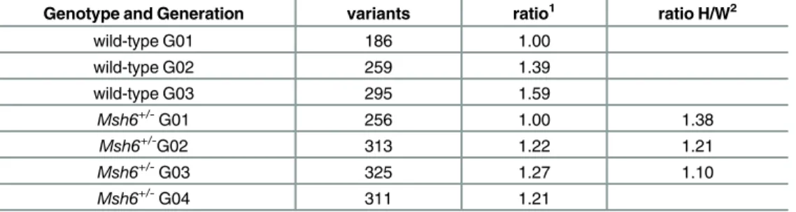
While this has the virtue of including more samples, coverage of each sample was on average 10X (compared to 20X for the individual analysis), so the number of mutations per exome ascertained was smaller. However, this sample depth still enabled comparison among cohorts. The variant number in each pool increased after each generation as previously observed, except for the fourth generation of ENU-treatedMsh6+/-mice (Table 3). However, also as previously observed, the increase in mutation frequency was lower than predicted.
An aim of this experiment was to test whether treating mice at least partially deficient in
Msh6would increase the mutagenesis frequency. In the individually analyzed mice, only one of the 3 generations showed an increase of mutation frequency in theMsh6+/-group, and this was small (Table 2). Again, the variability within a cohort was problematic. The analysis of pools of mice revealed a marked increase in the variant number for theMsh6+/-mice compared to the wild-type mice (Table 3). The mutation frequency was further increased for the second (G02) and third generations (G03); however, the ratio of mutations compared to the wild-type cohort decreased for each successive generation.
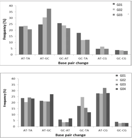
Examination of the type of mutations produced by the ENU treatment revealed a different spectrum in theMsh6+/-mice compared to wild-type. The most frequent base-pair alterations induced by ENU affected A-T base pairs, with A-T to T-A and A-T to G-C substitutions accounting for almost fifty percent of the mutations for both the wild-type andMsh6+/-ENU treated mice (Fig 3), which is consistent with previous results [27]. However, G-C to A-T tran-sitions were much reduced in theMsh6+/-mice compared to wild-type mice, while A-T to C-G transitions were elevated (Fig 3). Overall, wild-type mice have a similar fraction of A-T and G-C modifications, in contrast to theMsh6+/-mice that had an average of 75% A-T modifica-tions compared to 25% G-C modificamodifica-tions (p = 0.015,Table 4).
Discussion
In this study we demonstrate that serial administration of ENU to successive generations of
mice increases the ENU-induced mutation frequency in both wild-type andMsh6+/-mice. Our
initial analysis of 3 individual mice in each of 3 generations supported this observation; how-ever, we found considerable variation among individuals. We therefore analyzed pooled DNA obtained from up to 10 different mice from each generation, which confirmed this observation.
The increase we observed was less than expected based on Mendelian inheritance, which could be explained in a number of ways. For example, we used stringent parameters for whole exome sequencing analysis in order to increase the likelihood that we identifiedde novo ENU-induced variants; however, these may have excluded some true variants These parameters could be modified but the filtering of false positives would be less efficient. Another possibility
Table 3. Variant analysis for pooled mice.
Genotype and Generation variants ratio1 ratio H/W2
wild-type G01 186 1.00
wild-type G02 259 1.39
wild-type G03 295 1.59
Msh6
+/-G01 256 1.00 1.38
Msh6
+/-G02 313 1.22 1.21
Msh6+/-G03 325 1.27 1.10
Msh6+/-G04 311 1.21
1Ratio: the increase of variant present compared to the
first ENU injection (G01) 2Ratio H/W: the increase of variant present inMsh6
+/-compared to the same wild-type generation.
is that we are not accounting for loss due to lethality; however, given that even null mutations of important developmental genes are generally well tolerated in mice when heterozygous, this is unlikely to be an important factor. Of note, this is the case for individual mutant loci; it is possible that an accumulation of heterozygous mutants could affect viability or fertility.
Fig 3. ENU-induced mutation spectrum.(a) Frequencies of mutations in successive generation ofMsh6+/+
mice in the pooled samples. (b) Frequencies of mutations in the successive generation ofMsh6
+/-mice in the pooled samples.
doi:10.1371/journal.pone.0159377.g003
Table 4. Distribution of ENU-induced mutations in A-T vs. G-C base-pairs.
%A-T %G-C Avg %A-T Avg %G-C
wild-type G01 53 47
wild-type G02 61 39 59 41
wild-type G03 63 37
Msh6
+/-G01 73 27
Msh6+/-G02 69 31
Msh6
+/-G03 78 22 75 25
Msh6
+/-G04 78 22
We and others have hypothesized that a deficiency in the repair system that detects or cor-rects DNA mismatches would result in an increased ENU-induced mutation frequency. This is based on the biochemistry of the mutagen ENU, which can transfer its ethyl group to oxygen or nitrogen radicals present in DNA; this results in lesions that can cause mispairing during replication and eventually give rise to a single base pair substitution[28,29]. Three other stud-ies addressed this question in ES cells and mice [16], zebrafish [17] and rats [18].
In anMsh2null ES cell line, the background mutation rate (assayed by analysis of loss of HPRT activity) was elevated at least two orders of magnitude compared to wild-type cells, and the mutation rate after ENU treatment was further increased 2–3 fold [16]. In anMsh2 -defi-cient line that had 10% of wild-type activity, the mutation rate in untreated cells was close to background, and was increased 7–10 fold after ENU treatment. While this would seem promis-ing for a strategy of mutagenesis in an MMR deficient mouse, it is difficult to compare the dose regimen of ENU treatments forin vitroandin vivostudies, and it is possible the effective dose given in these studies would not be toleratedin vivo. Indeed, in our handsMsh6-/-mice did not tolerate even low doses of ENU.
InMsh6-/-rats, treatment with ¾ of a dose used in wild-type was modestly successful, elevat-ing the mutation rate to 0.8 mutations/Mb, a 1.5-fold increase compared to the mutation rate inMsh6-proficient animals [18].Msh6+/-rats were not tested. In contrast, in zebrafish there were no differences found for the mutation frequency for ENU-treated wild-type,Msh6+/-and
Msh6-/-lines [17]. The authors speculate that these results may indicate that the maximum mutation load for zebrafish has been reached with the currently used, highly optimized ENU mutagenesis protocol; it is notable that the mutation rate obtained of 6–7 mutations /Mb is 3–6 fold greater than that generally obtained in ENU-treated mice. Alternatively, the authors sug-gest that the MMR system in the zebrafish germ line may be saturated very rapidly, thereby having a limited effect on high-dose ENU mutagenesis.
In our analysis we see a small increase in mutation frequency in treatedMsh6+/-mice com-pared to wild-type for both individually analyzed and pooled cohorts. In the latter, which likely represents a more representative sample, after 3 serial treatments 325 variants were detected in 6 Mb obtained from 10 mice, for a mutation rate of 5.4 mutations/Mb per mouse. This is 1.1-fold the rate in an equivalent wild-type cohort, and 1.75-fold the frequency found in a wild-type cohort after a single treatment.
Our results also demonstrate that the common mutation both in wild-type andMsh6
+/-background were alterations of A-T base pairs, withMsh6+/-having a higher ratio of A-T tran-sitions. These results are consistent with what was observed in theMsh6-deficient rats and the
Msh2-deficient mice [16,18] where they also observed a decrease of A-T to G-C transitions. This phenomenon was associated with the fact that ethylated bases have been shown to direct misincorporation of bases when DNA polymerases succeed in trans-lesion synthesis [30,31]. Bypass of O2-ethylthymineT subsequently induced A-T to T-A transversions, whereas O4 -ethylthymine causes A-T to G-C transitions [30,31]. These DNA adducts contribute to the toxicity and mutagenicity of ENU and are speculated to be the target of MMR activity [16]. It has been suggested that MutSαpreferentially recognizes the O2-ethylthymineT lesion [18], which is consistent with our observation that transversions are elevated in the
Msh6+/-mutants at the expense of transitions.
ENU dose that is compatible with survival but is still mutagenic. However, it should also be noted that there are other DNA damage repair pathways that perform partially redundant functions with MMR. For example, the nucleotide excision repair pathway has been shown to mediate ENU-induced mutation repair in flies [32]. One could imagine mutagenizing mice car-rying multiple DNA repair defect mutations, although the husbandry task could potentially outweigh the benefit.
The results also suggest the possibility that our protocols do not succeed because we reached a ceiling on the level of mutations that mice can tolerate. While intriguing, this seems unlikely for a number of reasons. Firstly, one would require treating multiple additional generations to insure that the mutation frequency has truly peaked. Secondly, the mutations generated in our strategy are heterozygous (as we outcross at every generation), and these are generally well-tol-erated in mice, even when the homozygous phenotype is lethal. Lastly, we see no obvious effect on fertility, fecundity, gross morphology, or general viability, which one might expect if there were additive effects of multiple mutations. Determining what the upper limit of heterozygous mutational load might be in a mammalian system would be interesting, but we do not think this study conclusively addresses this.
The wealth of studies using ENU mutagenesis for a wide variety of phenotypic analyses demonstrates unequivocally that this approach is efficient, that these mutations can be readily mapped and that they can ultimately be identified using well-established approaches for posi-tional cloning [33]. However, given the development of powerful new methods for generating targeted mutations [34,35,36], it is necessary to examine the utility of an approach that requires positional cloning for mutation discovery.In this regard, the expedience of a genotype-driven approach must be balanced against that of a phenotype-driven method, in which one may obtain unbiased insight into the genetic basis of a trait. Many studies employing forward genetic analysis have provided unexpected insights and have identified mutations in unanno-tated genes whose function were otherwise unknown.
Importantly, the mutations induced by ENU often have functional consequences that are more informative than the null mutations that are commonly generated by genomic targeting. ENU induced mutations often create hypomorphic alleles, due either to“leaky”splice-site mutations or due to missense mutations that do not completely abolish gene function. Mis-sense mutations induced by ENU can also result in dominant“gain-of-function”effects, which can provide insight into protein structure-function and roles in specific developmental path-ways that are not revealed by characterization of null mutants.
In summary, the two strategies that we have described in this study do increase the ENU mutagenesis efficiency. However, the increase is small, and can arguably be equaled by simply expanding a screen that employs a standard treatment for a single generation. It is possible that the small increase reflects a biological limit on the number of heterozygous mutations that are tolerated in mice, but this would require additional analysis.
Supporting Information
S1 Fig. Pedigree from one family of mice used for the pooled sample analysis.The G0 through G04 generations are shown.Msh6+/-heterozygotes are shown as half-filled symbols. Mice used for DNA sample preparation are shown in red. For the total pool mice were obtained from multiple pedigrees, and relatedness of those used was not identical across generations. (TIFF)
S2 Fig. ENU induced point mutation frequencies in individual mice.G01, G02, and G03 samples of W (Msh+/+) and H (Msh+/-) mice are shown.
Author Contributions
Conceived and designed the experiments: JGL AT DRB. Performed the experiments: JGL AT RP JP. Analyzed the data: JGL AT GC DRB. Wrote the paper: JGL AT GC DRB.
References
1. Herron BJ, Lu W, Rao C, Liu S, Peters H, Bronson RT, et al. Efficient generation and mapping of reces-sive developmental mutations using ENU mutagenesis. Nat Genet. 2002; 30(2):185–9. PMID: 11818962
2. Noveroske JK, Weber JS, Justice MJ. The mutagenic action of N-ethyl-N-nitrosourea in the mouse. Mamm Genome. 2000; 11(7):478–83. PMID:10886009
3. Wyatt MD, Pittman DL. Methylating agents and DNA repair responses: Methylated bases and sources of strand breaks. Chem Res Toxicol. 2006; 19:1580–94. PMID:17173371
4. Arnold CN, Barnes MJ, Berger M, Blasius AL, Brandl K, Croker B, et al. ENU-induced phenovariance in mice: inferences from 587 mutations. BMC Res Notes. 2012; 5:577. Epub 2012/10/26. doi:10.1186/ 1756-0500-5-577PMID:23095377
5. Nguyen N, Judd LM, Kalantzis A, Whittle B, Giraud AS, van Driel IR. Random mutagenesis of the mouse genome: a strategy for discovering gene function and the molecular basis of disease. American journal of physiology Gastrointestinal and liver physiology. 2011; 300(1):G1–11. doi:10.1152/ajpgi. 00343.2010PMID:20947703
6. Andrews TD, Whittle B, Field MA, Balakishnan B, Zhang Y, Shao Y, et al. Massively parallel sequenc-ing of the mouse exome to accurately identify rare, induced mutations: an immediate source for thou-sands of new mouse models. Open Biol. 2012; 2(5):120061. Epub 2012/06/23. doi:10.1098/rsob. 120061PMID:22724066
7. Justice MJ, Carpenter DA, Favor J, Neuhauser-Klaus A, Hrabe de Angelis M, Soewarto D, et al. Effects of ENU dosage on mouse strains. Mamm Genome. 2000; 11(7):484–8. PMID:10886010
8. Beutler B, Du X, Xia Y. Precis on forward genetics in mice. Nat Immunol. 2007; 8(7):659–64. PMID: 17579639
9. Augustin M, Sedlmeier R, Peters T, Huffstadt U, Kochmann E, Simon D, et al. Efficient and fast targeted production of murine models based on ENU mutagenesis. Mammalian genome: official journal of the International Mammalian Genome Society. 2005; 16(6):405–13.
10. Sakuraba Y, Sezutsu H, Takahasi KR, Tsuchihashi K, Ichikawa R, Fujimoto N, et al. Molecular charac-terization of ENU mouse mutagenesis and archives. Biochemical and biophysical research communi-cations. 2005; 336(2):609–16. PMID:16139793
11. Fishel R. Mismatch Repair. The Journal of biological chemistry. 2015; 290(44):26395–403. doi:10. 1074/jbc.R115.660142PMID:26354434
12. Spies M, Fishel R. Mismatch repair during homologous and homeologous recombination. Cold Spring Harb Perspect Biol. 2015; 7(3):a022657. doi:10.1101/cshperspect.a022657PMID:25731766 13. Jiricny J. The multifaceted mismatch-repair system. Nat Rev Mol Cell Biol. 2006; 7(5):335–46. PMID:
16612326
14. Kadyrov FA, Dzantiev L, Constantin N, Modrich P. Endonucleolytic function of MutLalpha in human mismatch repair. Cell. 2006; 126(2):297–308. PMID:16873062
15. Li GM. New insights and challenges in mismatch repair: getting over the chromatin hurdle. DNA repair. 2014; 19:48–54. doi:10.1016/j.dnarep.2014.03.027PMID:24767944
16. Claij N, van der Wal A, Dekker M, Jansen L, te Riele H. DNA mismatch repair deficiency stimulates N-ethyl-N-nitrosourea-induced mutagenesis and lymphomagenesis. Cancer research. 2003; 63(9):2062– 6. PMID:12727820
17. Feitsma H, de Bruijn E, van de Belt J, Nijman IJ, Cuppen E. Mismatch repair deficiency does not enhance ENU mutagenesis in the zebrafish germ line. Mutagenesis. 2008; 23(4):325–9. doi:10.1093/ mutage/gen019PMID:18469325
18. van Boxtel R, Toonen PW, Verheul M, van Roekel HS, Nijman IJ, Guryev V, et al. Improved generation of rat gene knockouts by target-selected mutagenesis in mismatch repair-deficient animals. BMC geno-mics. 2008; 9:460. doi:10.1186/1471-2164-9-460PMID:18840264
19. Edelmann W, Yang K, Umar A, Heyer J, Lau K, Fan K, et al. Mutation in the mismatch repair gene Msh6 causes cancer susceptibility. Cell. 1997; 91(4):467–77. PMID:9390556
21. Li H, Durbin R. Fast and accurate short read alignment with Burrows-Wheeler transform. Bioinformat-ics. 2009; 25(14):1754–60. doi:10.1093/bioinformatics/btp324PMID:19451168
22. McKenna A, Hanna M, Banks E, Sivachenko A, Cibulskis K, Kernytsky A, et al. The Genome Analysis Toolkit: a MapReduce framework for analyzing next-generation DNA sequencing data. Genome research. 2010; 20(9):1297–303. doi:10.1101/gr.107524.110PMID:20644199
23. Li H, Handsaker B, Wysoker A, Fennell T, Ruan J, Homer N, et al. The Sequence Alignment/Map for-mat and SAMtools. Bioinforfor-matics. 2009; 25(16):2078–9. doi:10.1093/bioinformatics/btp352PMID: 19505943
24. Wang K, Li M, Hakonarson H. ANNOVAR: functional annotation of genetic variants from high-through-put sequencing data. Nucleic acids research. 2010; 38(16):e164. doi:10.1093/nar/gkq603PMID: 20601685
25. Quinlan AR, Hall IM. BEDTools: a flexible suite of utilities for comparing genomic features. Bioinformat-ics. 2010; 26(6):841–2. doi:10.1093/bioinformatics/btq033PMID:20110278
26. Dale RK, Pedersen BS, Quinlan AR. Pybedtools: a flexible Python library for manipulating genomic datasets and annotations. Bioinformatics. 2011; 27(24):3423–4. doi:10.1093/bioinformatics/btr539 PMID:21949271
27. Takahasi KR, Sakuraba Y, Gondo Y. Mutational pattern and frequency of induced nucleotide changes in mouse ENU mutagenesis. BMC molecular biology. 2007; 8:52. PMID:17584492
28. Justice MJ, Noveroske JK, Weber JS, Zheng B, Bradley A. Mouse ENU mutagenesis. Hum Mol Genet. 1999; 8(10):1955–63. PMID:10469849
29. Shibuya T, Murota T, Horiya N, Matsuda H, Hara T. The induction of recessive mutations in mouse pri-mordial germ cells with N-ethyl-N-nitrosourea. Mutat Res. 1993; 290(2):273–80. PMID:7694119 30. Klein JC, Bleeker MJ, Lutgerink JT, van Dijk WJ, Brugghe HF, van den Elst H, et al. Use of shuttle
vec-tors to study the molecular processing of defined carcinogen-induced DNA damage: mutagenicity of single O4-ethylthymine adducts in HeLa cells. Nucleic acids research. 1990; 18(14):4131–7. PMID: 2377457
31. Bhanot OS, Grevatt PC, Donahue JM, Gabrielides CN, Solomon JJ. In vitro DNA replication implicates O2-ethyldeoxythymidine in transversion mutagenesis by ethylating agents. Nucleic acids research. 1992; 20(3):587–94. PMID:1741292
32. Tosal L, Comendador MA, Sierra LM. In vivo repair of ENU-induced oxygen alkylation damage by the nucleotide excision repair mechanism in Drosophila melanogaster. Mol Genet Genomics. 2001; 265 (2):327–35. PMID:11361344
33. Stottmann RW, Beier DR. 2010. Using ENU mutagenesis for phenotype-driven analysis of the mouse. Methods Enzymol 477: 329–348. doi:10.1016/S0076-6879(10)77017-8PMID:20699149
34. Cong L, Ran FA, Cox D, Lin S, Barretto R, Habib N, Hsu PD, Wu X, Jiang W, Marraffini LA et al. 2013. Multiplex genome engineering using CRISPR/Cas systems. Science 339: 819–823. doi:10.1126/ science.1231143PMID:23287718
35. Mali P, Yang L, Esvelt KM, Aach J, Guell M, DiCarlo JE, Norville JE, Church GM. 2013. RNA-guided human genome engineering via Cas9. Science 339: 823–826. doi:10.1126/science.1232033PMID: 23287722