Predominantly Recognized by CD8
bright
Cells in multiple
Sclerosis Patients
Pernille B. Jørgensen1¤, Astrid H. Livbjerg1, Hans J. Hansen2, Thor Petersen2, Per Ho¨llsberg1,3*
1Department of Biomedicine, Aarhus University, Aarhus, Denmark,2Department of Neurology, MS Clinic, Aarhus University Hospital, Aarhus, Denmark,3Danish Neuroscience Center, Aarhus University and Aarhus University Hospital, Aarhus, Denmark
Abstract
Multiple sclerosis (MS) is associated with Epstein-Barr virus (EBV) infection, but impaired immune suppression may be part of the disease pathogenesis. CD8+T cells that are restricted by HLA-E exert an important immunoregulatory mechanism. To explore how EBV might interfere with immune regulation, we examined the expression of HLA-E and the frequency of CD8+ cells recognizing E, presenting either an EBV peptide from the BZLF1 protein or a signal sequence peptide from HLA-A2, in relapsing remitting (MS-RR), primary progressive (MS-PP) MS patients, and healthy controls (HC). Treatment with IFN-a
or EBV increased HLA-E expression on CD4+cells. However, only MS-PP had increased expression of HLA-E on resting CD4+ cells when compared with HC (p,0.005). CD8+ cells were divided into CD8bright and CD8dim cells by flow cytometry
analyses. MS-RR had significantly fewer CD8dimcells than HC (p
,0.003). Flow cytometry analyses were performed with HLA-E tetramers folded in the presence of the HLA-EBV or HLA-A2 peptide to identify HLA-HLA-E-interacting cells. MS-RR had increased frequency of CD8brightcells recognizing HLA-E/A2 (p = 0.006) and HLA-E/BZLF1 (p = 0.016). Conversely, MS-RR had fewer
CD8dimcells that recognized HLA-E/BZLF1 (p = 0.001), but this could be attributed to the overall lower number of CD8dim
cells in MS-RR. Whereas HLA-E/A2 was predominantly recognized by CD8dim cells, HLA-E/BZLF1 was predominantly recognized by CD8brightcells in MS-RR and MS-PP, but not in HC. As expected, HLA-E/A2 was also recognized by CD8-negative cells in a CD94-dependent manner, whereas HLA-E/BZLF1 was poorly recognized in all groups by CD8-CD8-negative cells. These data demonstrate that MS-RR patients have expanded their CD8brightcells recognizing HLA-E/BZLF1. Moreover, HLA-E/BZLF1 appears to be recognized by the immune system in a different manner than HLA-E/A2.
Citation:Jørgensen PB, Livbjerg AH, Hansen HJ, Petersen T, Ho¨llsberg P (2012) Epstein-Barr virus Peptide Presented by HLA-E is Predominantly Recognized by CD8brightCells in multiple Sclerosis Patients. PLoS ONE 7(9): e46120. doi:10.1371/journal.pone.0046120
Editor:Steven Jacobson, National Institutes of Health, United States of America
ReceivedAugust 9, 2011;AcceptedAugust 29, 2012;PublishedSeptember 25, 2012
Copyright:ß2012 Jørgensen et al. This is an open-access article distributed under the terms of the Creative Commons Attribution License, which permits unrestricted use, distribution, and reproduction in any medium, provided the original author and source are credited.
Funding:The study was supported by the Danish Multiple Sclerosis Society. The funders had no role in study design, data collection and analysis, decision to publish, or preparation of the manuscript.
Competing Interests:PBJ has nothing to declare; AHL has nothing to declare; HJH has received funding from Biogen Idec, Merck Serono, Bayer Schering, Sanofi-Aventis. TP has received funding or speaker honoraria from Biogen Idec, Merck Serono, Novartis, Bayer Schering, Sanofi-Aventis, Roche, and Genzyme; PH has received speaker honorarium from Biogen Idec. This does not alter our adherence to all the PLoS ONE policies on sharing data and materials..
* E-mail: [email protected]
¤ Current address: Department of Clinical Genetics, Vejle Hospital, Denmark.
Introduction
Multiple sclerosis (MS) is an organ-specific disease of the central nervous system with an early inflammatory reaction [1]. Environ-mental, genetic and possibly stochastic events are thought to be important for the development of disease, although the mecha-nisms are largely unknown. Based on clinical and sero-epidemi-ological observations, virus infection has been suspected to be involved. Nevertheless, it has been controversial whether or not different viruses could induce a common trigger or whether few or perhaps only one virus would be able to do this.
During the last decade, accumulating data suggest that Epstein-Barr virus (EBV) is strongly and possibly causally linked to MS [2– 4]. Virtually all MS patients, but not healthy controls, have seroconverted to EBV [5–9]. Importantly, 86–99% of children with MS have detectable antibodies to EBV, although only 64– 72% of healthy control children have seroconverted at a similar age [10–12]. Large prospective database analyses of individuals with infectious mononucleosis have demonstrated a significant
increase in later occurrence of MS [13,14], and prospective sero-epidemiological analyses have shown an increased titer of anti-EBV antibodies in individuals that later went on to develop MS [15]. Moreover, EBV infection is consistent with a number of known epidemiological features of MS [2,3].
NKG2A (inhibitory receptor) or CD94/NKG2C (activating receptor) on NK cells, which typically express no or low levels of CD8 (i.e., CD8 negative or CD8dimcells). Blocking CD94 by a monoclonal antibody allows the identification a small subset of CD8brightT cells that recognizes HLA-E by their T-cell receptor [24]. Presentation of HLA-E/peptide may induce cytotoxicity e.g. by clonal expansion of cytotoxic NK-T cells, which are alloreactive [25], or by eliciting GroEL-specific, cytotoxic CD8+
T cells that may cross-react with heat-shock protein (hsp) 60 in stressed, uninfected cells [25–27]. HLA-E may also provide a mechanism to suppress the HLA-E-expressing CD4+
T cells by HLA-E-restricted CD8+T cells [22,28]. In this case, the presented
peptide may be derived from the b-chain of the T-cell receptor variable region [29–32]. Perhaps the most convincing data demonstrating that HLA-E is a restriction element for suppressor CD8+T cells come from studies of Qa-1 in mice, the functional
equivalent gene to HLA-E. Qa-1-deficient mice develop exagger-ated secondary CD4+
T-cell responses after viral infection or immunization with foreign or self peptides [33].
HLA-E-restricted CD8+
T cells might be important in MS, since Qa-1-deficient animals have increased susceptibility to recurrence of proteolipid protein (PLP)-induced EAE [33]. Anti-myelin basic protein (MBP)- and anti-Anti-myelin oligodendrocyte glycoprotein (MOG)-reactive CD8+
T-cell clones restricted by HLA-E have been generated from MS patients, demonstrating that their expression of CD94 and NKG2A was increased during exacerbation, which may limit their capacity to inhibit autoreac-tive CD4+
T cells [34]. It has also been suggested that the treatment of MS patients with glatiramer acetate (Copaxone) in part exerts its effect by inducing cytotoxic CD8+ T cells that
directly kill CD4+
T cells [35].
To explore the mechanisms of how EBV might interfere with immune regulation in relapsing remitting (MS-RR) and primary progressive (MS-PP) MS patients as well as healthy controls (HC), we examined the expression of HLA-E and the frequency of CD8+
cells recognizing HLA-E. We made use of HLA-E tetramers presenting either an EBV peptide from the BZLF1 protein or a signal sequence peptide from HLA-A2.
Materials and Methods
Culture medium
All cells were cultured with RPMI 1640 (Invitrogen, Taastrup, Denmark) medium supplemented with 10 mM HEPES, strepto-mycin (0.2 g/l) and glutamine (0.292 g/l), 10% normal human serum (NHS) and 500 IE/ml IL-2 (Proleukin, Nomeco, Copenha-gen, Denmark).
Peripheral blood mononuclear cells
Blood was collected in Venoject vacutainers with citrate (Seelen, Holstebro, Denmark) following informed, written consent from 12
Isolation of CD4+cells from mononuclear cells was performed using magnetic bead separation as described by the manufacturer (Dynal Biotech, Invitrogen, Taastrup, Denmark).
Cytokine stimulation assays
Cytokine stimulation of HLA-E expression was performed on CD4+ T-cell clones that were generated and maintained as
previously described [36]. CD4+T-cell clones were incubated in
culture medium with 0, 5000 or to completely saturate the receptors 100,000 U/ml IFN-a (PBL Biomedical Laboratories, No. 11100-1) for 0, 4, 7, or 10 hours. After the incubation, cells were fixed with 1% paraformaldehyde for 5 min, centrifuged and resuspended in PBS. Cells were kept in the dark at 4uC until analysis by flow cytometry.
EBV infection
EBV containing supernatant was harvested from the producer cell line B95-8. PBMC were incubated for 30 hours at 26106 PBMCs/ml with one half volume of EBV-containing supernatant in the presence or absence of neutralizing anti-human IFN-a antibody (AHC4814, Invitrogen, Taastrup, Denmark) diluted 1:100. After incubation, the cells were stained with anti-HLA-E and anti-CD4 antibodies for flow cytometry analysis.
Flow cytometry analysis
Immunofluorescence analyses were performed by incubating the cells in RPMI+10% NHS on ice for 30 min light protected with either antibodies directly conjugated with FITC or PE or by unconjugated primary antibody followed by a secondary conju-gated antibody. Antibody concentration was determined by prior titration experiments. Cells were fixed in 1% paraformaldehyde prior to analysis. Expression of HLA-E was determined using anti-HLA-E (MEM-E/07, Abcam, Cambridge, UK) diluted 1:100 as primary antibody and PE-conjugated rabbit-anti-mouse (R0439, DAKO, Glostrup, Denmark) diluted 1:10 or FITC-conjugated goat-anti-mouse (F2772, Sigma-Aldrich) diluted 1:100 as second-ary antibody. CD4 expression was determined using FITC-conjugated anti-CD4 (F0766, DAKO) diluted 1:10. Two homo-tetramers of HLA-E were purchased from Baylor College of Medicine (MHC Tetramer Lab, Houston, TX). The tetramers were created by refolding HLA-E*01033 with the HLA-A2 leader peptide, VMAPRTLVL (referred to as HLA-E/A2), or with the EBV peptide, BZLF-139–47, SQAPLPCVL (referred to as HLA-E/
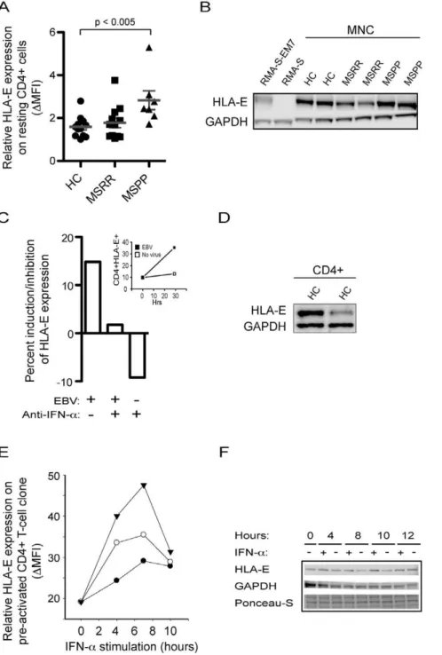
Figure 1. HLA-E expression.(A) Primary progressive MS patients have enhanced expression of HLA-E on CD4+
cells. Mononuclear cells from healthy controls (HC), relapsing-remitting MS patients (MS-RR) and primary progressive MS patients (MS-PP) were incubated with fluorophore-conjugated antibodies against HLA-E and CD4 and analysed by flow cytometry. Results are expressed as median of fluorescence intensity. (B) Expression of HLA-E on mononuclear cells from patients and controls. Western blot analysis of HLA-E expression on lysates from mononuclear cells of healthy controls and MS patients. Positive and negative control lysates are from RMA-S-EM7 and RMA-S cells. GAPDH is used as a loading control. Results are representative of two experiments. (C) EBV induces expression of HLA-E. Mononuclear cells were treated with EBV containing supernatant from B95-8 or medium in the presence or absence of neutralizing anti-IFN-aantibody. Results are expressed as induction/inhibition in percentage of uninduced cells by calculating [(MFItreated2MFIuntreated)/MFIuntreated]6100%. Insert shows flow cytometric analyses of CD4+HLA-E+ on the
mononuclear cells at 0 hr and 29 hrs incubation with EBV. Results are representative of 2 independent experiments. (D) Expression of HLA-E on CD4+ cells. Western blot analysis of HLA-E expression on lysates from CD4+
cold lysis buffer (Cell Signaling, #9803) supplemented with 1.0 mM PMSF (Sigma), 5.0 mM NaF and 150ml Complete Mini protease inhibitor cocktail (Roche, Hvidovre, Denmark). The lysates were then centrifuged at 20006g for 5 min at 4uC to
remove large cell debris and again at 160006gfor 10 minutes at 4uC. Lysates were frozen at270uC until they were analyzed by Western blotting. Protein concentration was determined by Bio-Rad Protein Assay (Bio-Bio-Rad Laboratories, Inc., Hercules, CA) and 30mg protein was added per lane on a Criterion XT Bis-Tris 10% gel (Bio-Rad). Proteins were separated under reducing conditions in MOPS buffer (Bio-Rad) and transferred to a nitrocellulose membrane (Amersham, Hillerød, Denmark) in 0.025 M Tris-HCl, 0.192 M Glycin, 20% v/v ethanol (pH 8.3) at 300 mA for 1.5 hours. Equal loading was assured by examining transferred protein in Ponceau-S (Sigma-Aldrich). To reduce non-specific binding, the nitrocellulose was incubated for 1 hour in 5% non-fat dry milk in TBS (0.01 M Tris-HCl; 0.14 NaCl; pH 7.4)+0.1% Tween 20 (Sigma) (TBST). Primary anti-HLA-E antibody (Abcam, MEM-E/02) was diluted 1:750, according to the manufacturer’s protocol. After 1 hour incubation at room temperature, the blot was washed 3 times 5 min in TBST and probed 1 hour at room temperature with the secondary horse radish peroxidase (HRP)-conjugated antibody diluted 1:2000 in 5% non-fatty milk in TBST. In addition to Ponceau-S staining, another loading control was made by probing with anti-GAPDH antibody (sc-25778, Santa Cruz Biotechnology, Santa Cruz, CA) diluted 1:200 according to the manufacturer’s protocol. Blots were developed using enhanced chemiluminescence SuperSignal West Pico Chemilumiscent Substrate (Pierce, Rockford, IL). Negative control lysates were derived from the mouse RMA-S cells, which lack HLA-E expression, and positive control lysates were derived from RMA-S cells transfected with human beta-2 microglobulin and HLA-E (RMA-S-EM7). These cell lines were a kind gift from Dr. Elisabeth Weiss, Germany [37].
Statistical analysis
Statistical analysis was performed using two-tailed Mann-Whitney U-test. Paired data with anti-CD94 blocking was analysed by two-tailed, paired t-test. Significant difference was defined as a p-value,0.05.
Results
Expression of HLA-E on resting CD4+cells from MS
patients
To address a possible role of HLA-E in MS, we initially examined its surface expression on resting CD4+
cells from healthy controls (HC), relapsing-remitting (MS-RR) and primary progres-sive (MS-PP) MS patients (Fig. 1A). Expression of HLA-E was generally low, but significantly increased on CD4+
cells from primary progressive MS patients when compared with healthy
number of HLA-E positive mononuclear cells by approximately 15% (Fig. 1C) and among these, CD4+ cells expressed HLA-E
(Fig. 1D) and demonstrated upregulated HLA-E expression upon EBV treatment (Fig. 1C, insert). Some of this induction may be attributed to the secretion of IFN-a, since the number of HLA-E positive mononuclear cells reverted to the level of uninduced cells in the presence of EBV and anti-IFN-aantibody. However, anti-IFN-ainhibited the expression of HLA-E in the absence of EBV indicating, that EBV may also induce HLA-E by a mechanism separate from IFN-a. Nevertheless, IFN-a may be a relevant inducer of HLA-E expression during virus infection.
Induction of HLA-E by IFN-a
HLA-E molecules are expressed at high levels in pre-activated CD4+
T cells, such as CD4+
T-cell clones. To further study the induction of HLA-E, we measured by flow cytometry the ability of type I and II interferons to induce surface expression of HLA-E. Since IFN-cinduces HLA-E gene transcription, we first confirmed that IFN-c induced expression of HLA-E in our system. As expected, the presence of 40, 80, or 160 ng/ml of IFN-cinduced a dose-dependent increase in HLA-E expression on isolated CD4+
T-cell clones 4 hours after addition of the cytokine (data not shown). To examine whether IFN-a was capable of inducing HLA-E expression, isolated CD4+
T-cell clones were incubated in the absence or presence of recombinant IFN-a for 4, 7, and 10 hours and the expression of HLA-E was measured by the median fluorescence intensity on flow cytometry. Recombinant IFN-ainduced expression of HLA-E by addition of 500 U/ml, an induction that could be further enhanced by the presence of very high concentration of IFN-a(Fig. 1E). The presence of IFN-aalso increased the total level of HLA-E molecules as measured by Western blotting analysis (Fig. 1F).
Recognition of HLA-E/A2 and HLA-E/BZLF1 by different CD8 cell subsets
HLA-E/peptide complexes may be recognized by either CD94/NKG2A/C (predominantly CD8 negative or CD8dimcells) or by the T-cell receptor (predominantly CD8bright cells). Therefore, the distribution of these CD8 cells was first character-ized (Fig. 2). Although the frequency of CD8+ (total) cells and
CD8bright cells were similar in HC, MS-RR and MS-PP, the presence of CD8dimcells was significantly reduced among the MS-RR patients (p = 0.003).
We then examined the binding to CD8+
Overall, the frequency of cells recognizing HLA-E/A2 was higher than the frequency of cells recognizing HLA-E/BZLF1. However, when divided into the CD8 total, CD8brightand CD8dim
subset, MS-RR had a significantly reduced binding of HLA-E/ BZLF1 to the CD8dimsubset (p = 0.001) when compared with HC, in contrast to the other cell subsets examined. Notably, the MS-RR patients had an increased recognition of HLA-E/BZLF1 by their CD8brightcells (p = 0.034) when compared with HC (Fig. 3A). Since the frequencies of the subsets were different between HC and MS-RR, the data were corrected for this by expressing the results as fraction of tetramer positive cells within the CD8+
cells (Fig. 3B). This indicated that the reduced recognition of HLA-E/ BZLF1 by CD8dim cells could in part be attributed to fewer CD8dimcells (the difference was no longer statistical significant), whereas the increased occurrence of CD8bright cells recognizing HLA-E/BZLF1 was even more pronounced after this correction (p = 0.016). The correction also indicated an increased fraction of CD8bright cells recognizing the HLA-E/A2 tetramer (p = 0.006). For both the MS-RR and MS-PP, the fraction of CD8dimcells was significantly lower than the CD8brightcells. This was not the case for HC (Fig. 3B). Together this indicated an altered recognition of HLA-E/peptide by MS-RR and MS-PP patients.
The CD8bright cells are thought to predominantly represent CD8+
T cells, whereas CD8dimcells are predominantly NK cells, or CD8+
T cells expressing alpha-alpha CD8 homodimers [38]. Although both T cells and NK cells may express the primary recognition molecule for HLA-E, CD94, blocking of CD94 may help elucidating the recognition of HLA-E by the T-cell receptor. This may be important, since cells recognizing HLA-E by the T-cell receptor complex may have different functions than T-cells recognizing HLA-E by the CD94 complex. When anti-CD94 was added to the cells, the recognition of the tetramer was uniformly reduced (Fig. 4A, B, C, D, F). The exception was CD8dim-cell recognition of HLA-E/BZLF1 in MS-RR and MS-PP, which was very low even without anti-CD94 (Fig. 4D). This may indicate that HLA-E/BZLF1 is poorly recognized by NK cells. In agreement with this notion, the recognition of HLA-E/BZLF1 was clearly found in the CD8bright population, where the frequency of HLA-E/BZLF1 tetramer positive cells was comparable to HLA-E/A2 positive cells. Importantly, after blocking CD94, the remaining population of CD8bright cells recognizing HLA-E/BZLF1 was significantly higher in MS-RR patients when compared with HC (p#0.01) (Fig. 4F). The similar comparison for recognition of the HLA-E/A2 complex was insignificant between these two groups (Fig. 4E), whereas MS-PP has significantly increased frequency of
CD8bright cells recognizing either tetramer when compared with HC.
Since the data suggested that HLA-E/BZLF1 was poorly recognized by NK cells, we examined the direct frequency of tetramer positive CD8-negative cells. Recognition of HLA-E by CD8-negative cells is thought to represent interaction with CD94/ NKG2A/C on NK cells. In agreement with the above observa-tion, CD8-negative cells predominantly bound HLA-E/A2 and this was completely inhibited by anti-CD94 antibodies (Fig. 5). In contrast, HLA-E/BZLF1 binding was much lower, and almost absent in the MS-RR and MS-PP groups. This further indicated that CD8+
T cells and not NK cells recognized HLA-E/BZLF1.
Discussion
Accumulating evidence suggest that EBV is associated with development of MS [2,3,39]. One, yet controversial, possibility is that EBV directly infects cells of the CNS, thereby provoking an immune reaction to virally presented peptides [16–18,40]. Another, and not necessarily mutually exclusive, possibility is that EBV infection participates in perturbation of the immunoregula-tory network that is responsible for controlling autoreactive immune responses. HLA-E-restricted CD8+
T cells are one of the suppressive cell types responsible for controlling autoreactive CD4+
T cells, but whether CD8+
T cells from MS patients recognize EBV presented by HLA-E has not previously been addressed.
HLA-E is the least polymorphic of all MHC class I proteins and has two predominant alleles in the Caucasian population. These alleles differ by only one amino acid at position 107 (arginine or glycine) giving rise to HLA-E*0101 (HLA-E107R) and HLA-E*0103 (HLA-E107G). Of these HLA-E107G is expressed at significantly higher levels than HLA-E107R on normal cells due to higher peptide binding abilities and stability on the cell surface. Thus, our tetramers used the HLA-E*0103/peptide complex for optimal recognition of the cells. We used a protocol from the MHC Tetramer Lab suggesting 1:200 dilution of the tetramer for staining. However, we cannot exclude the possibility that our staining was below saturation, since higher levels of binding was achieved at higher tetramer concentration. However, higher concentrations of tetramer might also cause higher levels of non-specific tetramer binding. Attempts to compete with HLA-E/ BZLF1 binding indicated that 28-fold excess of unlabelled tetramer was needed to obtain more than 50% inhibition. This Figure 2. MS-RR patients have reduced percentage of CD8dimcells.Mononuclear cells from HC, MS-RR, and MS-PP was incubated with
fluorophor-conjugated anti-CD8 antibody and examined by flow cytometry for the presence of CD8 total, CD8bright, and CD8dimcells. Comparisons
may indicate that the frequencies of tetramer binding cells are underestimated in our analyses.
The HLA-E/peptide complex may serve as a ligand for CD94/ NKG2A or C, which are NK cell inhibitory and activating receptors. CD94/NKG2A appears to bind ligand with higher affinity, which has led to the notion that HLA-E/peptide may protect the cell from NK-cell mediated killing [41]. However, association of other proteins with HLA-E may destroy the interaction with CD94/NKG2A. Stress-induced expression of uncommon peptides on HLA-E may therefore prevent the inhibitory signal and target the cell for NK-cell mediated lysis. An example of this is the HLA-E presentation of a peptide from
leader sequence of hsp60, which makes the HLA-E complex unrecognizable for the CD94/NKG2A inhibitory heterodimer [42]. Presentation of the EBV BZLF139–47peptide by HLA-E may
have similar consequences. In contrast to the HLA-E/A2 complex, we found a poor recognition of HLA-E/BZLF1 by CD8-negative cells, which include a large fraction of NK cells. Thus, we propose that HLA-E/BZLF1 may also have impaired affinity for CD94/NKG2A. Interestingly, the recognition of HLA-E/BZLF1 by CD8-negative cells was significantly reduced in the MS-RR group. We did not examine the proportion of NK cells within the lymphocytes in this study, so an explanation for the Figure 3. Increased frequency of cells recognizing HLA-E/BZLF1 by CD8brightcells in MS-RR patients.(A) Mononuclear cells from HC,
MS-RR, and MS-PP were incubated with tetramers of HLA-E/A2 and HLA-E/BZLF1 and anti-CD8 antibody. Tetramer positive cells are expressed as percentage of cells binding the tetramer within the lymphogate for CD8 total, CD8bright, and CD8dimcells. Mean
6SEM is indicated (B) The frequency of tetramer positive cells are expressed as a fraction of the indicated CD8 subset to correct for altered number of CD8+cells between the groups. SEM is indicated on the top of the bars. Comparisons are done using Mann-Whitney U-test.
reduced recognition could be a reduced number of NK cells, which of course in itself might be interesting.
Within recent years it has become clear that HLA-E/peptide is also recognized by CD8+
T-cells receptors in a manner comparable to peptides presented by classical MHC class I molecules [32,43]. In particular, HLA-E-restricted CD8+
T-cell recognition of peptides from pathogens may be an important mechanism of immune regulation during infections, as was first demonstrated for Mycobacterium tuberculosis [44,45]. Indeed,
pep-tides from several bacteria and viruses have been shown to bind to HLA-E [23,46]. Given the association between EBV and MS, we were interested in examining whether MS patients had a detectable CD8+
cell-response to the HLA-E/BZLF1 peptide derived from the BZLF-139–47[47], which has been shown to be
recognized by a CD8+
T-cell clone [48]. We demonstrate that CD8+
cells recognize HLA-E/BZLF1 in all the tested groups. Importantly, HLA-E/BZLF1 is recognized differently from HLA-E/A2. Whereas HLA-E/A2 is predomi-nantly recognized by CD8-negative cells (subset of NK cells) in a CD94-dependent manner, HLA-E/BZLF1 is predominantly recognized by CD8bright cells. These are expected to be T cells. The CD94 blocking precludes binding of the vast majority of HLA-E/A2 to both CD8+ and CD8-negative cells. Although
HLA-E/BZLF1 did bind CD8+
cells in a CD94-independent manner, some of the binding is clearly CD94-dependent. CD94/ NKG2 is the most prevalent NK-inhibitory receptor present on activated CD8+
T cells and it is possible that some of the CD8+
cell binding is through T-cell expressed CD94/NKG2 heterodimers. The HLA-E/BZLF1 tetramer binds poorly to the CD8-negative cells, although this subset is expected to contain an NK cell subset expressing CD94/NKG2. Nevertheless, some HC individuals apparently do have CD94-dependent binding to HLA-E/BZLF1 in the CD8-negative population.
MS-RR patients had normal levels of HLA-E on their CD4+
cells, whereas MS-PP had a slight increase in HLA-E expression when compared with HC. A number of factors may impact on HLA-E expression, most notably IFN-c[49]. We demonstrate that EBV and IFN-a, a cytokine that is induced by EBV and other viral infections [50,51], are also able to upregulate HLA-E. However, we do not know the mechanism for the altered expression of HLA-E on CD4+
cells from MS-PP patients. Our number of MS-PP patients is small and this finding should be reproduced on a larger cohort of MS-PP patients, although we do recognize that these patient samples are difficult to collect, since the MS-PP form of the disease only constitutes 10–15% of all MS patients. In addition, the antibody for HLA-E (MEM-E/07) is known to cross-react with HLA-B7 and to a lesser extent with other classical HLA class I Figure 4. Increased frequency of cells recognizing HLA-E/
BZLF1 by CD8brightcells in a CD94-independent manner in
MS-RR and MS-PP patients.Tetramer HLA-E/A2 binding to CD8 total (A), CD8bright(C), and CD8dim(E) and tetramer HLA-E/BZLF1 binding to CD8
total (B), CD8bright(D), and CD8dim(F) are all expressed as percentage
binding within lymphogate. Binding is analysed in the presence or absence of anti-CD94 blocking antibody. Comparisons between presence and absence of anti-CD94 are done using paired T-test. Other comparisons are done using Mann-Whitney U-test. The symbols indicating different p values are: * p#0.0001, ** p#0.001, *** p#0.005, **** p#0.003, ***** p#0.007,#p#0.01,##p#0.02,###
p#0.03,####p#0.04.
doi:10.1371/journal.pone.0046120.g004
Figure 5. Decreased binding of HLA-E/BZLF1 to CD8-negative cells in MS-RR patients.Tetramer binding of E/A2 (A) and HLA-E/BZLF1 (B) to CD8-negative cells is shown in percentage of lymphocytes (lymphogate). Binding is analysed in the presence or absence of anti-CD94 blocking antibody. Comparisons between presence and absence of anti-CD94 are done using paired T-test. Other comparisons are done using Mann-Whitney U-test. The sym-bols indicating different p values are: * p#0.0001, ** p#0.0008, *** p#0.0017, **** p#0.048.
E/BZLF1 tetramer in a CD94-independent manner. Nevertheless, it is possible that more EBV peptides may bind to HLA-E during infection. A careful examination of HLA-E-restricted responses to peptides from Mycobacterium tuberculosis found CD8+
T-cell responses to a number of these peptides [45].
Although we have characterized differences in the frequency of HLA-E/peptide reacting cells, we do not know whether they do suppress CD4+
T cells. Moreover, we speculate that regulatory
Neurology, Aarhus University Hospital, Aarhus. We thank Bettina Bundgaard for excellent technical assistance.
Author Contributions
Conceived and designed the experiments: PBJ PH. Performed the experiments: PBJ AHL. Analyzed the data: PBJ AHL PH. Contributed reagents/materials/analysis tools: HJH TP PH. Wrote the paper: PBJ PH. Contributed to writing of the manuscript: AHL TP.
References
1. Lassmann H, Bruck W, Lucchinetti CF (2007) The immunopathology of multiple sclerosis: an overview. Brain Pathol 17: 210–218.
2. Haahr S, Hollsberg P (2006) Multiple sclerosis is linked to Epstein-Barr virus infection. Rev Med Virol 16: 297–310.
3. Ascherio A, Munger KL (2007) Environmental risk factors for multiple sclerosis. Part I: the role of infection. Ann Neurol 61: 288–299.
4. Pender MP (2009) Preventing and curing multiple sclerosis by controlling Epstein-Barr virus infection. Autoimmun Rev 8: 563–568.
5. Sumaya CV, Myers LW, Ellison GW (1980) Epstein-Barr virus antibodies in multiple sclerosis. Arch Neurol 37: 94–96.
6. Sumaya CV, Myers LW, Ellison GW, Ench Y (1985) Increased prevalence and titer of Epstein-Barr virus antibodies in patients with multiple sclerosis. Ann Neurol 17: 371–377.
7. Bray PF, Bloomer LC, Salmon VC, Bagley MH, Larsen PD (1983) Epstein-Barr virus infection and antibody synthesis in patients with multiple sclerosis. Arch Neurol 40: 406–408.
8. Larsen PD, Bloomer LC, Bray PF (1985) Epstein-Barr nuclear antigen and viral capsid antigen antibody titers in multiple sclerosis. Neurology 35: 435–438. 9. Munch M, Riisom K, Christensen T, Moller-Larsen A, Haahr S (1998) The
significance of Epstein-Barr virus seropositivity in multiple sclerosis patients? Acta Neurol Scand 97: 171–174.
10. Alotaibi S, Kennedy J, Tellier R, Stephens D, Banwell B (2004) Epstein-Barr virus in pediatric multiple sclerosis. JAMA 291: 1875–1879.
11. Pohl D, Krone B, Rostasy K, Kahler E, Brunner E, et al. (2006) High seroprevalence of Epstein-Barr virus in children with multiple sclerosis. Neurology 67: 2063–2065.
12. Banwell B, Krupp L, Kennedy J, Tellier R, Tenembaum S, et al. (2007) Clinical features and viral serologies in children with multiple sclerosis: a multinational observational study. Lancet Neurol 6: 773–781.
13. Haahr S, Koch-Henriksen N, Moller-Larsen A, Eriksen LS, Andersen HM (1995) Increased risk of multiple sclerosis after late Epstein-Barr virus infection: a historical prospective study. Mult Scler 1: 73–77.
14. Nielsen TR, Rostgaard K, Nielsen NM, Koch-Henriksen N, Haahr S, et al. (2007) Multiple sclerosis after infectious mononucleosis. Arch Neurol 64: 72–75. 15. Levin LI, Munger KL, Rubertone MV, Peck CA, Lennette ET, et al. (2005) Temporal relationship between elevation of epstein-barr virus antibody titers and initial onset of neurological symptoms in multiple sclerosis. JAMA 293: 2496–2500.
16. Serafini B, Rosicarelli B, Franciotta D, Magliozzi R, Reynolds R, et al. (2007) Dysregulated Epstein-Barr virus infection in the multiple sclerosis brain. J Exp Med 204: 2899–2912.
17. Willis SN, Stadelmann C, Rodig SJ, Caron T, Gattenloehner S, et al. (2009) Epstein-Barr virus infection is not a characteristic feature of multiple sclerosis brain. Brain 132: 3318–3328.
18. Aloisi F, Serafini B, Magliozzi R, Howell OW, Reynolds R (2010) Detection of Epstein-Barr virus and B-cell follicles in the multiple sclerosis brain: what you find depends on how and where you look. Brain 133: e157.
19. Ransohoff RM (2007) Natalizumab for multiple sclerosis. N Engl J Med 356: 2622–2629.
20. Barun B, Bar-Or A (2012) Treatment of multiple sclerosis with anti-CD20 antibodies. Clin Immunol 142: 31–37.
21. Jiang H, Canfield SM, Gallagher MP, Jiang HH, Jiang Y, et al. (2010) HLA-E-restricted regulatory CD8(+) T cells are involved in development and control of human autoimmune type 1 diabetes. J Clin Invest 120: 3641–3650. 22. Jiang H, Wu Y, Liang B, Zheng Z, Tang G, et al. (2005) An affinity/avidity
model of peripheral T cell regulation. J Clin Invest 115: 302–312.
23. Rodgers JR, Cook RG (2005) MHC class Ib molecules bridge innate and acquired immunity. Nat Rev Immunol 5: 459–471.
24. Romagnani C, Pietra G, Falco M, Mazzarino P, Moretta L, et al. (2004) HLA-E-restricted recognition of human cytomegalovirus by a subset of cytolytic T lymphocytes. Hum Immunol 65: 437–445.
25. Davies A, Kalb S, Liang B, Aldrich CJ, Lemonnier FA, et al. (2003) A peptide from heat shock protein 60 is the dominant peptide bound to Qa-1 in the absence of the MHC class Ia leader sequence peptide Qdm. J Immunol 170: 5027–5033.
26. Imani F, Soloski MJ (1991) Heat shock proteins can regulate expression of the Tla region-encoded class Ib molecule Qa-1. Proc Natl Acad Sci U S A 88: 10475–10479.
27. Lo WF, Woods AS, DeCloux A, Cotter RJ, Metcalf ES, et al. (2000) Molecular mimicry mediated by MHC class Ib molecules after infection with gram-negative pathogens. Nat Med 6: 215–218.
28. Jiang H, Ware R, Stall A, Flaherty L, Chess L, et al. (1995) Murine CD8+T cells that specifically delete autologous CD4+T cells expressing V beta 8 TCR: a role of the Qa-1 molecule. Immunity 2: 185–194.
29. Panoutsakopoulou V, Huster KM, McCarty N, Feinberg E, Wang R, et al. (2004) Suppression of autoimmune disease after vaccination with autoreactive T cells that express Qa-1 peptide complexes. J Clin Invest 113: 1218–1224. 30. Jiang H, Braunstein NS, Yu B, Winchester R, Chess L (2001) CD8+T cells
control the TH phenotype of MBP-reactive CD4+T cells in EAE mice. Proc Natl Acad Sci U S A 98: 6301–6306.
31. Kumar V (2004) Homeostatic control of immunity by TCR peptide-specific Tregs. J Clin Invest 114: 1222–1226.
32. Li J, Goldstein I, Glickman-Nir E, Jiang H, Chess L (2001) Induction of TCR Vbeta-specific CD8+CTLs by TCR Vbeta-derived peptides bound to HLA-E. J Immunol 167: 3800–3808.
33. Hu D, Ikizawa K, Lu L, Sanchirico ME, Shinohara ML, et al. (2004) Analysis of regulatory CD8 T cells in Qa-1-deficient mice. Nat Immunol 5: 516–523. 34. Correale J, Villa A (2008) Isolation and characterization of CD8+regulatory T
cells in multiple sclerosis. J Neuroimmunol 195: 121–134.
35. Tennakoon DK, Mehta RS, Ortega SB, Bhoj V, Racke MK, et al. (2006) Therapeutic induction of regulatory, cytotoxic CD8+ T cells in multiple sclerosis. J Immunol 176: 7119–7129.
36. Hollsberg P, Ausubel LJ, Hafler DA (1994) Human T cell lymphotropic virus type I-induced T cell activation. Resistance to TGF-beta 1-induced suppression. J Immunol 153: 566–573.
37. Weiss EH, Cannich A, Sprinks M, Fernandez N, Ulbrecht M (1998) Unique biochemical properties of human leukocyte antigen-E allow for a highly specific function in immune recognition. Am J Reprod Immunol 40: 177–182. 38. Watanabe N, De Rosa SC, Cmelak A, Hoppe R, Herzenberg LA, et al. (1997)
Long-term depletion of naive T cells in patients treated for Hodgkin’s disease. Blood 90: 3662–3672.
40. Serafini B, Severa M, Columba-Cabezas S, Rosicarelli B, Veroni C, et al. (2010) Epstein-Barr virus latent infection and BAFF expression in B cells in the multiple sclerosis brain: implications for viral persistence and intrathecal B-cell activation. J Neuropathol Exp Neurol 69: 677–693.
41. Vales-Gomez M, Reyburn HT, Erskine RA, Lopez-Botet M, Strominger JL (1999) Kinetics and peptide dependency of the binding of the inhibitory NK receptor CD94/NKG2-A and the activating receptor CD94/NKG2-C to HLA-E. EMBO J 18: 4250–4260.
42. Michaelsson J, Teixeira de Matos C, Achour A, Lanier LL, Karre K, et al. (2002) A signal peptide derived from hsp60 binds HLA-E and interferes with CD94/NKG2A recognition. J Exp Med 196: 1403–1414.
43. Pietra G, Romagnani C, Falco M, Vitale M, Castriconi R, et al. (2001) The analysis of the natural killer-like activity of human cytolytic T lymphocytes revealed HLA-E as a novel target for TCR alpha/beta-mediated recognition. Eur J Immunol 31: 3687–3693.
44. Heinzel AS, Grotzke JE, Lines RA, Lewinsohn DA, McNabb AL, et al. (2002) HLA-E-dependent presentation of Mtb-derived antigen to human CD8+T cells. J Exp Med 196: 1473–1481.
45. Joosten SA, van Meijgaarden KE, van Weeren PC, Kazi F, Geluk A, et al. (2010) Mycobacterium tuberculosis peptides presented by HLA-E molecules are
targets for human CD8 T-cells with cytotoxic as well as regulatory activity. PLoS pathogens 6: e1000782.
46. Jensen PE, Sullivan BA, Reed-Loisel LM, Weber DA (2004) Qa-1, a nonclassical class I histocompatibility molecule with roles in innate and adaptive immunity. Immunol Res 29: 81–92.
47. Ulbrecht M, Modrow S, Srivastava R, Peterson PA, Weiss EH (1998) Interaction of HLA-E with peptides and the peptide transporter in vitro: implications for its function in antigen presentation. J Immunol 160: 4375–4385. 48. Garcia P, Llano M, de Heredia AB, Willberg CB, Caparros E, et al. (2002) Human T cell receptor-mediated recognition of HLA-E. Eur J Immunol 32: 936–944.
49. Ulbrecht M, Honka T, Person S, Johnson JP, Weiss EH (1992) The HLA-E gene encodes two differentially regulated transcripts and a cell surface protein. J Immunol 149: 2945–2953.
50. Lotz M, Tsoukas CD, Fong S, Dinarello CA, Carson DA, et al. (1986) Release of lymphokines after Epstein Barr virus infection in vitro. I. Sources of and kinetics of production of interferons and interleukins in normal humans. J Immunol 136: 3636–3642.