BIOMEDICAL SCIENCES
AND
CLINICAL INVESTIGATION
www.bjournal.com.br
www.bjournal.com.br
Braz J Med Biol Res, February 2010, Volume 43(2) 176-185
Rat Dlx5 is expressed in the subventricular zone and promotes
neuronal differentiation
H.F. Shu, F.Y. Gao, C.Q. Zhang, S.Y. Liu, Z.Y. Zhang, Y.C. Song, K.J. Qiu and H. Yang
Institutional Sponsors
Brazilian Journal of Medical and Biological Research (2010) 43: 176-185 ISSN 0100-879X
Rat Dlx5 is expressed in the subventricular
zone and promotes neuronal differentiation
H.F. Shu*, F.Y. Gao*
,a, C.Q. Zhang, S.Y. Liu, Z.Y. Zhang,
Y.C. Song, K.J. Qiu and H. Yang
Department of Neurosurgery, Xinqiao Hospital, Third Military Medical University, Chongqing, China
Abstract
The molecular mechanisms and potential clinical applications of neural precursor cells have recently been the subject of intensive study. Dlx5, a homeobox transcription factor related to the distal-less gene in Drosophila, was shown to play an im-portant role during forebrain development. The subventricular zone (SVZ) in the adult brain harbors the largest abundance of neural precursors. The anterior SVZ (SVZa) contains the most representative neural precursors in the SVZ. Further research is necessary to elucidate how Dlx5-related genes regulate the differentiation of SVZa neural precursors. Here, we employed immunohistochemistry and molecular biology techniques to study the expression of Dlx5 and related homeobox genes Er81 and Islet1 in neonatal rat brain and in in vitro cultured SVZa neural precursors. Our results show that Dlx5 and Er81 are also highly expressed in the SVZa, rostral migratory stream, and olfactory bulb. Islet1 is only expressed in the striatum. In cultured SVZa neural precursors, Dlx5 mRNA expression gradually decreased with subsequent cell passages and was completely lost by passage four. We also transfected a Dlx5 recombinant plasmid and found that Dlx5 overexpression promoted neuronal differentiation of in vitro cultured SVZa neural precursors. Taken together, our data suggest that Dlx5 plays an important role during neuronal differentiation.
Key words: Neural precursors; Dlx5; Green fluorescent protein; Recombinant plasmids
Introduction
Neural precursors are a population of self-renewing cells with the potential to differentiate into multiple lineages. Under appropriate conditions, these cells can undergo asymmetric cell division, giving rise to neurons and neuronal precur-sors that continue to self-renew (for areview, see Ref. 1). Therefore, these neural precursors can be used to replace injured neurons. However, the molecular mechanisms underlying neural precursor differentiation are unknown, limiting the clinical utilization of neural precursors. This research area is currently under intensive investigation. Elucidating the molecular mechanisms of neural precursor differentiation will greatly facilitate the clinical application of neural precursors to the treatment of central nervous system damage.
Populations of neural precursors are present in numer-ous areas of embryonic and adult mnumer-ouse brains, including the midbrain, hippocampus, subventricular zone (SVZ), and ganglionic eminence. Neural precursors are found in the
SVZ of human embryos and adults, but are concentrated in the lateral ventricles of the SVZ during adulthood (2,3). The anterior SVZ (SVZa) contains most of theneural pre-cursors in theSVZ. Starting from the embryonic stage and continuing under the regulation of dorsoventralsignaling, SVZa precursors migrate toward the core of the olfactory
bulb (OB) via a highly specific pathway, the rostral migratory
stream (RMS). The precursors from the central area then scatter toward the granule and peripheral layers, where they differentiate into mature OB interneurons (4). This process of directional migration, proliferation, and differentiation provides continual renewal of OB interneurons (5,6). This renewal is apparent in the neonatal stage and persists in adults (7). Therefore, SVZa neural precursors are an ideal model for the study of neuronal precursor migration and differentiation (8).
A multitude of molecules have been shown to regulate the migration and differentiation of SVZa neural precursors.
Correspondence: H. Yang, Department of Neurosurgery, Xinqiao Hospital, Third Military Medical University,
183 Xinqiao Main Street, Shapingba District, Chongqing 400037, China. Fax: +86-23-6521-8204. E-mail: [email protected]
*
These authors contributed equally to this study.aThe present address of F.Y. Gao is Department of Neurosurgery, Guizhou Provincial People’s Hospital, China.
Our previous studies have indicated that Pax6, BMPs, Wnt-1, and Mash1 play important roles in these processes (9). Recently, Dlx5 was shown to be involved in the development of the central nervous system, especially the forebrain (10). Distal-less homeobox (Dlx) genes belong to a family of highly conserved homeobox transcriptional factors homologous to the Drosophila distal gene (for areview, see Ref. 11) and are located on chromosomes in a paired manner. Three pairs have been cloned so far, Dlx1 and 2, Dlx3 and 4, and Dlx5 and 6, all of which are important during forebrain development (for reviews, see Refs. 11 and12). Among these, Dlx5 is crucially involved in the development of OB neurons. Studies using knock-out mice have shown that Dlx5 plays a critical role in the differentiation of OB interneurons and the axonal contact of OB receptor neurons (13). Dlx5 is also required for the maturation of SVZa precursors in the
OB (14). Furthermore, it specifically regulates differentiation
of the precursors related to olfactory system development and is therefore essential for neural development after birth (10). It has been hypothesized that Dlx5 may be involved in the migration and differentiation of SVZa precursors. Further studies are required to address the function and molecular mechanisms of Dlx5.
Unfortunately, given that Dlx5 knock-out is lethal, it is not feasible to study Dlx5 function in the neonatal stage, which happens to be the major period for OB interneu-ron maturation. Therefore, we have been using multiple techniques, such as tissue culture, immunohistochemistry
(IHC), recombinant plasmids, RT-PCR, and flow cytometry
(FCM), to study the expression and localization of Dlx5 and related homeobox proteins in the brains of neonatal rats. Furthermore, we studied the effect of Dlx5 on neuronal differentiation of cultured SVZa precursors.
Material and Methods
Ethics statement
All experiments were performed with the approval of the Animal Ethics Committee of the Third Military Medical University. All experimental procedures were conducted in accordancewith the National Institutes of Health “Guide for the Care and Use of Laboratory Animals” (NIH publica-tion No. 80-23, revised in 1996).All efforts were made to minimize animal suffering and to reduce the number of animals used.
Cell culture
According to the procedures described by Liu et al. (9) and Luskin et al. (15), neonatal Wistar rats (P0) were anesthe-tized with 0.3% pentobarbital and the SVZa and dorsolateral striatum brain areas were dissected out. We prepared the single cell suspension using mechanical dissociation. After counting living cells with Trypan blue, 1 x 10⁶ cells were
seeded into culture flasks and cultured in serum-free medium
(DMEM/F12, 1:1) supplemented with B27 (2%, Gibco, USA)
and bFGF (20 ng/mL, Sigma, USA). After the formation of primary clones, asingle cell suspension was prepared by mechanical dissociation of these primary clones and 1 x 10⁶
cells were again seeded into culture flasks. Cell passage
was performed by mechanical dissociation every 3-4 days.
Plasmid construction and transfection
The mouse Dlx5 (mDlx5) fragment was removed from pBluescriptBSK-mDlx5 (agift from Prof. Giovanni Levi) by
EcoRI and KpnI double digestion. The mDlx5 fragment was then ligated into a pEGFP eukaryotic expression vector (Clontech, Canada) to yield pEGFP-mDlx5, which encodes
a fusion protein containing enhanced green fluorescent
protein (EGFP) and Dlx5.
SVZa neural precursors (5 x 10⁶) from the fourth
pas-sage were transfected with 5 µg of the pEGFP-mDlx5 plasmid using Nucleofector™ (Amaxa, USA). Some cells were transfected with thepEGFP plasmid without mDlx5 as a negative control. Cells were then seeded onto6-well plates (1 x 10⁶ cells per well) and cultured in the presence
of 10% fetal bovine serum.
Immunohistochemistry staining
Cultured neural precursor cells from the third passage were treated with fetal bovine serum (FBS) for 2 h in order
to induce differentiation before immunofluorescent staining. Cells were collected and fixed in 4% paraformaldehyde
(PFA) at room temperature for 30 min. Following washing with PBS, the cells were plated onto poly-lysine-coated slides and allowed to dry in a 37°C incubator. Plated cells were incubated in PBS with 5% normal goat serum at room temperature for 30 min. Primary antibodies against
neuron-specific enolase (NSE) (1:100, Sigma), glial fibrillary
acidic protein (GFAP) (1:100, Sigma), 2’,3’-cyclic nucleotide 3’-phosphodiesterase (CNP; 1:100, Sigma), nestin (1:100, Santa Cruz, USA), Dlx5 (1:100, Santa Cruz), Er81 (1:100, Santa Cruz), or Islet1 (1:100, Santa Cruz) were then added. Primary antibody binding was detected following
incubation with fluorescently labeled secondary antibody
goat anti-rabbit IgG-Cy3 (1:100 dilution, Sigma) or goat anti-rabbit IgG-FITC (1:100 dilution, Sigma) at 37°C for 30 min. The slides were mounted with 50% glycerol and immediately observed under afluorescence microscope (Leica, Germany).
To analyze the distribution of Dlx5, Er81 and Islet1 proteins in the brain, neonatal rats were anesthetized with 0.3% pentobarbital and perfused with 10 mL saline and 50 mL pre-chilled 4% PFA. After perfusion, the brains were
dis-sected out, postfixed in 4% PFA for 24 h, and dehydrated
in 30% sucrose for 48 h. Transverse slices (20 µm) were then cut with a cryostat and collected on polylysine-coated slides. Primary antibodies against Dlx5 (1:200, Santa Cruz), Er81 (1:200, Santa Cruz) and Islet1 (1:200, Santa Cruz)
were used. Immunofluorescent staining of these slices was
178 H.F. Shu et al.
RNA extraction and RT-PCR
Total cellular RNA from the first four passages of cultured
SVZ neural precursors were extracted using RNArose kits
(HuaXun Inc., China). The final product of RNA was quanti
-fied by absorption and veri-fied by agarose gel electropho -resis.The PCR primer sequences and PCR conditions for rat Dlx5 and Er81 are listed in Table 1. Rat GAPDH was used as an internal standard.
Flow cytometry measurement
Differentially treated neural precursors were digested with 0.25% pancreatin, rinsed with D-Hank’s solution and centrifuged three timesat 250 g for 5 min. NSE
immuno-fluorescence staining was then performed. The samples
were fully mixed and 3 samples from each group were used for FCM detection (type PLUS of FACstar, Becton Dickinson, USA).
Statistical analysis
Data are reportedas means ± SD. Statistical analysis was performed by one-way ANOVA. Differences were
con-sidered to be statistically significant when P < 0.05.
Results
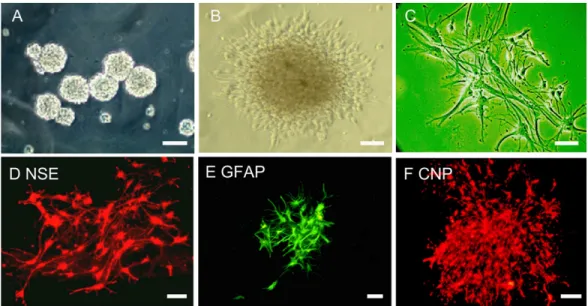
Culture and identification of neural precursors After 2 days of culture in serum-free medium, most of the primary cells isolated from theSVZa or the dorsolateral striatum died. However, some cells formed multicellular spheres and continued to proliferate. After cell passaging, the number of cells increased rapidly and the cell spheres expanded and became suspended in the medium to form neurospheres. As the cells proliferated, the number and size of neurospheres increased, and they tended to grow adher-ently (Figure 1A). After induction with 10% FBS for 2 h, the
Figure 1. In vitro culture of anterior subventricular zone neural precursors. The light microscope images show that the subventricular
zone neural precursors form neurospheres in suspension culture and rapidly increase in sizeand tend to grow together (A). After 2 h of induction with 10% fetal bovine serum, a light microscope image indicates that the neurosphere starts to adhere to the culture plate (B). The neurospheres shown in C are at the furthermost periphery. The images of immunohistochemical staining show that the
neurospheres were positive for the antibodies against NSE (D), GFAP (E), and CNP (F). NSE = neuron-specific enolase; GFAP = glial fibrillary acidic protein; CNP = 2’,3’-cyclic nucleotide 3”-phosphodiesterase. Magnification bars are A = 50 µm, B and F = 40 µm, C-E
= 20 µm.
Table 1. Primer sequence and reaction conditions for PCR experiments.
Genes Sense primer (5’→3’) Anti-sense primer (5’→3’) Renaturation temperature Amplification length
mDlx5 AGAGTCCCAAGCATCCGATCC CCAGCACAACACTGTAGTCCC 56.8°C 897 bp
rDlx5 TTATGCGGACTACGGCTACG GGCAGGTGGGAATTGATTGA 52.3°C 543 bp
Er81 AAGGGTCCCAGGCAGTTCTAT GATGCTCTTCAGGCTCAATCA 52.9°C 229 bp
neurospheres started to adhere to the plate and to differenti-ate (Figure 1B). The peripheral edges of the neurospheres formed short and branchless radial protrusions (Figure 1C). After two additional days of culture, these protrusions became longer and thinner, with more and more branches. Small neurospheres were almost completely differentiated.
Immunostaining results confirmed the expression of GFAP,
NSE, and CNP (Figure 1D-F). These results suggested that cultured neurospheres were capable of differentiating
in vitro into neuronal cell types, thus highlighting a basic feature of neural precursors.
Expression of Dlx5, Er81 and Islet1 in neurospheres from the SVZa and dorsolateral striatum
Immunostaining revealed that these neurospheres from theSVZa were positive for Er81, Dlx5, and nestin proteins (Figure 2A-C) but negative for Islet1 expression (Figure 2D).
However, the neurospheres from the dorsolateral striatum were positive for Er81, Islet1, and nestin proteins (Figure 2A’,C’,D’) but negative for Dlx5 expression (Figure 2B’).
Localized expression of Dlx5, Er81, and Islet1 in SVZ-RMS-OB
Stenman et al. (16) have previously analyzed the expression of Dlx5/6, Er81, and Islet1 at embryonic day E12.5, E16.5 and in P0 mice. Similar to their reports, the IHC results of the present study revealed that Er81 (Fig-ure 3A-C,E,F) and Dlx5 (Fig(Fig-ure 3A’-C’,E’,F’) were widely distributed in various brain areas of neonatal rats (P0), such as the SVZa-RMS-OB, hippocampus, and subcortex. Islet1 was not expressed in the SVZa-RMS-OB, but was expressed in both the dorsolateral and ventral striatum (Figure 3G,H). However, the expression pattern for Er81 and Dlx5 observed here was different from Stenman’s results (16). We observed that Er81 was also widely expressed in both the dorsolateral and ventral striatum (Figure 3D), while Stenman et al. (16) reported that Er81 was observed in scattered striatal neurons. In addition, our data showed that Dlx5 was negatively expressed in the striatum (Figure 3C’-D’), while Stenman et al. (16) showed that Dlx proteins were positively expressed in the striatum. Thus, we think that the difference in the expression pattern of Dlx proteins in the striatum might be caused by multiple factors. For example, the Stenman study used specimens and Dlx an-tibodies from different sources that might have contributed to the differences observed. Furthermore, the expression
pattern of Dlx proteins in Stenman’s study may reflect
the distribution of Dlx1, Dlx2, Dlx3, Dlx4, and Dlx6 in the
developing brain, while in the present study it specifically reflected the SVZa-RMS-OB location of the Dlx5 protein
in the newborn rat brain.
The effects of mDlx5 on the morphology and differentiation of SVZa neural precursors
Green fluorescence first appeared 4 h after
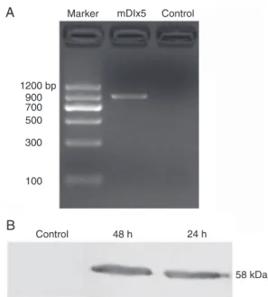
pEGFP-mDlx5 transfection and dramatically increased at 8 h, peaking 24 to 48 h post-transfection and lasting several days. At 24 h post-transfection, total cellular RNA was ex-tracted, and mDlx5 mRNA could be detected by RT-PCR (Figure 4A: the product is 897 bp). At 24 and 48 h post-transfection, the target 58-kDa protein could be detected by Western blot (Figure 4B), indicating that the transfected GFP-mDlx5 fusion protein was expressed robustly. These results indicated that we could use this system to study the role of Dlx5 in SVZa neural precursors further.
The transfected SVZa neural precursors were cultured for 24 h and then stained with NSE-Cy3. As shown in Fig-ure 5, the transfected cells were GFP-positive (FigFig-ure 5A) and differentiated to protrusion-bearing cells (Figure 5B). The most GFP-positive cells were positive for NSE-Cy3 (Figure 5C-E), indicating that the neural precursors that expressed mDlx5 successfully differentiated into neurons.
Figure 2. Expression of nestin (A, A’), Dlx5 (B, B’), Er81 (C, C’), and Islet1 (D, D’) in the neurospheres from anterior subventricu-lar zone (SVZa) and striatum (Str). The neurospheres from the SVZa are positive for nestin (A), which is a precursor cell marker. They are also positive for Dlx5 (B) and Er81 (C), but negative for Islet1 (D). However, the neurospheres from the striatum are posi-tive for nestin (A’), Er81 (C’) and Islet1 (D’) but negaposi-tive for Dlx5
180 H.F. Shu et al.
However, not all GFP-positive cells were positive for NSE (data not shown). As a negative control experiment, some cells were transfected with an empty pEGFP plasmid that did not contain mDlx5 (Figure 5F). The NSE-staining re-sults indicated that only a few scattered GFP-positive cells
expressed NSE (Figure 5G,H).
In passages 1 through 3 of the in vitro cultured SVZa neural precursors, expression of endogenous Dlx5 (rat Dlx5) mRNA could be detected by RT-PCR (Figure 6, left panel). However, endogenous Dlx5 could no longer be detected in
Figure 3. Expression of Er81 (A-F), Dlx5 (A’-F’), and Islet1 (G-H) at P0. Er81 proteins are widely expressed in various brain areas including OB (A), RMS (B), SVZa (C), striatum (D), hippocampus (E), and subcortex (F). Similarly, Dlx5 are also located in theOB (A’), RMS (B’), SVZa (C’), hippocampus (E’), and subcortex (F’), but not in the striatum (C’,D’). Islet1 was not expressed in the SVZa, RMS and OB but was expressed in the striatum (Figure 3G,H). OB = olfactory bulb; RMS = rostral migratory stream; SVZa = anterior
passage 4 precursors (Figure 6, middle panel). In contrast, Er81 mRNA was detected in different passages of SVZa precursors (Figure 6, middle panel). When the exogenous mDlx5 gene was transfected into passage 4 precursors, the expression of mDlx5 and of endogenous rat Dlx5 mRNA appeared after 24 h of culture (Figure 6, right panel).
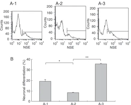
Next, we used FCM analysis to determine the extent of neuronal differentiation in precursor populations express-ing different amounts of Dlx5. As shown in Figure 7, 19.2 ± 0.8% of the passage 3 precursors with endogenous Dlx5 mRNA could differentiate to NSE-positive neurons. In passage 4 precursors, which did not have Dlx5 mRNA,
a significantly smaller fraction of cells (only 8.3 ± 0.4%; P < 0.05) differentiated to NSE-positive cells. Intriguingly, in
the same background, the neuronal differentiation rate was enhanced to 35.7 ± 0.6% when mDlx5 was overexpressed. Therefore, we conclude that overexpression of Dlx5 (P
< 0.05) significantly promoted neuronal differentiation of
SVZa precursors.
Discussion
In this study, we showed that the precursors from SVZa were positive for Dlx5 and Er81 but negative for Islet1, while the precursors from the dorsolateral striatum were positive for Er81 and Islet1 but negative for Dlx5. Similar to that reported in a previous investigation (16), the present IHC data indicated that Dlx5 and Er81 are widely expressed
Figure 4. Expression of mouse Dlx5 (mDlx5) in anterior subven-tricular zone (SVZa) precursors transfected with pEGFP-mDlx5. Agarose gelelectrophoresis analysis of RT-PCR showed the ex-pression of mDlx5 RNA in SVZa precursors 24 h after transfec-tion (A). Lane M is the marker, the middle lane (mDlx5) is from cells transfected with pEGFP-mDlx5, and the right lane (control) is from cells without transfection of pEGFP-mDlx5. B, Western blot analysis showed the expression of mDlx5 proteins in SVZa precursors at 24 and 48 h after transfection.
Figure 5. Expression of GFP and NSE in
ante-rior subventricular zone (SVZa) neural precur-sors transfected with pEGFP-mDlx5. A, After 24 h oftransfection with pEGFP-mDlx5, the majority of SVZa neurospheres expressed GFP. B, Repre-sentative GFP-positive SVZa neural precursors. Double-staining with GFP (C) and NSE-Cy3 (D) re-veals that most of GFP-positive neurospheres/cells were differentiated into neurons (E). However, cells tranfected with the pEGFP plasmid without mDlx5 (F) show low rates of NSE staining (G,H). GFP =
182 H.F. Shu et al.
Figure 6. Expression of Dlx5 and Er81 mRNA in anterior subventricular zone (SVZa) precursors. Passage 3 (left panel) and passage 4 (middle panel) of the cultured SVZa precursors were analyzed. Er81 and endogenous Dlx5 (rDlx5) mRNA detection by RT-PCR fol-lowed by agarose electrophoresis analysis. The products are 229 and 543 bp, respectively. Right panel, Expression of endogenous Dlx5 (rDlx5) and transfected Dlx5 (mDlx5) mRNAs by RT-PCR followed by agarose gelelectrophoresis analysis. After transfection with pEGFP-mDlx5, mDlx5 (897 bp) and rDlx5 expression can be detected in passage 4 precursors. Rat GAPDH was used as an internal standard; the product is 289 bp. GAPDH = glyceraldehyde 3-phosphate dehydrogenase.
Figure 7. Determination of percent neuronal differentiation in subventricular zone (SVZ) precursors with different Dlx5 levels. A, Flow
cytometry measurement was used to quantify NSE-positive cells after the anterior SVZ (SVZa) neural precursors differentiated. The cell populations are induced to differentiate from passage 3 of SVZ precursors (A-1) and passage 4 of SVZa precursors (A-2). Passage 4 of SVZ precursors were transfected with pEGFP-mDlx5 (A-3). B, Quantification and comparison of the data in A. One-way ANOVA
confirmed that the differences between A-1 and A-2, A-2 and A-3 groups were significant (*P < 0.01, **P < 0.001). NSE =
across brain areas, such as SVZa-RMS-OB, hippocampus, and subcortex. Islet1, on the other hand, is not expressed in these areas but is localized in the striatum. Interestingly, we observed that Dlx5 was negative but Er81 was positively expressed in the striatum area, again differing from the previous report (16). In addition, the expression of endog-enous Dlx5 mRNA in cultured SVZa neural precursors was gradually down-regulated and wascompletely lost by the fourth passage. Exogenous Dlx5 transfected into passage 4 precursors could stabilize thelevels of Dlx5 and promote the neuronal differentiation of cultured SVZa neural precur-sors. These data demonstrated that Dlx5 plays an important role during neuronal differentiation. Further investigation is required to elucidate the underlying mechanism.
The expression of Dlx5 in SVZa-RMS-OB and SVZa neural precursors
Wichterle et al. (17) studied the migration pathway of embryonic lateral ganglionic eminence (LGE) cells and found that LGE cells continuously migrate to the OB and become the major source of OB interneurons. However, not all LGE cells migrate to the OB; some cells migrate to the striatum and become striatal projection neurons. These results suggest that there are at least two populations of neural precursors in the LGE. Further studies by Stenman et al. (16) showed that only the cells in the dorsolateral LGE migrate to the OB and differentiate into OB interneurons. These cells have distinct molecular characteristics: they are positive for Dlx and Er81 protein but negative for Islet1. Most of the cells in the ventral lateral LGE migrate toward the striatum and eventually differentiate into striatal interneurons and projection neurons, which are positive for Dlx and Islet1 but negative for Er81. Because the SVZa neural precursors also directionally migrate to the OB and differentiate there, we hypothesized that SVZa neural precursors exhibit the
same molecular features as LGE dorsolateral cells. Specifi -cally, we hypothesized that they are also positive for Dlx and Er81 and negative for Islet1.
In the present investigation, we studied the expression of Dlx5 in SVZa-RMS-OB by IHC and found that Dlx5 is widely expressed in a multitude of areas, including the SVZa-RMS-OB, hippocampus, and subcortex. However, Er81 is also widely expressed, not only in these areas but also in the striatum. Hence, Dlx5 and Er81 expression is high and overlaps in the SVZa-RMS-OB. On the other hand, Islet1 is not expressed in the SVZa-RMS-OB, but is expressed in the striatum. The expression of Dlx5 in SVZa-RMS-OB suggests a close relationship between the Dlx5 gene and the process of migration and differentiation of OB interneurons.
It also confirms our hypothesis that SVZa precursors and
LGE dorsolateral cells possess similar molecular features, i.e, they are positive for Dlx and Er81 and negative for Islet1. Previous studies have shown that Dlx5 mRNA is highly expressed in forebrain subcortex GABA and dopamine neurons as well as in precursors of GABA neurons, but is
low in differentiated neurons (11). Dlx5 appears to be pref-erentially expressed in neural precursors and stem cells. Therefore, the dorsolateral area of the LGE in embryonic stages may be similar to the postnatal SVZa structure. Furthermore, the SVZa neural precursors in neonates may originate from the dorsolateral area of the LGE.
Neural precursors isolated and cultured from the SVZa exhibited features of neural stem cells. Our data demon-strate that they continuously divide and proliferate, form suspended neurospheres in serum-free medium, show positive staining for the stem cell marker nestin (for a review, see Ref. 18), and can differentiate into neural cell types. These results suggest that, indeed, we
success-fully isolated neural precursors. Immunostaining confirmed
that these precursors are positive for Dlx5 and Er81 and negative for Islet1. We performed immunostaining studies on dorsolateral striatal neural precursors to compare the molecular characteristics of SVZa neural precursors and neural precursors from other brain areas. We found that these cells are Islet1 and Er81 positive but Dlx5 negative. These data suggested that detecting Dlx5, Er81, and Islet1 proteins in cultured SVZa neural precursors can facilitate exclusion of the precursors from other regions (at least the dorsolateral striatum). Islet1 and Er81 have been used as markers for striatal and OB interneurons, respectively (16), although their functions are still largely unknown. Our studies showed that Dlx5 is closely involved in the differentiation of OB interneurons from SVZa precursors and may play an important role in SVZa neural precursor differentiation.
Effect of Dlx5 on the neuronal differentiation of SVZa neural precursors
After migration to the OB through the RMS, SVZa neural precursors terminally differentiated into OB interneurons. This process is regulated by multiple signaling events at both the transcriptional and post-transcriptional levels. Long et al. (14) showed that Dlx5 knock-out mice had few OB interneurons, suggesting that Dlx5 plays a major role in the differentiation of OB interneurons and inthe synaptogenesis of OB receptor neurons. The neural precursors isolated from the telencephalon of Dlx5-/- mice lost the ability to differen-tiate into neurons. Therefore, we speculated that Dlx5 is related to the migration and neuronal differentiation of SVZ precursors. Dlx5-/- mice are neonatal lethal and therefore
cannot be studied further. Hence, we studied the role of Dlx5 in neuronal differentiation of SVZa neural precursors using recombinant plasmid and in vitro culture methods.
Green fluorescence protein is a small stable protein
that does not interfere with the function of cells and shows no toxicity, even when overexpressed (19). It is a widely
used marker for live fluorescent labeling. Here, we used
the EGFP vector to construct the plasmid pEGFP-mDlx5. This permitted us to directly monitor mDlx5 expression in SVZa neural precursors and todetect the cells expressing
-184 H.F. Shu et al.
References
1. Imayoshi I, Sakamoto M, Ohtsuka T, Kageyama R. Continu-ous neurogenesis in the adult brain. Dev Growth Differ 2009; 51: 379-386.
2. Quinones-Hinojosa A, Sanai N, Gonzalez-Perez O, Garcia-Verdugo JM. The human brain subventricular zone: stem cells in this niche and its organization. Neurosurg Clin N Am 2007; 18: 15-20, vii.
3. Ayuso-Sacido A, Roy NS, Schwartz TH, Greenfield JP,
Boockvar JA. Long-term expansion of adult human brain subventricular zone precursors. Neurosurgery 2008; 62: 223-229.
4. Ventura RE, Goldman JE. Dorsal radial glia generate ol-factory bulb interneurons in the postnatal murine brain. J Neurosci 2007; 27: 4297-4302.
5. Pencea V, Luskin MB. Prenatal development of the rodent rostral migratory stream. J Comp Neurol 2003; 463: 402-418.
6. Lledo PM, Merkle FT, Alvarez-Buylla A. Origin and function of olfactory bulb interneuron diversity. Trends Neurosci 2008; 31: 392-400.
7. Alvarez-Buylla A. Mechanism of migration of olfactory bulb interneurons. Semin Cell Dev Biol 1997; 8: 207-213. 8. Haughey NJ, Liu D, Nath A, Borchard AC, Mattson MP.
Disruption of neurogenesis in the subventricular zone of
adult mice, and in human cortical neuronal precursor cells in culture, by amyloid beta-peptide: implications for the pathogenesis of Alzheimer’s disease. Neuromolecular Med 2002; 1: 125-135.
9. Liu SY, Zhang ZY, Song YC, Qiu KJ, Zhang KC, An N, et al. SVZa neural stem cells differentiate into distinct lineages in response to BMP4. Exp Neurol 2004; 190: 109-121. 10. Perera M, Merlo GR, Verardo S, Paleari L, Corte G, Levi G.
Defective neuronogenesis in the absence of Dlx5. Mol Cell Neurosci 2004; 25: 153-161.
11. Panganiban G, Rubenstein JL. Developmental functions of the Distal-less/Dlx homeobox genes. Development 2002; 129: 4371-4386.
12. Merlo GR, Zerega B, Paleari L, Trombino S, Mantero S, Levi G. Multiple functions of Dlx genes. Int J Dev Biol 2000; 44: 619-626.
13. Levi G, Puche AC, Mantero S, Barbieri O, Trombino S, Paleari L, et al. The Dlx5 homeodomain gene is essential for olfactory development and connectivity in the mouse. Mol Cell Neurosci 2003; 22: 530-543.
14. Long JE, Garel S, Depew MJ, Tobet S, Rubenstein JL. DLX5 regulates development of peripheral and central components of the olfactory system. J Neurosci 2003; 23: 568-578.
pression of mDlx5-GFP fusion protein in transfected SVZa
neural precursors by fluorescence microscopy. Because the
molecular weight (MW) of GFP is approximately 27 kDa and that of rat Dlx5 is 31 kDa, the fusion protein should be approximately 58 kDa. Our Western blot data demonstrated that the MW of the fusion protein matches the expected MW
(Figure 5B), thus further confirming that the recombinant
plasmid can be used to study the role of Dlx5 in the migra-tion and differentiamigra-tion of SVZ neural precursors.
The technique of live fluorescent labeling provided a
very useful tool for analyzing the morphology of cells in the central nervous system. Here we studied the morphological changes in SVZa neural precursors following overexpres-sion of GFP-mDlx5, demonstrating that the transfected cells differentiate into neuron-like cells. IHC staining for
NSE confirmed that most of the Dlx5-transfected cells dif -ferentiated into neurons. We further found that the expres-sion of endogenous Dlx5 decreased with increasing cell passage and became undetectable by RT-PCR in passage 4 cells. The Er81 expression level was unchanged during subsequent passages. Consistently, when the passage 4 precursors were induced to differentiate, less than 10% of cells successfully differentiated into neurons. This leads us to conclude that Dlx5 levels may play an important role during the neuronal differentiation of SVZa neural precur-sors. This conclusion is further supported by the previous report that the Dlx genes encode homeodomain transcrip-tion factors related to Drosophila distal-less gene, and their mammalian homologs, Dlx1, Dlx2, Dlx5, and Dlx6,
are expressed in the subcortical forebrain and are involved in the initiation of GABAergic neuron differentiation and neuronal migration (10,11).
We have provided multiple lines of evidence demon-strating that Dlx5 plays an important role in the neuronal differentiation of SVZa neural precursors. First, Dlx5 and Er81 are expressed in SVZa neural precursors; this ex-pression pattern is one of the molecular features of SVZa precursors. In the in vitro cultured SVZa neural precursors, Dlx5 expression is gradually lost and can be regained by exogenous Dlx5 transfection, suggesting the existence of auto-regulation and external cue regulation. Taken together, these results demonstrate that Dlx5 plays an important role during neuronal differentiation.
Acknowledgments
The authors thank Prof. Giovanni Levi (Co-Director of the Laboratory of Physiology, Museum National d’Histoire Naturelle, France) for his gift of pBluescriptBSK-mDlx5 plasmid. Sincere thanks are dueto Ms. Wei Sun and Ms. Li-Ting Wang (senior experimental division and assistant laboratory technician, respectively, Central Laboratory, Third Military Medical University, Chongqing, China) for their as-sistance with the confocal laser scanning microscopy. The Chinese National Natural Science Foundation (#30130110, #30600638) and Chongqing Natural Science Foundation
15. Luskin MB, Zigova T, Soteres BJ, Stewart RR. Neuronal pro-genitor cells derived from the anterior subventricular zone of the neonatal rat forebrain continue to proliferate in vitro and express a neuronal phenotype. Mol Cell Neurosci 1997; 8: 351-366.
16. Stenman J, Toresson H, Campbell K. Identification of two distinct progenitor populations in the lateral ganglionic eminence: implications for striatal and olfactory bulb neuro-genesis. J Neurosci 2003; 23: 167-174.
17. Wichterle H, Turnbull DH, Nery S, Fishell G, Alvarez-Buylla
A. In utero fate mapping reveals distinct migratory pathways and fates of neurons born in the mammalian basal forebrain. Development 2001; 128: 3759-3771.
18. Kempermann G, Jessberger S, Steiner B, Kronenberg G. Milestones of neuronal development in the adult hippocam-pus. Trends Neurosci 2004; 27: 447-452.