BIOMEDICAL SCIENCES
AND
CLINICAL INVESTIGATION
www.bjournal.com.br
www.bjournal.com.br
Braz J Med Biol Res, March 2010, Volume 43(4) 367-376
Participation of neuronal nitric oxide synthase in experimental
neuropathic pain induced by sciatic nerve transection
M. Chacur, R.J.B. Matos, A.S. Alves, A.C. Rodrigues, V. Gutierrez, Y. Cury and L.R.G. Britto
Institutional Sponsors
Brazilian Journal of Medical and Biological Research (2010) 43: 367-376 ISSN 0100-879X
Participation of neuronal nitric oxide synthase
in experimental neuropathic pain induced
by sciatic nerve transection
M.
Chacur
1,2, R.J.B. Matos
1, A.S. Alves
1, A.C. Rodrigues
3,
V. Gutierrez
4, Y.
Cury
4and L.R.G.
Britto
11Departamento de Fisiologia e Biofísica, 2Departamento de Anatomia, Instituto de Ciências Biomédicas, 3Departamento de Análises Clínicas e Toxicológicas, Faculdade de Ciências Farmacêuticas,
Universidade de São Paulo, São Paulo, SP, Brasil
4Laboratório de Patofisiologia, Instituto Butantan, São Paulo, SP, Brasil
Abstract
Nerve injury leads to a neuropathic pain state that results from central sensitization. This phenomenom is mediated by NMDA receptors and may involve the production of nitric oxide (NO). In this study, we investigated the expression of the neuronal isoform of NO synthase (nNOS) in the spinal cord of 3-month-old male, Wistar rats after sciatic nerve transection (SNT). Our attention was focused on the dorsal part of L3-L5 segments receiving sensory inputs from the sciatic nerve. SNT resulted in the
development of neuropathic pain symptoms confirmed by evaluating mechanical hyperalgesia (Randall and Selitto test) and
allodynia (von Frey hair test). Control animals did not present any alteration (sham-animals). The selective inhibitor of nNOS, 7-nitroindazole (0.2 and 2 µg in 50 µL), blocked hyperalgesia and allodynia induced by SNT. Immunohistochemical analysis showed that nNOS was increased (48% by day 30) in the lumbar spinal cord after SNT. This increase was observed near the central canal (Rexed’s lamina X) and also in lamina I-IV of the dorsal horn. Real-time PCR results indicated an increase of nNOS mRNA detected from 1 to 30 days after SNT, with the highest increase observed 1 day after injury (1469%). Immunoblotting
confirmed the increase of nNOS in the spinal cord between 1 and 15 days post-lesion (20%), reaching the greatest increase (60%) 30 days after surgery. The present findings demonstrate an increase of nNOS after peripheral nerve injury that may
contribute to the increase of NO production observed after peripheral neuropathy.
Key words: Allodynia; Neuropathic pain; Neuronal nitric oxide synthase; Peripheral neuropathy; Sciatic nerve transection; Spinal cord
Introduction
Neuropathic pain is initiated by a primary lesion or dysfunction of the nervous system. In spite of extensive investigations, the mechanism of neuropathic pain is still
unclear. Neuropathic pain is much more difficult to treat
than nociceptive pain. The therapeutic approaches include the use of gabapentin, opioids (mainly morphine) and, more recently, pregabalin. However, these treatments are not very effective. Thus, neuropathic pain could interfere with the quality of life of patients, representing a serious clinical problem.
Animal models are relevant for the understanding of the mechanisms and pathophysiology of human neuropathic pain. Among these models, transection of the rat sciatic nerve produces abnormalities in pain sensation reminiscent
of those observed in humans, and it represents a good model to study both nerve regeneration and neuropathic pain.
Several lines of evidence have indicated that nitric oxide (NO) is an important mediator of nerve injury and may be responsible for pain-linked behavior in animals. Peripheral nerve injury may result in an increased excitability of spinal cord neurons through activation of nociceptive afferents, leading to activation of N-methyl-D-aspartate receptors and subsequent spinal production of NO (1). Therefore, an altered expression of spinal NO synthase (NOS) and its enhanced catalytic activity may contribute to neuroplasticity after nerve injury and may play an important role in central sensitization. However, pharmacological evidence regard-ing the role of spinal NO in the development of nerve
injury-Correspondence: M. Chacur, Departamento de Anatomia, ICB III, USP, Av. Prof. Lineu Prestes, 2415, 05508-900 São Paulo, SP, Brasil. Fax: +55-11-3091-7426. E-mail: [email protected]
evoked allodynia or hyperalgesia has been conflicting.
NO is a biological messenger molecule synthesized by three isoforms of NOS, which diffuses from the site of production across cellular membranes. All three isoforms of NOS are present in the nervous system, namely the constitutive neuronal (nNOS) and endothelial isoforms (eNOS), and the inducible isoform (iNOS) (2). nNOS is
up-regulated after peripheral inflammation, iNOS is mainly
absent in neural tissues under normal conditions, but is
up-regulated in inflammation, while eNOS is present in
the brain vasculature. nNOS is present in the spinal cord
of rats, predominantly in the superficial dorsal horn (Rexed
laminae I and II), in the area around the central canal (lamina X) and in the intermediolateral nucleus column of the thoracic, upper lumbar, and sacral segments (3). Strong up-regulation of NOS expression has been detected in large neurons of laminae III and IV after sciatic nerve transection (SNT) (4).
The effects of nerve transection on neuronal nitrergic activity have been extensively investigated, and the data are contradictory. Indeed, some investigators observed that treatment with NOS inhibitors interferes with allodynia and hyperalgesia (5,6) induced by rat nerve injury, whereas others have reported that nNOS inhibitors have no effect on the hyperalgesia induced by nerve injury (7,8).
In light of these conflicting results and despite the fact
that in vivo studies suggest an important role for spinal cord NO in the transmission of pain signals, the possible partici-pation of NO after SNT and its effects on the lumbar spinal cord have not been evaluated. The aim of the present study was to investigate nNOS expression in the lumbar spinal cord of adult rats after SNT using immunohistochemistry, Western blot, and real-time PCR assays together with the analysis of nociceptive behavior. Attention was focused on the dorsal part of the cord (L3-L5 segments), which receives sensory inputs from the sciatic nerve (4). In addition, we also evaluated the effect of the administration of 7-nitroindazole (7-NI), a selective nNOS inhibitor, on the development of hyperalgesia and allodynia after SNT.
Material and Methods
Animals
Male Wistar rats weighing 200 to 250 g (approximately 3 months of age) were used. They were housed individually and maintained on a 12-h light/dark cycle. All procedures were approved by the Animal Care Committee of the Uni-versity of São Paulo (protocol #071/2005).
Surgical procedure
Animals were anesthetized with intramuscular injec-tions of ketamine (5 mg/100 g body weight) and xylazine (1 mg/100 g body weight), both from Sespo Ltda. (Brazil). The right sciatic nerve of the animal was exposed at the mid-thigh level and a short segment (5 mm) of the nerve was
removed. Sham-operated animals (N = 20) were subjected to an identical surgical procedure but not SNT and served as controls. Animals used for immunohistochemistry (N = 32), immunoblotting (N = 24) and real-time PCR (N = 24) were anesthetized and perfused and killed by cervical dis-location at 1, 7, 15, and 30 days after SNT and the lumbar segments of the spinal cord were removed by laminectomy.
Prior to sacrifice, all rats were tested for sensitivity to noxious
mechanical stimulation of the hind paw using the Randall and Selitto and von Frey tests (described below).
Intrathecal injections
Lumbar puncture was performed as described
previ-ously (9). Briefly, a sterile 23-gauge hypodermic needle
with the plastic hub removed was attached to a sterile polyethylene tube (PE-50, Becton Dickinson, USA) and the open end of the PE-50 was attached to a 50-µL Hamilton syringe for drug delivery. Puncture was made by inserting the tube between lumbar vertebrae L5 and L6. Successful
subarachnoid entry was confirmed by immediate manifesta
-tion of a characteristic tail flick response, and later by visual
inspection of Evan’s blue dye after euthanasia.
In order to evaluate the involvement of nNOS, 7-NI (0.2 and 2 µg in 50 µL, N = 5; Sigma-Aldrich, USA), a selective nNOS inhibitor, or vehicle (50 µL distilled water, N = 5) was injected into the spinal cord byintrathecal route, 60 min before behavioral testing in each experimental group (at 1, 7, 15, and 30 days post-lesion). The drug solution was prepared fresh just before the injection, by dissolving the drug in distilled water. Behavioral signs of mechanical hyperalgesia and allodynia were measured before and 1, 2, 4, and 6 h after the injections. The dose used was based on our previous studies (9) that indicated that 0.2 and 2.0
µg/rat 7-NI were sufficient to block neuronal nNOS, allowing
mechanical hyperalgesia.
Upon completion of behavioral testing and euthanasia,
correct catheter placement was confirmed visually and only
results from rats in which the Evans Blue dye was restricted to and uniformly distributed in the intrathecal space with no apparent leakage were included in the analysis.
Evaluation of pain threshold
Peripheral nerve injury was induced by specific transec -tion of the sciatic nerve, which innervates the plantar side of the rat hind paw. The development of neuropathic pain symptoms in the hind paw was evaluated by testing me-chanical hyperalgesia (Randall and Selitto test) (10) and allodynia (von Frey hair test) (11). Both sensory tests were used because of literature data showing that SNT induces hyperalgesia and allodynia of the ipsilateral hind paw.
Mechanical hyperalgesia (Randall and Selitto test). An Ugo-Basile pressure apparatus (10) was used to assess pressure pain thresholds prior to surgery and again at dif-ferent times thereafter. Testing was blind in regard to group
Participation of nitric oxide in neuropathic pain 369
to the right hind paw. The force needed to induce paw withdrawal was recorded as the pain threshold. To reduce stress, the rats were habituated to the testing procedure the day before the experiment (12).
Low threshold mechanical allodynia (von Frey test). The von Frey test (11) was used to assess low-threshold mechanical pain thresholds prior to surgery and again at different times thereafter. Testing was blind in regard to group designation. This test was performed as described
in detail (9). Briefly, a logarithmic series of 10 calibrated Semmes-Weinstein monofilaments (von Frey hair test,
Stoelting, USA) were applied to the middle of the plantar surface of the right hind paw, for a maximum of 10 s to determine the threshold intensity of the stiffness stimulus required to elicit a paw withdrawal response. Log stiffness of the hairs is determined by log10 (mg x 10) and ranges from 3.61 (0.407 g) to 5.18 (15.136 g). Baseline assess-ment was initiated with the 0.2041-g hair. In the event of paw withdrawal, the same hair was again presented 60-90 s later. If the response was again elicited, the 0.407
g monofilament was presented. In the absence of a paw
withdrawal response to the 0.407 g stimulus, the next
stronger monofilament was presented (0.692 g). The monofilament that elicited a clear response was recorded,
and was presented once again 30-60 s later. If the animal withdrew its paw in two consecutive trials with the same stiffness value, no further von Frey hairs were tested. How-ever, in the absence of a response to the initial 0.2041-g
monofilament, presentation of monofilaments continued
in ascending order until two consecutive responses were
elicited from the same monofilament. All single responses
were recorded, but assessment was complete only after two consecutive responses were elicited from the same
monofilament. Failure to respond to the strongest stimulus
(15.136 g) was considered to be the cut-off value. Response to the weakest stimulus (0.407 g) was considered to be the lower cut-off value for that time. To reduce stress, rats were habituated to the experimental environment daily for 4 days before the experiments. Behavioral responses were used to calculate the 50% paw withdrawal threshold (absolute
threshold), by fitting a Gaussian integral psychometric function using a maximum-likelihood fitting method. This fitting method allows parametric analyses (13).
RNA isolation, cDNA synthesis and real-time PCR
Tissue from the ipsilateral and contralateral side of the lumbar spinal cord was homogenized in 1 mL TRIzol (In-vitrogen, USA) with a homogenizer (Kinematica Polytron® PT 2100, USA) and total RNA was isolated following the manufacturer’s suggested protocol. Total RNA (1 µg) was reverse transcribed into cDNA using 200 U M-MLV reverse transcriptase and oligo-dT as a primer (Invitrogen).
The real-time PCR system consisted of 250-nM primers, 50 ng cDNA samples, and 1X SYBR® Green PCR Master Mix (Applied Biosystems, USA). Using the Rotor-Gene
3000 Real-time PCR detection system (Corbett Research, Australia), PCR cycling conditions were set as follows: after initial activation at 50°C for 2 min and 95°C for 10 min, 40 cycles of 95°C for 15 s and 60°C for 1 min; then melt curve analysis was performed by heating samples from 65° to 99°C (1°C increment changes at 5-s intervals). All sample measurements were performed in duplicate. The following primers were used: nNOS sense: 5’-GTC GCATTCAACAGCGTCTC-3; nNOS antisense: 5’-CCC AAAGGCACAGAAGTGG-3’ and GAPDH sense: 5’-GA TGCTGGTGCTGAGTATGTCG-3’; GAPDH antisense: 5’-GTGGTGCAGGATGCATTGCTGA-3’, which resulted in 163- and 197-bp amplicons, respectively (14).
Sample cycle threshold values were determined from
plots of normalized fluorescence versusPCR cycle number
during exponential amplification. Standard curves for all primer amplifications were generated by plotting average
cycle threshold values against the logarithm of the quantity of target template molecules.
All quantitations were normalized to the endogenous control (GAPDH). The relative quantitation value of each target gene was analyzed using a comparative CT method (15). The following formula (2-ΔΔCT) was used to calcu-late the relative amount of the transcript in the sample and normalized to an endogenous reference (GAPDH): 2-ΔΔCT, where
Δ
CT is the difference in CT between the gene of interest and GAPDH, andΔΔ
CT for the sample = meanΔ
CT of the sample - meanΔ
CT of the control sample (used as calibration).Immunoblotting
Naive and sham rats and rats subjected to transection
of the sciatic nerve were sacrificed by cervical dislocation
and the ipsilateral and contralateral sides of the lumbar spinal cord were collected and homogenized in an extrac-tion buffer containing 100 mM Tris, pH 7.4, 10 mM EDTA, 2 mM PMSF, and 10 µg/mL aprotinin. After extraction, the homogenates were centrifuged at 11.5g for 20 min and the protein concentration of the supernatant was determined using the Bradford protein assay with albumin as standard (Bio-Rad, USA) (16). Samples containing 100 µg protein were loaded on a 6.5% acrylamide gel and electrotransferred to nitrocellulose membranes using a Trans-Blot cell system (Bio-Rad). After transfer, the membranes were treated for 4 h at room temperature with a blocking solution containing 5% powdered milk, washed and incubated overnight at 4°C with a mouse monoclonal antibody against nNOS (1:2000, Sigma, USA). The membranes were then washed and incubated for 2 h at room temperature with a peroxidase-conjugated anti-mouse antibody diluted 1:10,000
(Amer-sham Biosciences, UK). β-actin was used as an endogenous control (1:1000, Sigma). The specifically bound antibody
Immunohistochemistry and image analysis
After the appropriate survival time, the animals were anesthetized deeply with ketamine and xylazine and per-fused through the heart with phosphate-buffered saline and 4% paraformaldehyde in 0.1 M phosphate buffer (PB), pH 7.4. The lumbar spinal cord (L3-L5) was then removed and
post-fixed for 4 h in 4% paraformaldehyde. The spinal cord
was transferred to a 30% sucrose solution in PB to ensure cryoprotection, which lasted for 48 h, and transverse sections (30 µm) were obtained with a sliding microtome adapted for
cryosectioning. Free-floating sections were incubated with a
mouse monoclonal antibody against nNOS (Sigma) diluted 1:1000 in PB containing 0.3% Triton X-100 and 5% normal goat serum. Incubations with the primary antibody were conducted overnight at 24°C. The sections were then washed three times for 10 min each in PB and incubated with a biotinylated goat anti-mouse serum (Vector, USA) diluted 1:200 in PB for 2 h at 24°C. The sections were washed again in PB as above and incubated with the avidin-biotin-peroxidase complex (ABC Elite, Vector). After washing, the sections were reacted with 0.05% 3-3’-diaminobenzidine and 0.01% hydrogen peroxide in PB.
Intensification was conducted with 0.05% osmium tetroxide
in water. The sections were mounted on gelatinized slides, dehydrated, cleared, and coverslipped. The material was analyzed under a light microscope and digital images were collected. Controls for immunostaining included the omis-sion of the primary antibody and its replacement withnormal mouse serum, which completely eliminated the staining. It should be stressed that the antibody used here has been extensively tested and characterized (14). All lumbar spinal cords processed for immunohistochemistry were subjected to quantitative analysis, which involved 10 sections from each lumbar spinal cord processed from each experimental or control rat. Each section was labeled to avoid repeated counts. Data are reported as the numbers of nNOs-positive cells. For the comparative analyses of the number of cells, we considered the
controls for each period studied as 100%. Data are reported as means ± SEM.
Statistical analysis
Data were analyzed statistically by analysis of variance (ANOVA) and sequential differences between means were tested by Tukey contrast analysis at P < 0.05 (17). Data from the von Frey test were analyzed as the interpolated 50% threshold (absolute threshold) in log base 10 of stimulus
intensity (monofilament stiffness in milligrams x 10).
Results
Evaluation of hyperalgesia and allodynia
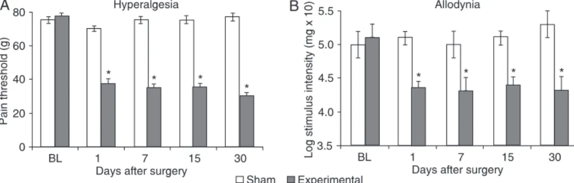
Rats submitted to SNT developed a protective behav-ior characterized by guarding of the ipsilateral hind paw, which is a sign of neuropathic pain. The SNT induced
a significant decrease of the mechanical pain threshold
(Figure 1A), as measured by the Randall and Selitto test, and reduced the withdrawal threshold as measured by the von Frey test (Figure 1B). These alterations were detected as early as on day 1 and persisted at least 1 month after surgery. Sham-operated animals (taken as control) did not present any alteration of the withdrawal threshold (Figure
1). No significant differences in paw withdrawal frequency
were observed between measurements or at any time post-transection on the contralateral side of injury (data not shown).
Effect of intrathecal administration of an nNOS
inhibitor onhyperalgesia and allodynia induced by
sciatic nerve transection
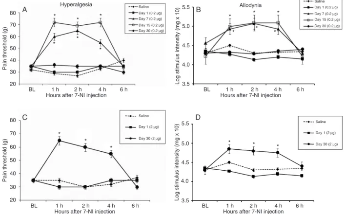
The intrathecal administration of 7-NI (0.2 µg in 50 µL), a selective inhibitor of the neuronal isoform of NOS,
significantly inhibited hyperalgesia (Figure 2A) and allo -dynia (Figure 2B) induced by nerve SNT on days 7 and 15
Participation of nitric oxide in neuropathic pain 371
post-lesion. To confirm these results, we also administered a
high dose (2 µg in 50 µL) of the same inhibitor (Figure 2C and D). This treatment decreased hyperalgesia and allodynia on day 30 after SNT. This inhibition was observed from 1
to 4 h after treatment, disappearing after 6 h. No significant
differences were observed on day 1 after transection for either dose used (Figure 2).
Administration of 7-NI per se did not change the pain threshold (pain threshold = 70 ± 2.1, 72 ± 4.1, 68 ± 3.2, and 75 ± 3.3 g; low-mechanical threshold (log mg x 10) = 5.05 ± 0.2, 4.95 ± 0.4, 4.88 ± 0.2, and 5.00 ± 0.5 g at 1, 2, 4, and 6 h after treatment, respectively).
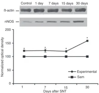
nNOS mRNA expression evaluated by real-time PCR
Semi-quantitative real-time PCR was used to detect nNOS mRNA expression in lumbar spinal cord tissue. GAPDH was determined as a suitable endogenous control (average CT = 17.28 ± 0.28).
Compared with the ipsilateral side of sham animals
(taken as controls), nNOS transcript levels were significantly
increased after all post-lesion periods tested (Figure 3).
Maximum induction of 1469.0 ± 35.6% was observed on day 1 post-lesion. Seven to 30 days after lesion, the nNOS gene was still up-regulated: 488.0 ± 21.9% by day 7 (P < 0.001), 448.0 ± 59.0% by day 15 (P < 0.001), and 453.2
± 31.1% by day 30 (P < 0.001). No significant differences
were observed between the contralateral and ipsilateral sides from experimental animals (1.00 ± 1.22 vs 0.50 ± 1.27 by day 7, 1.00 ± 1.12 vs 1.46 ± 1.47 by day 15, and 1.00 ± 0.55 vs 0.93 ± 0.99 by day 30).
The contralateral side from sham-operated and the contralateral side from naive animals (taken as controls) ex-hibited similar nNOS mRNA (1.00 ± 0.73 vs 1.29 ± 0.20).
Immunoblotting
We identified a single labeled nNOS-positive band
of about 150 kDa in extracts of both sides of the lumbar spinal cord of both sham and experimental animals after SNT. Figure 4 shows an increase of nNOS protein levels on the ipsilateral side from experimental animals when compared to the ipsilateral side from sham rats, taken as 100%. Densitometric analysis revealed that the increase
of nNOS expression started on the first day after lesion:
21.0 ± 2.1% by day 1 (P < 0.01), 22.0 ± 0.6% by day 7 (P < 0.01), 19.0 ± 2.3% by day 15 (P < 0.01), and reaching the greatest increase after 30 days (58.0 ± 2.7%, P < 0.001).
β-actin was used as an endogenous control, and no differ-ences were observed between control and experimental sides at any of the times tested (Figure 4).
The contralateral side from sham-operated and from naive animals (taken as controls) exhibited the same nNOS protein levels (data not shown).
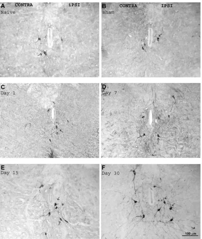
Immunohistochemistry
We examined the expression of nNOS in dorsal lumbar rat spinal cord sections (L3-L5) from transected rats. Mea-surements were performed of neurons of the ipsilateral (right) and contralateral (left) sides in relation to the transected nerve, on the ipsilateral (right) side of sham-operated rats, and on the right side of control (naive) animals.
nNOS immunoreactivity was detected in the lumbar spinal cord under normal conditions. A moderate number of cells labeled for nNOS was observed on both sides of the lumbar spinal cord of naive (Figure 5A) and sham rats (Figure 5B), and on the contralateral side (taken as a control for the operated animals, Figures 5 and 6A,B) of the lum-bar spinal cord of experimental rats. These neurons were located in the dorsal horn of the spinal cord (Figure 6) as well as around the central canal (Figure 5). It is important to point out that the dorsal part of the spinal cord was analyzed. It receives sensory inputs from the sciatic nerve, since the ventral part consisting of sciatic nerve motor neurons was not the objective of this study.
After transection of the sciatic nerve, there was a marked increase in the number of lumbar cells that were nNOS-immunoreactive on the ipsilateral side (lesion side) compared to control (Figure 7). This increase was observed around the central canal, lamina X (Figure 5C-F), and also in lamina I-IV of the dorsal horn (Figure 6). In addition,
there was also an increase in the density of labeled cells in the lumbar area encompassing the dorsal and central portions of the lumbar spinal cord that started on day 1 after
SNT (quantification in Figure 7). One day after transection,
there was an increase of approximately 19.0 ± 0.4% (P < 0.01) in the number of nNOS-positive neurons compared to control, and this appeared to increase thereafter to 22.0 ± 0.3% by day 7 (P < 0.01) and to 35.0 ± 1.0% by day 15 (P < 0.01), reaching the greatest increase after 30 days (48.0 ± 0.6%, P < 0.001) (Figure 7). No differences in nNOS im-munoreactivity were observed in sham-operated animals when compared to the contralateral side of experimental rats (taken as a control for the operated animals).
Discussion
In this study, we investigated the regulation of spinal nNOS during the development of neuropathic pain states induced by SNT. Nerve transection induces the develop-ment of hyperalgesia and allodynia, detected here from day 1 to day 30 post-lesion, in agreement with previous observations (18,19).
To investigate the involvement of spinal NO in the nociceptive effect induced by nerve transection, we admin-istered 7-NI, a selective inhibitor of the neuronal isoform of NOS, by an intrathecal route. This inhibitor was able to block hyperalgesia and allodynia induced on days 7, 15 and
Figure 3. Expression of neuronal nitric oxide synthase (nNOS) mRNA in the lumbar spinal cord after lesion. Data are reported as means ± SEM for 6 rats. *P < 0.001 compared to sham animals (one-way ANOVA followed by the Tukey test).
Participation of nitric oxide in neuropathic pain 373
30 after SNT. On the other hand, we did not observe any behavioral effect of the nNOS inhibitor on day 1 after injury. These results suggest that NO played an important role in the neuropathic pain process but did not participate in the nociceptive phenomena in the early periods after SNT.
The role of NO in several systems remains controversial and complex. It is well demonstrated that there is an increase of NOS associated with hyperalgesia caused by nerve injury. Furthermore, a large body of evidence supports a role of spinal NO in the development and maintenance of hype-ralgesia, and also shows that nNOS inhibition decreases hyperalgesia in models of acute and chronic pain (20,21). Our results agree with the literature and also point out the importance of the activity of the spinal neuronal isoform of NOS to the nociceptive effect induced by nerve injury.
Despite this evidence, it is important to stress that, in our study, inhibition of nociceptive pain induced by intrathecal administration of the nNOS inhibitor could also result from inhibition of peripheral nNOS. In fact, several data have
Figure 6. Digital images of neuronal nitric oxide synthase (nNOS) immunostaining in the dorsal horn of the rat spinal cord after sciatic nerve transec-tion (SNT). The images show nNOS-immunoreactive cells (arrows) in the dorsal horn of the spinal cord (laminae I-IV) and around the central canal (contralateral - CONTRA, lamina X). Panels A and C show whole sections of the lumbar spinal cord at 15 and 30 days after SNT, respectively. Pan-els B and D show the ipsilateral (IPSI) side of the spinal cord of rats sacrificed 15 and 30 days post-lesion, respectively, at higher magnification.
Participation of nitric oxide in neuropathic pain 375
References
1. Meller ST, Dykstra C, Gebhart GF. Production of endoge-nous nitric oxide and activation of soluble guanylate cyclase are required for N-methyl-D-aspartate-produced facilitation
of the nociceptive tail-flick reflex. Eur J Pharmacol 1992; 214: 93-96.
2. Dawson TM, Dawson VL. Nitric oxide: action and pathologi-cal roles. Neuroscientist 1995; 1: 7-18.
3. Rogerio F, Teixeira SA, de Souza Queiroz L, De Nucci G,
Langone F. Expression of neuronal isoform of nitric oxide synthase in spinal neurons of neonatal rats after sciatic nerve transection. Neurosci Lett 2001; 307: 61-64. 4. Lukacova N, Davidova A, Kolesar D, Kolesarova M,
Schreiberova A, Lackova M, et al. The effect of N-nitro-L-arginine and aminoguanidine treatment on changes in constitutive and inducible nitric oxide synthases in the spinal cord after sciatic nerve transection. Int J Mol Med 2008; 21:
indicated that, because the spinal cord cells are enveloped by the subarachnoid space, drugs injected by the intrathecal route could act not only spinally, but also at the periphery, affecting the afferent nerve roots reaching the dorsal root ganglia or being absorbed via the pre-synaptic membrane (22,23). Furthermore, several lines of evidence have dem-onstrated that NO produced in response to nociceptive stimulation acts as a retrograde messenger, enhancing presynaptic activity, and intensifying pain signaling (24).
Taken together, our results are consistent with the literature, reinforcing the notion of an important role of NO in the development of neuropathic pain.
The demonstration that nNOS is relevant for the nocicep-tive phenomena observed after nerve transection led us to evaluate the expression of this enzyme isoform by using in vitro assays (Western blot and real-time PCR).
The results herein described provide the first evidence
that, in the rat lumbar spinal cord, nNOS is up-regulated at the messenger RNA level after SNT. The mRNA of nNOS was detectable in all injuried groups and was up-regulated at all times tested, starting in the early stages after nerve
injury. This finding is similar to data reported by Verge et al.
(25), whodemonstrated that the content of nNOS mRNA in dorsal root ganglia after SNT is increased 2 days after surgery and persisted for more than 2 months.
Interestingly, the present study reports a marked increase of nNOS mRNA 1 day after transection of the sciatic nerve, with smaller increases thereafter. Biochemi-cal evidence indicated that nNOS expression regulation is complex and the increase in its expression may occur via transcriptional or post-transcriptional processes by
modifications of mRNA stability (26,27). Therefore, the rapid
increase of nNOS mRNA herein observed could result from post-transcriptional phenomena.
Based on these results, we also examined the protein expression of the nNOS isoform in the lumbar spinal cord tissue after SNT. An increase of the neuronal NOS isoform protein level determined by Western blot was observed at all times post-lesion. This suggests that the expression of nNOS increases following induction of chronic pain, which in turn would presumably lead to an increase in the produc-tion of NO. It should be noted that despite the observaproduc-tion that nNOS mRNA and protein levels were both increased during all periods studied, the up-regulation of mRNA did
not correlate very closely to protein up-regulation. Our data also demonstrated an increase in the number of neurons that expressed nNOS immunoreactivity in the lumbar spinal cord after transection of the sciatic nerve. This immunoreactivity was located in neurons present at the dorsal horn (lamina II-III) and around the central canal (lamina X) of the lumbar spinal cord.
An increase in NOS has also been detected, by other authors,in the superficial cord laminae I and II, around the central canal and in the intermediolateral nucleus (28) in both adult and neonatal animals (29). In addition, Luka-cova et al. (4) reported an ipsilateral increase of nNOS in laminae I to IV of the rabbit lower lumbar spinal cord after SNT. Therefore, our results are in good agreement with
these findings.
The present results reveal an increase of nNOS protein and mRNA in the spinal cord after SNT, from 1 to 30 days post-lesion, as well as the involvement of nNOS in the nociceptive effect induced by SNT. These results suggest that NO, produced by nNOS, might play an important role in nociception after SNT. The mechanisms involved in the nociceptive effect of NO were not presently investigated, but experimental evidence has indicated that NO modulates the activity of pro-nociceptive kinases (30,31) and might be required, for example, for activation of protein kinase A (32).
Also, studies have shown that mice deficient in NO-sensitive
guanylyl cyclase exhibited a reduced nociceptive behavior
in models of inflammation and neuropathic pain, suggesting
that the NO-guanylyl cyclase pathway is involved in the central sensitization of neuropathic pain (33). Furthermore, NO modulates the release of spinal neurotransmitters (34), which might contribute to the central sensitization observed after nerve lesions. Although the exact mechanisms of NO involvement in neuropathic pain are not clear, the present data permit us to suggest the involvement of nNOS in the nociceptive process in the spinal cord of axotomized rats, including a possible role in neuropathic pain.
Acknowledgments
413-421.
5. Yoon YW, Sung B, Chung JM. Nitric oxide mediates behav-ioral signs of neuropathic pain in an experimental rat model.
Neuroreport 1998; 9: 367-372.
6. Meller ST, Pechman PS, Gebhart GF, Maves TJ. Nitric oxide mediates the thermal hyperalgesia produced in a model of neuropathic pain in the rat. Neuroscience 1992; 50: 7-10. 7. Calcutt NA, Chaplan SR. Spinal pharmacology of tactile
allodynia in diabetic rats. Br J Pharmacol 1997; 122: 1478-1482.
8. Luo ZD, Chaplan SR, Scott BP, Cizkova D, Calcutt NA, Yaksh TL. Neuronal nitric oxide synthase mRNA upregula-tion in rat sensory neurons after spinal nerve ligaupregula-tion: lack of a role in allodynia development. J Neurosci 1999; 19: 9201-9208.
9. Chacur M, Milligan ED, Sloan EM, Wieseler-Frank J, Bar-rientos RM, Martin D, et al. Snake venom phospholipase A2s (Asp49 and Lys49) induce mechanical allodynia upon peri-sciatic administration: involvement of spinal cord glia,
proinflammatory cytokines and nitric oxide. Pain 2004; 108: 180-191.
10. Randall LO, Selitto JJ. A method for measurement of
anal-gesic activity on inflamed tissue. Arch Int Pharmacodyn Ther
1957; 111: 409-419.
11. Chaplan SR, Bach FW, Pogrel JW, Chung JM, Yaksh TL. Quantitative assessment of tactile allodynia in the rat paw.
J Neurosci Methods 1994; 53: 55-63.
12. Chacur M, Gutierrez JM, Milligan ED, Wieseler-Frank J, Brit-to LR, Maier SF, et al. Snake venom components enhance pain upon subcutaneous injection: an initial examination of spinal cord mediators. Pain 2004; 111: 65-76.
13. Harvey LO. Efficient estimation of sensory thresholds. Be-hav Res Methods Instrum Comput 1986; 623-632. 14. Chacur M, Matos RJ, Batista SS, Kihara AH, Britto LR.
Dif-ferential regulation of the neuronal isoform of nitric oxide synthase in the superior colliculus and dorsal lateral genicu-late nucleus of the adult rat brain following eye enucleation.
Int J Dev Neurosci 2006; 24: 461-468.
15. Livak KJ, Schmittgen TD. Analysis of relative gene expres-sion data using real-time quantitative PCR and the 2(-Delta Delta C(T)) Method. Methods 2001; 25: 402-408.
16. Bradford MM. A rapid and sensitive method for the quantita-tion of microgram quantities of protein utilizing the principle of protein-dye binding. Anal Biochem 1976; 72: 248-254. 17. Sokal RR, Rohlf FJ. Biometry. New York: Co WHF; 1981.
18. Dowdall T, Robinson I, Meert TF. Comparison of five different
rat models of peripheral nerve injury. Pharmacol Biochem Behav 2005; 80: 93-108.
19. Li L, Qin H, Shi W, Gao G. Local Nogo-66 administration reduces neuropathic pain after sciatic nerve transection in rat. Neurosci Lett 2007; 424: 145-148.
20. Guan Y, Yaster M, Raja SN, Tao YX. Genetic knockout and pharmacologic inhibition of neuronal nitric oxide synthase attenuate nerve injury-induced mechanical hypersensitivity in mice. Mol Pain 2007; 3: 29.
21. Yaksh TL. Spinal systems and pain processing:
develop-ment of novel analgesic drugs with mechanistically defined
models. Trends Pharmacol Sci 1999; 20: 329-337. 22. Khasar SG, Levine JD. Neonatal capsaicin attenuates
mechanical nociception in the rat. Neurosci Lett 1996; 205: 141-143.
23. Gendron L, Lucido AL, Mennicken F, O’Donnell D, Vincent JP, Stroh T, et al. Morphine and pain-related stimuli enhance cell surface availability of somatic delta-opioid receptors in rat dorsal root ganglia. J Neurosci 2006; 26: 953-962. 24. Yamamoto T, Shimoyama N. Role of nitric oxide in the
development of thermal hyperesthesia induced by sciatic nerve constriction injury in the rat. Anesthesiology 1995; 82: 1266-1273.
25. Verge VM, Xu Z, Xu XJ, Wiesenfeld-Hallin Z, Hokfelt T. Marked increase in nitric oxide synthase mRNA in rat dorsal root ganglia after peripheral axotomy: in situ hybridization and functional studies. Proc Natl Acad Sci U S A 1992; 89: 11617-11621.
26. Boissel JP, Zelenka M, Godtel-Armbrust U, Feuerstein TJ, Forstermann U. Transcription of different exons 1 of the human neuronal nitric oxide synthase gene is dynamically
regulated in a cell- and stimulus-specific manner. Biol Chem
2003; 384: 351-362.
27. Newton DC, Bevan SC, Choi S, Robb GB, Millar A, Wang Y, et al. Translational regulation of human neuronal nitric-oxide synthase by an alternatively spliced 5’-untranslated region leader exon. J Biol Chem 2003; 278: 636-644.
28. Fiallos-Estrada CE, Kummer W, Mayer B, Bravo R, Zimmer-mann M, Herdegen T. Long-lasting increase of nitric oxide synthase immunoreactivity, NADPH-diaphorase reaction and c-JUN co-expression in rat dorsal root ganglion neurons following sciatic nerve transection. Neurosci Lett 1993; 150: 169-173.
29. Rogerio F, Teixeira SA, Junior HJ, Maria CC, Vieira AS, de Rezende AC, et al. mRNA and protein expression and ac-tivities of nitric oxide synthases in the lumbar spinal cord of neonatal rats after sciatic nerve transection and melatonin administration. Neurosci Lett 2006; 407: 182-187.
30. Minamino T, Kitakaze M, Node K, Funaya H, Hori M. Inhibi-tion of nitric oxide synthesis increases adenosine producInhibi-tion via an extracellular pathway through activation of protein kinase C. Circulation 1997; 96: 1586-1592.
31. Studer RK, DeRubertis FR, Craven PA. Nitric oxide sup-presses increases in mesangial cell protein kinase C,
transforming growth factor beta, and fibronectin synthesis
induced by thromboxane. J Am Soc Nephrol 1996; 7: 999-1005.
32. Aley KO, McCarter G, Levine JD. Nitric oxide signaling in pain and nociceptor sensitization in the rat. J Neurosci 1998; 18: 7008-7014.
33. Schmidtko A, Tegeder I, Geisslinger G. No NO, no pain? The role of nitric oxide and cGMP in spinal pain processing.
Trends Neurosci 2009; 32: 339-346.