BIOMEDICAL SCIENCES
AND
CLINICAL INVESTIGATION
www.bjournal.com.br
www.bjournal.com.br
Institutional Sponsors
The Brazilian Journal of Medical and Biological Research is partially financed by
Braz J Med Biol Res, October 2009, Volume 42(10) 902-911
+ +
Ventricular performance and Na -K ATPase activity are reduced
early and late after myocardial infarction in rats
Brazilian Journal of Medical and Biological Research (2009) 42: 902-911 ISSN 0100-879X
Ventricular performance and Na
+
-K
+
ATPase
activity are reduced early and late after
myocardial infarction in rats
I. Stefanon
1, J.R. Cade
1, A.A. Fernandes
1, R.F. Ribeiro Junior
1,
G.P. Targueta
1, J.G. Mill
1and D.V. Vassallo
1,21Departamento de Ciências Fisiológicas, Universidade Federal do Espírito Santo, Vitória, ES, Brasil 2Departamento de Ciências Fisiológicas, Escola Superior de Ciências da
Santa Casa de Misericórdia de Vitória, Vitória, ES, Brasil
Abstract
Myocardial infarction leads to compensatory ventricular remodeling. Disturbances in myocardial contractility depend on the active transport of Ca2+ and Na+, which are regulated by Na+-K+ ATPase. Inappropriate regulation of Na+-K+ ATPase activity leads to excessive loss of K+ and gain of Na+ by the cell. We determined the participation of Na+-K+ ATPase in ventricular performance early and late after myocardial infarction. Wistar rats (8-10 per group) underwent left coronary artery ligation (infarcted, Inf) or sham-operation (Sham). Ventricular performance was measured at 3 and 30 days after surgery using the Langendorff technique. Left ventricular systolic pressure was obtained under different ventricular diastolic pressures and increased extracellular Ca2+ concentrations (Ca2+
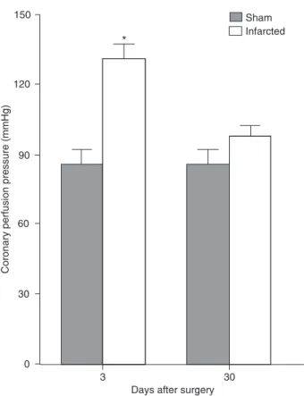
e) and after low and high ouabain concentrations. The baseline coronary perfusion pressure increased 3 days after myocardial infarction and normalized by 30 days (Sham 3 = 88 ± 6; Inf 3 = 130 ± 9; Inf 30 = 92 ± 7 mmHg; P < 0.05). The inotropic response to Ca2+
e and ouabain was reduced at 3 and 30 days after myocardial infarction (Ca2+ = 1.25 mM; Sham 3 = 70 ± 3; Inf 3 = 45 ± 2; Inf 30 = 29 ± 3 mmHg; P < 0.05), while the Frank-Starling mechanism was preserved. At 3 and 30 days after myocardial infarction, ventricular Na+-K+ ATPase activity and contractility were reduced. This Na+-K+ ATPase hypoactivity may modify the Na+, K+ and Ca2+ transport across the sarcolemma resulting in ventricular dysfunction.
Key words: Heart failure; Ouabain; Na+-K+ ATPase; Myocardial infarction; Calcium handling
Introduction
Correspondence: I. Stefanon, Departamento de Ciências Fisiológicas, Universidade Federal do Espírito Santo, Centro de Ciências da Saúde, Av. Marechal Campos, 1468, 29040-093 Vitória, ES, Brasil. Fax: +55-27-3335-7340. E-mail: [email protected]
Research supported by CNPq and FAPES/FUNCITEC.
Received November 28, 2008. Accepted June 26, 2009. Available online September 4, 2009.
Myocardial infarction induces a progressive geometric, structural, and functional remodeling of both ventricles (1-4). Several adaptive changes resulting from the myocardium infarction, including myocyte hypertrophy (5), increased de-position of extracellular matrix components (6), and chamber enlargement (7,8), seem to depend either on hemodynamic disturbances determined by the loss of contractile tissue (9) or on the neurohumoral activation triggered by myocardial ischemia (10-12).
Studies in intact and isolated preparations have sug-gested that contractile abnormalities may depend on changes in transient calcium concentrations (13) and on the decreased Ca2+ sensitivity in the contractile machinery
of the ventricular myocardium (4,14). The influences of the coronary vasculature and its perfusion on the diastolic properties of the heart and on cardiac contraction have
also been well established. Coronary perfusion affects the diastolic ventricular properties through changes in the lengthwise stiffness of the coronary vasculature. Under low oxygen conditions, an increase in perfusion enhances cardiac muscle contractility via the increased oxygen supply (15). However, the best described disturbances in myocardial contractility are intimately dependent on the active transport of ions, namely Ca2+ and Na+, which are
regulated by SERCA and Na+-K+ ATPase. Na+-K+-ATPase
is a heteromeric protein consisting of α and β subunits. While the α subunit contains the amino acids involved in catalytic function, ion transport and cardiac glycoside bind-ing, the function of the β subunit is not yet fully understood, although it is essential for the normal activity of the enzyme and is involved in the transport of the functional Na+-K+
Na+-K+ ATPase at a site formed in the extracellular part of
the catalytic α subunit by the H1-H2, H3-H4, and H5-H6 loops. Reversible interaction of cardiotonic steroids with this site may induce a conformational change in the Na+/K+
ATPase protein. In the active Na+-K+ ATPase, cardiotonic
steroids are fixed leading to the enzyme’s inactivation. In cardiac myocytes, ouabain induces an intracellular increase of Na+ concentration, resulting from inhibition of the Na+
-K+ ATPase, subsequently increasing the intracellular Ca2+
concentration via Na+/Ca2+ exchanger inhibition, causing a
positive inotropic effect (17). Thus, inappropriate regulation of Na+-K+ ATPase activity, which leads to excessive loss of
K+ from the cell and gain of Na+ into the cell, could result in
disturbances in Ca2+ signaling and contractility.
The Na+-K+ ATPase has a binding site for endogenous
cardiac glycosides (18,19). It has been proposed that an increase in endogenous ouabain levels could be involved in the pathogenesis of congestive heart failure (20). Fur-thermore, although inhibition of Na+-K+ ATPase by cardiac
glycosides is well documented and increases in intracel-lular Na+ and Ca2+ have been observed in the presence
of cardiotonic steroids, a direct connection between a reduction in Na+-K+ ATPase activity and contractility in the
heart failure following myocardial infarction (MI) has not been demonstrated.
Since levels of endogenous digitalis-like factor increase in the plasma of humans (18,19)after MI and during heart failure, we designed this study to assess the exogenous effect of ouabain on heart contractility performance early and late after myocardial infarction inrats. Since rodents exhibit an ouabain-insensitive α1-isoenzyme (21), we perfused a
higher concentration of ouabain in the isolated heart (50 nM) than usually observed in human plasma during heart failure (18). Our hypothesis was that a decrease in Na+-K+
ATPase activity could contribute to the reduced myocardial contractility occurring early and late after MI.
Material and Methods
Animals and coronary artery occlusion
Adult male Wistar rats aged 8-12 weeks at the beginning of the study were used. They were housed under controlled temperature and light conditions with free access to stan-dard rat chow and water. Care and use of the laboratory animals were in accordance with NIH guidelines and the local Ethics Committee at EMESCAM (Escola Superior de Ciências da Santa Casa de Misericórdia de Vitória; Protocol #03/2007).
Animals were anesthetized with ether and a thoracotomy was performed at the level of the fourth left intercostal space to exteriorize the heart. The anterior descending branches of the left coronary artery were occluded between the tip of the left atrium appendage and the right ventricle outflow tract with 6-0 mononylon thread (3). After coronary ligature the chest was rapidly closed and spontaneous respiratory
movements were resumed. Most of the animals developed an anterolateral transmural infarction involving 20-40% of the left ventricular surface. Sham-operated rats, used as control, were similarly treated except for the coronary artery ligation. The mortality rate was approximately 30%, with most cases occurring within 30 min of coronary ligation. After recovery from anesthesia, the animals were kept in collective cages in animal care and control facility of the department.
Hemodynamic and left ventricular function
Surgicalinstrumentation was performed using aseptic surgical procedures. Rats were anesthetized with chloral hydrate (0.3 g/kg, ip) and the carotid artery was cannulated with a polyethylene catheter (PE 50, Clay-Adams, USA), and filled with heparin in saline (50 U/mL). The cannulas were advanced into the left ventricular chamber and connected to pressure transducers (TSD 104A) and a Biopac System (MP 100; USA) to measure ventricular pressure. After sta-bilization the pressure waves were recorded for 1 to 3 min. The following parameters were analyzed: left ventricular systolic and end-diastolic pressures (LVSP and LVEDP, respectively), their first derivative (positive and negative, dP/dtmax and dP/dtmin, respectively) and heart rate.
Isolated heart perfusion
Groups of infarcted and Sham-operated rats were assessed at 3 and 30 days after surgery. The rats were treated with heparin (50 IU, ip) and anesthetized with pen-tobarbital sodium (40 mg/kg body weight, ip). Ten minutes later, the heart was excised, mounted in an isolated organ chamber and perfused according to the Langendorff tech-nique at a constant flow (10 mL/min) with Krebs-Henseleit bicarbonate-buffered solution containing: 120 mM NaCl, 5.4 mM KCl, 1.2 mM CaCl2, 2.5 mM MgSO4, 1.2 mM Na2SO4,
2.0 mM NaH2PO4, 24 mM NaHCO3, and 11 mM glucose.
This solution was filtered through a 0.8-µm nitrocellulose filter (Millipore; Sigma, USA) and aerated with 5% CO2 and
95% O2, pH 7.4, at 35°C. The left atrium was opened and a
soft distensible balloon mounted at the tip of a rigid plastic tube was inserted into the left ventricular cavity through the atrioventricular valve. The balloon was connected via a Y piece to a pressure transducer (Gould P23XL, USA) and to a syringe so that the diastolic pressure of the left ventricle cavity could be adjusted to predetermined values by injecting water into the balloon. The developed pressure was registered with a chart recorder (FUNBEC, RG 300, Brazil).
904 I. Stefanon et al.
pressure transducer (Gould P23XL) to the inflow of the aortic pressure tube. Since coronary flow was maintained constant throughout the experiment (10 mL/min), changes of CPP depended on changes of coronary resistance. Ventricular function curves were obtained by measuring the LVSP de-veloped while diastolic pressure was increased from 0 to 30 mmHg in steps of 5 mmHg. This curve was obtained under control conditions and repeated 30 min after the addition of 50 nM ouabain (Sigma) to the perfusion solution. In order to determine the inotropic response to Ca2+, the diastolic
pressure was set at 5 mmHg and the steady-state LVSP was measured at increasing Ca2+ concentrations (0.62,
1.25, and 2.5 mM) before and after the addition of 50 nM ouabain (30 min) to the perfusion solution.
At the end of experimental protocol, the inotropic re-sponse to increasing concentrations of ouabain (1, 10, and 100 µM) was tested in an environment with an extracel -lular Ca2+ concentration of 1.25 mM. The concentration of
ouabain used here was significantly higher than the plasma concentrations of ouabain or the dose-delivered pharmaco-logical treatment. It was only used as a tool to understand the functional participation of Na+-K+ ATPase during the
initial and late phase after myocardial infarction.
During the protocols performed in this study, experi-ments without ouabain were used as the control condition to be compared with ouabain perfusion. The only exception was in Figure 3. In this case, the CPP and LVSP responses were analyzed in the presence of increasing ouabain con-centrations (1, 10, and 100 µM). In these experiments, we considered the low dose of ouabain as a baseline condition (control, using 50 nM ouabain). The justification for this was our interest in determining the effects of the increased ouabain concentration compared to the lowest concentration (50 nM ouabain) that is closest to the endogenous plasma ouabain concentration.
Determination of infarct size
The presence of infarction was initially determined by gross observation of the heart. At the end of each experi-ment the ventricles were separated, blotted and weighed and their wet weights were normalized to the respective body weight. The infarct size 3 days after coronary ligation was evaluated by staining four coronal sections of the left ventricle in 1% tetrazolium chloride for 5 min (22). After staining, the infarcted tissue was dissected from the surviv-ing muscle under microscopic visualization. Both fragments were blotted and weighed and the scar size was reported as the percentage of the left ventricle not stained during the staining procedure. In the groups sacrificed 30 days after surgery, the fibrous scar was separated from the remaining left ventricle muscle. The outlines of both fragments were drawn on graph paper and the areas were measured using the cross-point method (3). The interventricular septum was considered to be part of the left ventricle. Only hearts with a fibrotic scar covering more than 30% and less than 40%
of the left ventricular endocardial surface were included in this study.
Na+-K+ ATPase activity
The enzymatic material was extracted from the left ventricle by the method of Velema and Zaagsma (23) with the following modifications: the left ventricle was homog -enized in a solution containing 20 mM Tris-HCl and 1 mM EDTA, pH 7.5. The homogenized tissue was centrifuged at 10,000 g for 20 min and the precipitate was discarded. The same volume of solution was added to the supernatant and centrifuged at 10,500 g again for 1 h. The precipitate was resuspended in 20 mM Tris-HCl and 1 mM EDTA, pH 7.5, in a final volume of 400 µL. Na+-K+ ATPase activity was
assayed by measuring Pi liberation from 3 mM ATP in the presence of 125 mM NaCl, 3 mM MgCl2, 20 mM KCl and
50 mM Tris-HCl, pH 7.5. The enzyme was preincubated for 5 min at 37°C and the reaction was initiated with the addition of ATP. Incubation times and protein concentration were chosen in order to ensure that the measurements were made in the linear part of the reaction. The reaction was stopped by the addition of 200 µL 10% trichloroacetic acid. Controls containing added enzyme preparation after the addition of trichloroacetic acid were used to correct for nonenzymatic hydrolysis of the substrate. All samples were run in triplicate. Specific activity is reported as nmol Pi released·min-1·mg protein-1 unless otherwise stated. The
specific activity of the enzyme was determined in the pres-ence and abspres-ence of 50 nM ouabain. Protein concentration was measured by the method of Bradford (24) using bovine serum albumin as standard.
Statistical analysis
Data are reported as means ± SEM. Continuous vari-ables were compared by one-way and two-way ANOVA followed by the Tukey test to determine differences between groups. Relationships between variables were evaluated by univariate linear regression analysis and comparisons between the two groups were made using the Student t -test. A P value of < 0.05 was considered to be statistically significant.
Results
increased LVEDP. Subsequent hypertrophic growth was observed in both chambers. In the present study, coronary artery occlusion did not affect normal body growth because body weight was not different among the groups. The left ventricle weight-to-body weight ratio did not change in either infarct groups; however, right ventricle hypertrophy was observed at 30 days after coronary occlusion.
Coronary perfusion pressure and ventricular performance
Figure 1 shows that the hearts of the 3-day infarcted (Inf) group perfused with normal Krebs solution (1.25 mM Ca2+)
presented increased CPP when compared to the respective Sham group. One interesting feature is that CPP tended to decrease towards control values in the 30-day group. This behavior was maintained at lower (0.62 mM) and higher (2.5 mM) extracellular Ca2+. In the Sham groups, CPP was
constant throughout the observation period.
CPP was measured after extracellular Ca2+ was
in-creased from 0.62 to 2.5 mM for both groups at 3 and 30 days after left coronary occlusion (Figure 2, top). The baseline CPP was higher in the Inf group at 3 days but re-turned to normal at 30 days (Sham 3 = 88 ± 6; Inf 3 = 130 ± 9; Inf 30 = 92 ± 7 mmHg; P < 0.05). Coronary perfusion pressure variation produced by an increase in extracellular Ca2+ from 0.62 to 2.5 mM before and after 50 nM ouabain
perfusion was similar between groups at 3 days (Sham before ouabain: 15.4 ± 4.3 vs after ouabain: 43.6 ± 8.5 mmHg; Inf before ouabain: 32.6 ± 6 vs after ouabain: 38.4 ± 10 mmHg) and at 30 days (Sham before ouabain: 12.5 ± 8 vs after ouabain: 33.8 ± 9 mmHg; Inf before ouabain:
24.6 ± 3 vs after ouabain: 40.6 ± 4 mmHg) as determined by two-way ANOVA.
Figure 2 shows that extracellular calcium enhanced the LVSP at 3 and 30 days after infarction or Sham operation. However, the infarcted hearts developed a smaller LVSP compared to the Sham group. Second implication of these results is that the inotropic response to the increase in extracellular Ca2+ was depressed in both infarcted groups.
The reduced ventricular performance was observed by a smaller increment in the relative LVSP when extracellular Ca2+ was increased from 0.62 to 2.5 mM. The percent
changes in LVSP increased by 300% in the Sham group at 3 days compared to 138% in the Inf group. A similar reduc-tion of the inotropic response to Ca2+ was also observed
at 30 days.
Ouabain responsiveness
Ouabain (50 nM) perfused for 30 min did not produce an inotropic effect in the Inf group but altered the ventricular performance of the Sham groups at 3 and 30 days (Figure 2, bottom). In the Sham groups, 3 and 30 days after MI, the increment of extracellular Ca2+ concentration increased
LVSP. However, when 50 nM ouabain was used, a posi-tive inotropic effect was obtained only at 0.62 mM Ca2+. At
1.25 mM, the perfusion of Ca2+ plus ouabain produced an
increase in LVSP similar to that observed in the ouabain-free condition. At 2.5 mM Ca2+ plus ouabain, the increase
of the LVSP was less than that observed in the absence of ouabain.
Figure 3 (top) presents the changes in CPP at increas-ing concentrations of ouabain. In both groups only 100
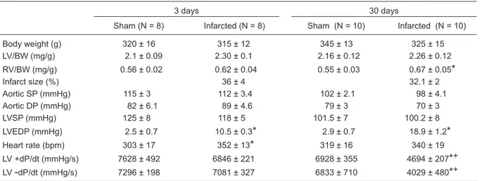
Table 1. Characteristics of the animal groups.
3 days 30 days
Sham (N = 8) Infarcted (N = 8) Sham (N = 10) Infarcted (N = 10)
Body weight (g) 320 ± 16 315 ± 12 345 ± 13 325 ± 15
LV/BW (mg/g) 2.1 ± 0.09 2.30 ± 0.1 2.16 ± 0.12 2.26 ± 0.12
RV/BW (mg/g) 0.56 ± 0.02 0.62 ± 0.04 0.55 ± 0.03 0.67 ± 0.05
*
Infarct size (%) 36 ± 4 32.1 ± 2
Aortic SP (mmHg) 115 ± 3 112 ± 3.4 102 ± 2.1 98 ± 4.1
Aortic DP (mmHg) 82 ± 6.1 89 ± 4.6 79 ± 3 70 ± 3
LVSP (mmHg) 125 ± 8 118 ± 5 101.5 ± 7 100.2 ± 8
LVEDP (mmHg) 2.5 ± 0.7 10.5 ± 0.3
*
2.9 ± 0.7 18.9 ± 1.2*
Heart rate (bpm) 303 ± 17 352 ± 13
*
319 ± 16 340 ± 19LV +dP/dt (mmHg/s) 7628 ± 492 6846 ± 221 6928 ± 355 4694 ± 207
*
+LV
-
dP/dt (mmHg/s) 7296 ± 198 7081 ± 327 6833 ± 710 4029 ± 480*
+906 I. Stefanon et al.
µM ouabain increased CPP. Although at 3 days CPP was larger in the Inf than in the Sham group, the CPP increase occurred in parallel in both groups. Even at 30 days, when the baseline CPP was similar, ouabain produced the same CPP response in both groups. Figure 3 (bottom) shows the changes in LVSP as ouabain concentration was increased in the perfusion solution from 50 nM to 100 µM. In both groups the inotropic effect of ouabain was observed only at 100 µM. The baseline LVSP in the Inf group was lower compared to the Sham group and remained lower when the ouabain concentration was increased. The absolute change of LVSP in response to 100 µM ouabain was smaller in the Inf groups (Sham 3 days = 25.7 ± 3.5 vs Inf 3 days = 15.3 ± 2.5 mmHg; Sham 30 days = 26.2 ± 5 vs Inf 30 days = 11.3 ± 2.4 mmHg; P < 0.05).
Frank-Starling mechanism of the heart
The Frank-Starling mechanism of the heart was used to determine the intrinsic inotropic response of the cardiac muscle after MI (Figure 4). To evaluate the effects of the increase of LVEDP, the experiments were performed using
0.62 mM Ca2+. This Ca2+ concentration was used because,
at least at 3 days, both the Sham and Inf groups presented similar LVSP values at the beginning of the ventricular function curve protocol (Figure 2). Thus, any difference in LVSP achieved after increases of diastolic pressure could be interpreted as being dependent on the ventricular stretch. At 3 days after surgery, under low Ca2+ concentration, the
increase of diastolic pressure from 0 to 30 mmHg produced a similar inotropic effect in both groups. Ouabain (50 nM) perfused for 30 min produced a further parallel upward
Figure 1. Changes in coronary perfusion pressure in Sham and infarcted hearts 3 and 30 days after left coronary occlusion during perfusion with 1.25 mM extracellular Ca2+. Diastolic pressure was maintained constant at 5 mmHg. Data are reported as means ± SEM for 8-10 rats in each group.
*
P < 0.05 compared to 3-day Sham group (two-way ANOVA and Tukey test).Figure 2. Changes in coronary perfusion pressure (CPP) and left ventricular systolic pressure (LVSP) in response to extracellular Ca2+ in Sham-operated rat hearts before (filled circles) and af -ter 50 nM ouabain (open circles), and infarcted rat hearts before
(filled squares) and after 50 nM ouabain (open squares) at 3 and
Figure 3. Coronary perfusion pressure (CPP) and left ventricle isovolumic systolic pressure (LVSP) response to increased
oua-bain concentration (1, 10 and 100 µM) at 3 and 30 days after
surgery. C = control using 50 nM ouabain. Diastolic pressure was maintained constant at 5 mmHg at 0.62 mM extracellular Ca2+. In the infarcted group, baseline CPP was higher than in the Sham group at 3 days, but equal at 30 days. Data are reported as means ± SEM for 8-10 rats in each group.
*
P < 0.01 for infarcted vs Sham; +P < 0.05 compared to its own control (two-way ANOVA plus Tukey test).Figure 4.Top, Left ventricle systolic pressure curves (LVSP) obtained at different diastolic pressures (DP). Sham-operated
before (filled circles) and after 50 nM ouabain (open circles), and infarcted hearts before (filled squares) and after 50 nM
908 I. Stefanon et al.
displacement of the ventricular function curve only in the Sham group. However, at 30 days LVSP was reduced in the Inf group for all diastolic pressures in the presence and absence of 50 nM ouabain. The relative increase of LVSP (Figure 4, bottom) was calculated considering the values obtained at 5 mmHg as 100%. The normalized ventricular function curves were similar in both groups at 3 and 30 days after surgery.
CPP remained unchanged during the diastolic pressure increase for both groups, even during ouabain perfusion (50 nM).
Na+-K+ ATPase activity
Maximal Na+-K+ ATPase activity, estimated as nmol Pi
released·min-1·mg protein-1 in the presence and absence of
50 nM ouabain was reduced after MI (Figure 5). At 3 days, the activity was 69% lower in Inf animals compared to Sham animals, whereas at 30 days the activities were 60% lower in Inf animals compared to Sham animals.
Discussion
In the present study, we analyzed the effects of ouabain on coronary resistance and myocardial contractility at 3 and 30 days after MI in rats. Coronary resistance increased 3 days after MI at all calcium concentrations and normalized by 30 days. Ouabain did not affect these responses. MI reduced the force developed, the calcium responsiveness and the inotropic effects of ouabain, while the Frank-Starling mechanism of the heart was preserved. It is well-known that ouabain increases LVSP by inhibiting Na+-K+ ATPase, which
leads to an increase of intracellular Na+ and, indirectly, in
intracellular Ca2+. Different Na+-K+ ATPase subunit isoforms
are present in the heart. Na+-K+ ATPase-α
1 is predominant,
although there are variable amounts of Na+-K+ ATPase-α 2 in
the adult ventricular myocytes of most species (25). Despa and Bers (25) demonstrated that the functional density of Na+-K+ ATPase-α
2 is 4.5 times higher in the T-tubules
than in the sarcolemma, whereas Na+-K+ ATPase-α 1 is
almost uniformly distributed between the T-tubules and sarcolemma. The intracellular Ca2+ increase is believed
to be the mechanism responsible for the enhancement of myofibrillar force development.
In this study, we indirectly measured the coronary resis-tance after MI to extracellular Ca2+, evaluated before and
after a low ouabain concentration. Our study demonstrated that the baseline CPP was higher at 3 days but normalized at 30 days after left coronary occlusion (Figure 1). We raised two possibilities to account for this. First, a high CPP in the acute phase could be associated with a reduction in the coro-nary resistance distribution. If we assume that the corocoro-nary circulation is a parallel circuit, then the total resistance is equal to the individual resistance in each resistive element divided by the element number. Thus, the occlusion of one of these elements, for the same flow, would increase the
total resistance. In order to test this hypothesis, we occluded the coronary circulation in vitro in a similar way to that used to produce MI. CPP increased by approximately 15 mmHg (results not shown), while the average CPP increment at 3 days after MI was approximately 25 mmHg. Thus, the increase in total coronary resistance could contribute, at least in part, to the high CPP in the acute phase after MI. However, after 30 days, the CPP was virtually normal and at 90 days it was normal (results not shown). This normal-ization could be due the angiogenesis process installed at this time (26). An increase in capillary density and in the capillary/myocyte ratio in the surviving myocardium might explain the CPP normalization. The second possibility arises from a change in coronary resistance. We subsequently tested the influence of extracellular Ca2+ and ouabain on
coronary resistance. In our studies, the elevation of ex-tracellular Ca2+ from 0.62 mM to 2.5 mM increased CPP
in all groups, although the increase was higher in the Inf group when compared with the Sham group. In contrast, a decreased contractile response to the same extracellular Ca2+ concentration was observed in the left ventricular at
3 and 30 days after MI. A low concentration of ouabain did not affect coronary resistance at 3 days and did not seem to affect coronary resistance under the conditions studied here. Another point to consider is that coronary pressure-flow relationships are strongly determined by autoregulation. Furthermore, flow reserve depends on autoregulation, and the supply-to-demand ratio assumes that flow is related to muscle metabolism and is negligible in systole. Also, CPP depends on vasomotor tone and cardiac contractility and thus on the mechanical properties of the vasculature and the cardiac muscle (15).
In the present study, Na+-K+ ATPase activity was reduced
at 3 and 30 days after MI compared to Sham-operated animals. We also observed a large increase in Na+-K+
ATPase activity in the Sham-operated animals compared to control. Our interpretation of this phenomenon is based on the fact that the control group was used in our study as a non-operated group of rats, although the best control for an infarction is the Sham group. The reason why a Sham group is widely used as a control for MI is the fact that these animals undergo the entire surgical procedure without having the coronary artery occluded. After 3 days the animals may present some inflammatory response due to the surgical procedure, which would be absent in the control group. Thus, Sham animals are a better control for MI than rats not subjected to a surgical procedure (control). The inflammatory response may induce a slight increase in cardiac Na+-K+ ATPase activity since during the
inflam-matory response proinflaminflam-matory molecules may activate enzymes. An increase in vascular mRNA for inducible nitric oxide synthase was demonstrated in the acute phase of MI (27). In fact, results from our laboratory have demonstrated that the inducible nitric oxide synthase isoform was not detected in the tail artery of control rats not subjected to surgery (27), but was increased in Sham animals 3 days after surgery (28). Another study demonstrated that pros-taglandins, key inflammatory mediators, induce vascular smooth muscle relaxation by stimulating Na+-K+ ATPase
activity (29). Thus, the increase of cardiac Na+-K+ ATPase
activity early in the Sham group could be associated with the inflammatory response following the surgery.
The inhibition of Na+-K+ ATPase by glycosides initiates
an increase in the force dependent on the decrease in Ca2+ extrusion by the Na+-Ca2+ exchanger, which in turn
increases the cellular content of Ca2+ (30). In this study,
ouabain induced an increase in the contraction force only in the presence of low extracellular calcium (0.62 mM). In the presence of high concentrations of extracellular calcium, ouabain induced a reduction in force in the Sham group but not in the Inf group. It is possible that in the Sham group the ouabain-induced negative inotropic effect in the presence of high intracellular calcium could be due to the condition of calcium overload as previously demonstrated in rat heart in the presence of high calcium concentrations (31,32). However, the ouabain-induced negative inotropic effect on high intracellular calcium was not seen in the Inf group, a fact probably due to a reduced Na+-K+ ATPase
activity in these animals. It has been well established that length-dependent positive inotropic effects on the myocar-dium are dependent on the myofibrillar responsiveness as well as on the internal Ca2+ handling after each
excitation-contraction cycle. The positive inotropic response induced by increased left ventricle diastolic pressure did not change after ouabain perfusion in the Inf group at 3 and 30 days.
It is possible that myocytes from the Inf group were not able to properly use the available intracellular Ca2+, which
could be due to a reduction of Ca2+ responsiveness and
impairment of Na+-K+ ATPase activity. This hypothesis is
corroborated by our previous results showing a reduced left ventricular Ca2+-responsiveness after MI. This has
been explained by a change in intracellular Ca2+ handling
(31) or may be due to a reduction in myofilament intracel-lular Ca2+ responsiveness (14,33). This can be explained
by the reduction of myocardial ouabain binding sites and Na+-K+ ATPase activity observed after experimental MI (34)
after heart failure in dogs (35) and in rats (36). Messenger RNA levels of phospholemman, a member of the FXYD family of small single span membrane proteins with puta-tive ion-transport regulatory properties, were increased in postinfarction (MI) rat myocytes (37). Indeed, it has been proposed that, in addition to reduced expression of Na+-K+
-ATPase (25,30,34-36), overexpression of phospholemman may also account for the depressed Na+-K+ ATPase activity
observed in post-infarction animals (37).
These results were different in failing human hearts, where increased inotropic response and toxic effects of ouabain have been observed (38). Furthermore, in-creased plasma levels of endogenous ouabain have been described as a consequence of MI (18) and have been interpreted as a homeostatic response to the diminished cardiac output of patients with heart failure. It is probable that in these situations the physiological role of ouabain is global rather then limited to the myocardium. This may explain why a tissue distal from the heart is also affected. This is the case for skeletal muscle from rats subjected to MI in which ouabain sites were reduced even though their affinity was not different (36,39). Furthermore, our results demonstrate that the inotropic response to ouabain was Ca2+-dependent in the Sham group. The positive inotropic
response in the Sham group was only seen in the presence of low extracellular Ca2+ concentrations. This is considered
to be a pharmacological action of digitalis. However, when extracellular Ca2+ was increased to 2.5 mM, a reduction in
the positive inotropic effect was detected (Figure 2). This situation suggests a condition known as calcium overload, in which the contraction force is reduced by a mechanism involving reduced ATP production in myocytes (40). In our study some infarcted hearts (data not shown) did not survive perfusion with a high extracellular Ca2+
concentra-tion associated with 50 nM ouabain, probably due to the development of calcium overload.
In conclusion, the reduced ventricular Na+-K+ ATPase
910 I. Stefanon et al.
References
1. Pfeffer MA, Pfeffer JM, Fishbein MC, Fletcher PJ, Spadaro J, Kloner RA, et al. Myocardial infarct size and ventricular function in rats. Circ Res 1979; 44: 503-512.
2. Anversa P, Beghi C, Kikkawa Y, Olivetti G. Myocardial infarc-tion in rats. Infarct size, myocyte hypertrophy, and capillary growth. Circ Res 1986; 58: 26-37.
3. Mill JG, Stefanon I, Leite CM, Vassallo DV. Changes in per-formance of the surviving myocardium after left ventricular infarction in rats. Cardiovasc Res 1990; 24: 748-753. 4. Stefanon I, Martins MA, Vassallo DV, Mill JG. Analysis of
right and left ventricular performance of the rat heart with chronic myocardial infarction. Braz J Med Biol Res 1994; 27: 2667-2679.
5. Capasso JM, Li P, Zhang X, Anversa P. Heterogeneity of ventricular remodeling after acute myocardial infarction in rats. Am J Physiol 1992; 262: H486-H495.
6. Cleutjens JP, Verluyten MJ, Smiths JF, Daemen MJ. Colla-gen remodeling after myocardial infarction in the rat heart. Am J Pathol 1995; 147: 325-338.
7. Fletcher PJ, Pfeffer JM, Pfeffer MA, Braunwald E. Left ventricular diastolic pressure-volume relations in rats with healed myocardial infarction. Effects on systolic function. Circ Res 1981; 49: 618-626.
8. Pfeffer JM, Pfeffer MA, Mirsky I, Steinberger CR, Braunwald E. Progressive ventricular dilatation and diastolic wall stress with myocardial infarction and failure. Circulation 1982; 66: II-66.
9. Goldsmith SR, Francis GS, Cowley AW Jr, Goldenberg IF, Cohn JN. Hemodynamic effects of infused arginine vaso-pressin in congestive heart failure. J Am Coll Cardiol 1986; 8: 779-783.
10. Gottlieb SS, Rogowski AC, Weinberg M, Krichten CM, Hamilton BP, Hamlyn JM. Elevated concentrations of en-dogenous ouabain in patients with congestive heart failure. Circulation 1992; 86: 420-425.
11. McAlpine HM, Morton JJ, Leckie B, Rumley A, Gillen G, Dargie HJ. Neuroendocrine activation after acute myocardial infarction. Br Heart J 1988; 60: 117-124.
12. Hodsman GP, Kohzuki M, Howes LG, Sumithran E, Tsunoda K, Johnston CI. Neurohumoral responses to chronic myocar-dial infarction in rats. Circulation 1988; 78: 376-381. 13. Litwin SE, Morgan JP. Captopril enhances intracellular
calcium handling and beta-adrenergic responsiveness of myocardium from rats with postinfarction failure. Circ Res 1992; 71: 797-807.
14. Li P, Hofmann PA, Li B, Malhotra A, Cheng W, Sonnenblick
EH, et al. Myocardial infarction alters myofilament calcium
sensitivity and mechanical behavior of myocytes. Am J Physiol 1997; 272: H360-H370.
15. Westerhof N, Boer C, Lamberts RR, Sipkema P. Cross-talk between cardiac muscle and coronary vasculature. Physiol Rev 2006; 86: 1263-1308.
16. Schwinger RH, Bundgaard H, Muller-Ehmsen J, Kjeldsen K. The Na, K-ATPase in the failing human heart. Cardiovasc Res 2003; 57: 913-920.
17. Schoner W, Scheiner-Bobis G. Endogenous and exogenous cardiac glycosides and their mechanisms of action. Am J Cardiovasc Drugs 2007; 7: 173-189.
18. Bagrov AY, Kuznetsova EA, Fedorova OV. Endogenous
digoxin-like factor in acute myocardial infarction. J Intern Med 1994; 235: 63-67.
19. Bagrov AI, Kuznetsova EA, Fedorova OV. [Endogenous digoxin-like factor in myocardial infarction]. Klin Med 1996; 74: 15-17.
20. Bagrov AY, Shapiro JI. Endogenous digitalis: pathophysi-ologic roles and therapeutic applications. Nat Clin Pract Nephrol 2008; 4: 378-392.
21. Blanco G, Mercer RW. Isozymes of the Na-K-ATPase: het-erogeneity in structure, diversity in function. Am J Physiol 1998; 275: F633-F650.
22. Vivaldi MT, Kloner RA, Schoen FJ. Triphenyltetrazolium staining of irreversible ischemic injury following coronary artery occlusion in rats. Am J Pathol 1985; 121: 522-530.
23. Velema J, Zaagsma J. Purification and characterization of
cardiac sarcolemma and sarcoplasmic reticulum from rat ventricle muscle. Arch Biochem Biophys 1981; 212: 678-688.
24. Bradford MM. A rapid and sensitive method for the quantita-tion of microgram quantities of protein utilizing the principle of protein-dye binding. Anal Biochem 1976; 72: 248-254. 25. Despa S, Bers DM. Functional analysis of Na+/K+-ATPase
isoform distribution in rat ventricular myocytes. Am J Physiol Cell Physiol 2007; 293: C321-C327.
26. Xie Z, Gao M, Batra S, Koyama T. The capillarity of left ven-tricular tissue of rats subjected to coronary artery occlusion. Cardiovasc Res 1997; 33: 671-676.
27. Rossoni LV, Salaices M, Miguel M, Briones AM, Barker LA, Vassallo DV, et al. Ouabain-induced hypertension is accom-panied by increases in endothelial vasodilator factors. Am J Physiol Heart Circ Physiol 2002; 283: H2110-H2118. 28. Sartorio CL, Pinto VD, Cutini GJ, Vassallo DV, Stefanon I.
Effects of inducible nitric oxide synthase inhibition on the rat tail vascular bed reactivity three days after myocardium infarction. J Cardiovasc Pharmacol 2005; 45: 321-326. 29. Lockette WE, Webb RC, Bohr DF. Prostaglandins and
potas-sium relaxation in vascular smooth muscle of the rat. The role of Na-K ATPase. Circ Res 1980; 46: 714-720.
30. Schwinger RH, Wang J, Frank K, Muller-Ehmsen J, Brixius K, McDonough AA, et al. Reduced sodium pump alpha1, alpha3, and beta1-isoform protein levels and Na+,K+-ATPase
activity but unchanged Na+-Ca2+ exchanger protein levels in
human heart failure. Circulation 1999; 99: 2105-2112. 31. Sjaastad I, Bentzen JG, Semb SO, Ilebekk A, Sejersted OM.
Reduced calcium tolerance in rat cardiomyocytes after myo-cardial infarction. Acta Physiol Scand 2002; 175: 261-269. 32. Tsuji T, Ohga Y, Yoshikawa Y, Sakata S, Abe T, Tabayashi
N, et al. Rat cardiac contractile dysfunction induced by Ca2+
overload: possible link to the proteolysis of alpha-fodrin. Am J Physiol Heart Circ Physiol 2001; 281: H1286-H1294. 33. Novaes MA, Stefanon I, Mill JG, Vassallo DV. Contractility
changes of the right and ventricular muscle after chronic myocardial infarction. Braz J Med Biol Res 1996; 29: 1683-1690.
34. Maixent JM, Lelievre LG. Differential inactivation of inotropic and toxic digitalis receptors in ischemic dog heart. Molecular basis of the deleterious effects of digitalis. J Biol Chem 1987; 262: 12458-12462.
Reductions of myocardial Na-K-ATPase activity and ouabain binding sites in heart failure: prevention by nadolol. Am J Physiol 1993; 265: H2086-H2093.
36. Barr DJ, Green HJ, Lounsbury DS, Rush JW, Ouyang J. Na+-K+-ATPase properties in rat heart and skeletal muscle
3 mo after coronary artery ligation. J Appl Physiol 2005; 99: 656-664.
37. Zhang XQ, Moorman JR, Ahlers BA, Carl LL, Lake DE, Song J, et al. Phospholemman overexpression inhibits Na+-K+-ATPase in adult rat cardiac myocytes: relevance to
decreased Na+ pump activity in postinfarction myocytes. J Appl Physiol 2006; 100: 212-220.
38. Shamraj OI, Grupp IL, Grupp G, Melvin D, Gradoux N, Kremers W, et al. Characterisation of Na/K-ATPase, its iso-Characterisation of Na/K-ATPase, its iso-forms, and the inotropic response to ouabain in isolated fail-ing human hearts. Cardiovasc Res 1993; 27: 2229-2237. 39. Pickar JG, Mattson JP, Lloyd S, Musch TI. Decreased [3H]
ouabain binding sites in skeletal muscle of rats with chronic heart failure. J Appl Physiol 1997; 83: 323-327.