The Brazilian Journal of
INFECTIOUS DISEASES
w w w . e l s e v i e r . c o m / l o c a t e / b j i d
Original article
Desmodus rotundus
and
Artibeus
spp. bats might present
distinct rabies virus lineages
Willian Oliveira Fahl
a,∗, Pedro Carnieli Jr.
a, Juliana Galera Castilho
a,
Maria Luiza Carrieri
a, Ivanete Kotait
a, Keila Iamamoto
a, Rafael Novaes Oliveira
a,
Paulo Eduardo Brandão
baInstituto Pasteur, São Paulo, SP, Brazil
bDepartment of Preventive Veterinary Medicine and Animal Health, School of Veterinary Medicine, Universidade de São Paulo, São Paulo,
SP, Brazil
a r t i c l e
i n f o
Article history:
Received 18 May 2012 Accepted 26 July 2012
Available online 10 November 2012
Keywords:
Rabies Bat
Artibeusspp.
Desmodus rotundus
Phylogeny
Molecular epidemiology
a b s t r a c t
In Brazil, bats have been assigned an increasing importance in public health as they are important rabies reservoirs. Phylogenetic studies have shown that rabies virus (RABV) strains from frugivorous batsArtibeusspp. are closely associated to those from the vam-pire batDesmodus rotundus, but little is known about the molecular diversity of RABV in
Artibeusspp. The N and G genes of RABV isolated fromArtibeusspp. and cattle infected byD. rotunduswere sequenced, and phylogenetic trees were constructed. The N gene nucleotides tree showed three clusters: one forD. rotundusand two forArtibeusspp. Regarding putative N amino acid-trees, two clusters were formed, one forD. rotundusand another forArtibeus
spp. RABV G gene phylogeny supported the distinction betweenD. rotundusandArtibeusspp. strains. These results show the intricate host relationship of RABV’s evolutionary history, and are invaluable for the determination of RABV infection sources.
© 2012 Elsevier Editora Ltda. All rights reserved.
Introduction
Rabies is a worldwide neglected fatal encephalitis,1,2 which
is listed amongst the ten major infectious causes of human deaths worldwide, estimated at 55,000 per year.3This infection
is efficiently preventable by vaccination,4but treatment costs
of rabid or exposed patients, diagnostic procedures, and vac-cines make it a significant challenge for public health systems in endemic countries.5,6
The disease is caused by rabies virus (RABV) (fam-ily Rhabdoviridae, genus Lyssavirus),7 an enveloped virus
with a length of 110-250 nm and a diameter of 75 nm,
∗ Corresponding autho at:Av. Paulista 393, 01311-001, São Paulo, SP, Brazil. E-mail address:[email protected](W.O. Fahl).
usually transmitted by the saliva of an infected mammal. The RABV genome has a negative-stranded non-segmented ssRNA with 11,932-nucleotides that encodes the five structural proteins N (nucleoprotein), P (phosphoprotein), M (matrix), G (envelope glycoprotein), and L (large, dependent RNA-polymerase).8,9Between 2004 and 2005, 62 people died in the
Brazilian Amazon of rabies transmitted byDesmodus rotundus
vampire bat, a primary reservoir of rabies in Latin America.10,11
Regarding frugivorous bats of the genusArtibeus, rabies has been reported in the species A. fimbriatus, A. jamaicensis, A. lituratus, and A. planirostris.12 Artibeus spp. bats have been assigned an increasing importance in public health, as they are considered a rabies reservoir for humans in urban areas
in Brazil, which is aggravated by the increasing population of these bats and the fact that they share roosts with D. rotundus.12–14
From 2003 to 2008, the Instituto Pasteur in Brazil tested 18,007 non-hematophagous bats for rabies, 252 of which were found to be positive. Also, from 2005 and 2007, 56 out of 160 non-hematophagous bats that tested positive for rabies were classified asA. lituratus.
Phylogenetic studies based on the N gene have suggested that RABV lineages fromArtibeussp. are not divergent from those fromD. rotundus,15 all belonging to the same genic16
and antigenic variant 3 (AgV3).17Nonetheless, these studies
have provided only inconclusive results, as they were based on a very restricted sampling regarding geographic area and sample number, mainly in the case ofArtibeusspp.In addi-tion, these studies were based on a single gene sequences for phylogenetic reconstructions.
The ability to determine the source of infection and the epidemiology of rabies cycles are paramount for accurate decision-making in public health, mainly regarding vacci-nation strategies and animal population control. This study aimed to evaluate the possibility of distinction between RABV genetic lineages related toD. rotundusandArtibeusspp. bats based on N and G genes sequences.
Materials and methods
Rabies virus strains
TwentyArtibeusspp. RABV strains were obtained from first-passage isolates in mice inoculated with 20% suspensions ofA. lituratusandArtibeusspp. central nervous systems (CNS); 15D. rotundus-related strains were obtained directly from naturally infected cattle brain tissues (Table 1). These 35 strains were collected in nine municipalities from São Paulo State, South-eastern Brazil (Fig. 1), between 2004 and 2005. All samples were diagnosed positive for rabies by direct immunofluorescence test (DIFT) targeted to the viral nucleoprotein.18
Fig. 1 – A map of São Paulo State (minor map) and Brazil (larger map), showing the cities (in black) were the 35 bats used in this study were collected. A) Catanduva, B) Ribeirão Preto, C) Altinópolis, D) Santo Antônio da Alegria, E) Monte Mor, F) Garc¸a, G) Marília, H) Platina, and I) Paraguac¸ú Paulista.
The nucleoprotein and the glycoprotein sequences gener-ated in this study have been assigned the GenBank accession numbers JF682392 - JF682426 and JF682427-JF682461, respec-tively.
Amplification and sequencing of the nucleoprotein and glycoprotein genes
Total RNA from the 35 RABV isolates CNS samples tested and the from positive and negative controls were extracted with TRIzolTM (Invitrogen – Carlsbad, CA, USA) method, following
the manufacturer’s instructions. The challenge virus standard (CVS) fixed strain of RABV isolated in mice brain and nucle-ase free-water were used as positive and negative controls, respectively.
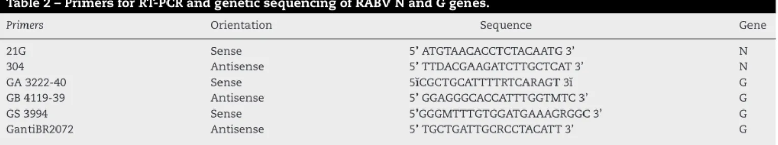
Reverse transcription polymerase chain reaction (RT-PCR) to partially amplify the N and G genes was performed accord-ing to the protocol described by Carnieli et al.,19 using the
primers described by Orciari et al.20for the N gene and those
described by Sato et al.21for the G gene (Table 2). The PCR
products were purified from the PCR reactions using the QIAquickTMGel Extraction KitTM(Qiagen – Valencia, CA, USA),
according to the manufacturer’s instructions. The products with nonspecific bands were purified using 1% agarose gel and the QIAquick® kit. After the purification step, the DNA sam-ples were visually quantified in 2% agarose gel with Low Mass DNA Ladder (Invitrogen – Carlsbad, CA, USA), following the manufacturer’s instructions.
The DNA sequencing reaction mixture consisted of 4L
of BigDye 3.1TM(Applied Biosystems – Foster City, CA, USA),
3.2 pmol of both sense and antisense primer for each gene in separate reactions, 30–60 ng of target DNA and DNase-free water to a final reaction of 10L. The reaction was performed
in a Mastercycler Gradient thermal cyclerTM(Eppendorf, NY,
USA) with 35 cycles at 96◦C for 10s, 50◦ C for 5s, and
60◦C for 4 min, with a ramp of 1◦C/s between each
tem-perature. Sequencing reaction products were purified with SephadexTMG-50 fine beads (GE Healthcare Biosciences) in
96-well multiscreen HV plates. After purification, the sequences were resolved in an ABI-3130TMAutomatic Sequencer (Applied
Biosystems – Foster City, CA, USA).
Phylogenetic analyses
To construct the genealogic trees, nucleotide and putative amino acids sequences for the 35 RABV strains of N and G genes were aligned by the CLUSTAL/W multiple alignment algorithm method using the BioEdit program,22and then by
manually checking the alignments for each set of aligned sequences. A score was assigned to each of the nucleotides shown on the electropherograms for each of the sequencing reactions using the online Phred application. Only positions with a Phred score > 20 were used.23The final sequence for
each strain was obtained using the Contig Assembly Program (CAP) in BioEdit v.5.0.922and submitted to BLASTn for
homol-ogy confirmation.
Table 1 – GenBank accession numbers for the reference sequences of N gene and G gene used in phylogenetic analysis in this study showing strain, the specific-host from which the AgV3 RABV lineages were isolated, city, and year in which the samples were obtained.
GenBanknumber Strain City Specific-host Year
N gene G gene
JF682392 JF682427 05/Art3250 Catanduva Artibeusspp. 2005
JF682393 JF682428 05/Art4578 Monte Mor Artibeus lituratus 2005
JF682394 JF682429 05/Art3598 Ribeirão Preto Artibeus lituratus 2005
JF682395 JF682430 05/Art3738 Ribeirão Preto Artibeus lituratus 2005
JF682396 JF682431 05/Art4850 Ribeirão Preto Artibeusspp. 2005
JF682397 JF682432 05/Art4932 Ribeirão Preto Artibeusspp. 2005
JF682398 JF682433 05/Art5459 Ribeirão Preto Artibeus lituratus 2005
JF682399 JF682434 05/Art6734 Ribeirão Preto Artibeus lituratus 2005
JF682400 JF682435 05/Art6956 Ribeirão Preto Artibeusspp. 2005
JF682401 JF682436 05/Art7045 Ribeirão Preto Artibeusspp. 2005
JF682402 JF682437 05/Art7436 Ribeirão Preto Artibeus lituratus 2005
JF682403 JF682438 05/Art8688 Ribeirão Preto Artibeus lituratus 2005
JF682404 JF682439 05/Art8921 Ribeirão Preto Artibeus lituratus 2005
JF682405 JF682440 05/Art10509 Ribeirão Preto Artibeusspp. 2005
JF682406 JF682441 05/Art7270 Paraguac¸ú Paulista Artibeus lituratus 2005 JF682407 JF682442 05/Art7547 Paraguac¸ú Paulista Artibeus lituratus 2005 JF682408 JF682443 05/Art8456 Paraguac¸ú Paulista Artibeus lituratus 2005 JF682409 JF682444 05/Art11206 Paraguac¸ú Paulista Artibeus lituratus 2005
JF682410 JF682446 05/Art8639 Marília Artibeus lituratus 2005
JF682413 JF682445 05/Art7848 Marília Artibeusspp. 2005
JF682411 JF682460 05/Bov6314 Garc¸a Bovine 2005
JF682412 JF682461 05/Bov7535 Platina Bovine 2005
JF682414 JF682447 05/Bov451 Altinópolis Bovine 2005
JF682415 JF682449 05/Bov3924 Altinópolis Bovine 2005
JF682416 JF682451 05/Bov10339 Altinópolis Bovine 2005
JF682417 JF682450 04/Bov8967 Altinópolis Bovine 2004
JF682418 JF682448 04/Bov2196 Altinópolis Bovine 2004
JF682419 JF682452 05/Bov2579 Santo Antônio da Alegria Bovine 2005
JF682420 JF682453 04/Bov3441 Santo Antônio da Alegria Bovine 2004
JF682421 JF682454 04/Bov3833 Santo Antônio da Alegria Bovine 2004
JF682422 JF682455 04/Bov4698 Santo Antônio da Alegria Bovine 2004
JF682423 JF682456 04/Bov6930 Santo Antônio da Alegria Bovine 2004
JF682424 JF682457 04/Bov11044 Santo Antônio da Alegria Bovine 2004
JF682425 JF682458 04/Bov11817 Santo Antônio da Alegria Bovine 2004
JF682426 JF682459 04/Bov11818 Santo Antônio da Alegria Bovine 2004
Additionally, 38 homologous sequences recovered from Gen-Bank were included in the phylogenetic trees of the N and G genes (29 and none, respectively) and European batLyssavirus 1(another specie in theLyssavirus) was used as an outgroup.
The minimum, maximum, and mean nucleotide (with the MCL model) and amino acids (with the Poisson correction) identities for the clusters for the N and G gene sequences were calculated using Excel (©1985–2003 Microsoft Corpora-tion) based on the identity matrices calculated with the BioEdit program. The changes in the amino acids observed in the
samples analyzed were studied using Mega 4.1 (© 1993–2008)24
and BioEdit v.7.0.022programs.
Results
After editing, the N gene was 1,281-nucleotides long, located between nucleotides 68 and 1,350 in relation to the N gene CVS reference strain (GenBank number AF406696) and had a putative amino acid sequence with 427 amino acids.
Table 2 – Primers for RT-PCR and genetic sequencing of RABV N and G genes.
Primers Orientation Sequence Gene
21G Sense 5’ ATGTAACACCTCTACAATG 3’ N
304 Antisense 5’ TTDACGAAGATCTTGCTCAT 3’ N
GA 3222-40 Sense 5ˇıCGCTGCATTTTRTCARAGT 3ˇı G
GB 4119-39 Antisense 5’ GGAGGGCACCATTTGGTMTC 3’ G
GS 3994 Sense 5’GGGMTTTGTGGATGAAAGRGGC 3’ G
61
04/Bov3441
FJ649166.10B
04/Bov2196 04/Bov11818 04/Bov4698 04/Bov11044 04/Bov8967 05/Bov3924 04/Bov6930 05/Bov451 05/Bov2579 05/Bov10339
04/Bov3833 04/Bov11817
05/Art7436 05/Art8688 05/Art7270 05/Art8456 05/Art3738 05/Art8921
GQ160933.1 GQ160953.1 GQ160958.1 GQ160912.1 GQ160951.1 GQ160959.1 GQ160942.1 FJ649181.1 GQ160923.1 GQ160944.1 FJ649161.1 FJ649176.1 GQ160954.1 FJ649180.1 GQ160925.1 FJ649183.1 GQ160943.1
GQ160940.1
05/Art7045 05/Art4850 05/Art8639 05/Art7848 05/Bov6314
GQ160934.1
05/Art4578
AB117972.1
05/Bov7535 05/Art5459
AB117969.1
05/Art7547 05/Art4932
AB117970.1 AB117971.1
05/Art6956 05/Art11206 05/Art6734 05/Art10509
AF406696.1 AB263324.1
AB083798.1 AB263328.1 AB263332.1 EU636794
EU626551
66 79 100
61 99
0.01
Desmodus rotundus
Artibeus sp.
EBLV1
CVS
Dog-related 05/Art3250
05/Art3598
Fig. 2 – RABV N amino acids tree showing clusters specific toDesmodus rotundusandArtibeusspp. bats. The tree was built with the Neighbor-joining distance algorithm and the Poisson correction with 1,000 bootstrap replicates and European bat
Lyssavirus(EBLV-1) as outgroup. The bar represents the number of substitutions per site.
Regarding the G gene, sequences were 1,571-nucleotides long, located between nucleotides 8 and 1,579 in relation to the G gene CVS reference strain (GenBank number FJ979833) and had a putative amino acid sequence with 520 amino acids.
The N gene nucleotide phylogenetic tree showed three clusters for the 35 RABV strains included in the study; one cluster forD. rotundusstrains and two for theArtibeusspp. strains. Regarding N protein amino acids tree, the 35 strains segregated in only two clusters, one forD. rotundusand one formed mainly byArtibeusspp. strains (Fig. 2). Nucleotides and amino acids identities for the N region under analysis between
D. rotundusandArtibeusspp. sequences ranged from 97.4% to 98.7% and 98.1% to 99.7%, respectively.
The G gene nucleotide phylogenetic tree showed two clus-ters for the 35 RABV strains included in the study; one cluster
forD. rotundusstrains and one for theArtibeusspp. strains. Regarding G amino acids tree, the 35 strains segregated in two clusters, one forD. rotundusand one formed byArtibeusspp. strains (Fig. 3). Nucleotides and amino acids identities for the G region under analysis betweenD. rotundusandArtibeusspp. sequences ranged from 97.0% to 99.1% and 96.1% to 99.0%, respectively.
Discussion
EBLV1
CVS
05/Art359805/Art3738
05/Art5459
05/Art6734
05/Art8921
05/Art6956
05/Art4850
05/Art4932
05/Art7436
05/Art7848
05/Art10509
05/Art8456
05/Art11206
AB247423.2
AB110664.1
05/Art7045
05/Art8688
05/Art8639
05/Art7547
05/Bov7535
05/Art3250
05/Art7270
05/Art4578
05/Bov6314
AB247422.2
AB247421.2
04/Bov8967
05/Bov2579
04/Bov11817
04/Bov3833
05/Bov451
05/Bov10339
04/Bov11818
04/Bov4698
04/Bov11044
04/Bov2196
04/Bov6930
05/Bov3924
04/Bov3441
FJ979833.1
AB247410.2
AB247412.2
AB247415
AB247418.2
EU636794.1
EU626551.1
86 100 100 100
85 79
92 96
87
100 70
0.05
45
36
Dog-related
Desmodus rotundus
Artibeus
sp.
investigation, RABV N and G phylogenies of strains recovered from these bats showed the existence of viral lineages that can be accurately attributed toD. rotundusorArtibeusspp. bats, a previously unknown fact.
RABV lineages heterogeneity expressed phylogenetically as host-specific lineages is a widely documented epidemio-logical phenomenon,16,19,25,26and is influenced by geographic
barriers rather than by species barriers only.27 Accordingly,
the twoArtibeus spp. clusters found for N gene sequences might also represent regional sub lineages of RABV, a fact already described forD. rotundusRABV lineages in Brazil.28,29
These observations agree with the proposition that, regarding rabies in bats, host species are as important as geographic variations.30Regarding viral lineages, genetic variations that
occur within a host species are different from those that occur in another, and this variation, coupled with the host’s geo-graphical isolation, may explain the RABV genetic differences reported in this work.31
Seven strains (05/Art7270, 05/Art8456, 05/Art7436, 05/Art8921, 05/Art8688, 05/Art3738, and 08IacriSP3577B) were classified in theArtibeusspp. group for the N nucleotides tree; however they segregated into theD. rotunduscluster of the N amino acids tree. This fact suggests the possibility that
Artibeusspp. strains are still under an adaptation process after the spill-over event fromD. rotundus, as already suggested by Kissi,31who have already experimentally reported this type of
stepped adaptation of RABV. This phenomenon can occur as a consequence of the predominance of synonymous mutations over non-synonymous nucleotides mutations, which leads to greater differences among the nucleotide sequences than among the amino acids sequences.32,33
Regarding the Bov7525 and Bov6314 strains, in cattle, both segregated in theArtibeusspp. RABV cluster; this was unex-pected, as cattle rabies is related to that ofD. rotundusand not to that of frugivorous bats.34,35 The most plausible
explana-tion is that these two strains have been transmitted from an
Artibeusspp. to aD. rotundus, and then from this bat to the cattle in a rare class of spill-over transmission.
Kobayashi et al.,16 analyzing the N gene of the RABV
isolated from frugivorous bats, insectivorous bats, and D. rotundus, reported lineages associated withArtibeusspp. bats (frugivorous),D. rotundus, and insectivorous bats, suggesting that there are species-specific lineages.26However, they have
not provided significant results to distinguish between RABV isolated fromD. rotundusand Artibeusspp., possibly due to a restricted number of samples and sampling area. In the present study, this problem was compensated by the inclu-sion of 35 RABV isolates of bats (20 from Artibeusspp. and 15 fromD. rotundus) from a broad geographical area of São Paulo. In addition, this study also analyzed the complete G gene, which provides a more specific distinction between the genetic sequences.
Different hosts pose different challenges for rabies con-trol. This is more complex in bats due to the large number of species, the different ecologic niches that they occupy, and the impossibility to vaccinate these animals. For instance, the population of D. rotundus in Latin America can be legally controlled with vampiricide anticoagulants applied on cattle or on the bats themselves.34 Conversely,
non-hematophagous bats are under legal protection, and only now
the knowledge of rabies epidemiology in these bats species is increasing.35,36
In this context, the results obtained in this study are valu-able, because based on the partial amino acid sequences for the N gene it is possible to differentiate RABV strains from
Artibeusspp. andD. rotundusfor the purpose of defining the infection sources in molecular epidemiology. These results show the close host relationship of RABV transitions, and have an invaluable application for determining the sources of rabies infections transmitted mainly to dogs and cats in urban cen-ters.
In conclusion, for rabies virus isolates related to frugiv-orous bats of the Artibeus spp. and to the vampire batD. rotundus, the phylogeny based on sequences of the N and G genes shows segregation patterns in genus-specific agreement in each of these bats. Data from this study suggest that a lin-eage of RABV is possibly being established in theArtibeusspp. genus.
Conflict of interest
All authors declare to have no conflict of interest.
Acknowledgements
The authors are grateful to CAPES (W.O. Fahl’s PhD fellowship) and CNPq (P.E. Brandão’s PQ-2 fellowship).
r e f e r e n c e s
1. Fooks AR, Brookes SM, Johnson N, Mcelhinney LM, Hutson AM. European bat lyssaviruses: an emerging zoonosis. Epidem and Infect. 2003;131:1029–39.
2. Dodet B. Preventing the incurable: Asian rabies, experts advocate rabies control. Vaccine. 2006;24:3045–9.
3. World Health Organization. World survey of rabies: n. 34 for the year 1998. Geneva: WHO; 2000.
4. Briggs D, Hanlon CA. World rabies day 7: focusing attention on a neglected disease. Vet Rec. 2007;161:288–9.
5. Kaplan G, Turner GS, Warrel D. Rabies: the facts. 2nded. Oxford; New York: Oxford University Press; 1986. 6. Who Expert Consultation On Rabies, 2004. Geneva:
Switzerland. WHO Expert consultation on rabies: First report. Geneva: WHO; 2005. 87 p.(Technical report series, 931). 7. Fauquet CM, Mayo MA, Maniloff J, Desselberger U, Ball LA.
Virus taxonomy classification and nomenclature of viruses Eighth Report of the International Committee on the Taxonomy of Viruses. Amsterdam: Elsevier Acad Press; 2005. 8. Tordo N, Poch O, Ermine A, Keith G. Primary structure of
leader RNA and nucleoprotein genes of the rabies genome: segmented homology with VSV. Nucl Assim Res.
1986;14:2671–83.
9. Tordo N, Poch O, Ermine A, Keith G, Rougeon F. Walking along the rabies genome: is the large G-L intergenic region a remnant gene? Proc Natl Acad of Sci. 1986;83:3914–8. 10. Da Rosa ES, Kotait I, Barbosa TF, et al. Bat-transmitted human
rabies outbreaks, Brazilian Amazon. Emerg Infect Dis. 2006;12:1197–202.
in Augusto Correa municipality, Brazilian Amazon. Virol. 2008;370:228–36.
12. Sodré MM, Gama AR, Almeida MF. Updated list of bat species positive for rabies in Brazil. Rev Inst Med Trop. 2010;52:75–81. 13. Uieda W, Hayashi MM, Gomes LH, Silva MMS. Espécies de
quirópteros diagnosticados com raiva no Brasil. Bull Inst Pasteur. 1996;1:17–35.
14. Cunha EMS, Lara MCCSH, Nassar AFC, Sodré M, Amaral LVF. Isolamento do vírus da raiva emArtibeus fimbriatusno estado de São Paulo, Brasil. Rev Saude Publica. 2005;39:683–4. 15. Shoji Y, Kobayashi Y, Sato G, et al. Genetic characterization of
rabies viruses isolated from frugivorous bat (Artibeusspp.) in Brazil. J Vet Med Sci. 2004;66:1271–3.
16. Kobayashi Y, Sato G, Shoji Y, et al. Molecular epidemiological analysis of bat rabies viruses in Brazil. J Vet Med Sci. 2005;67:647–52.
17. Albas A, Souza EA, Lourenc¸o RA, Favoretto SR, Sodré MM. Antigen profile of rabies virus isolated from different species of non-hematophagous bats in the region of Presidente Prudente, State of São Paulo. Rev Soc Bras Med Trop. 2009;42:15–7.
18. Dean DJ, Abelseth MK, Atanasiu P. The fluorescent antibody test. In: Meslin FX, Kaplan MM, Koprowski H, editors. Laboratory techniques in rabies. 4thed. Geneva: World Health Organization; 1996. p. 88–95.
19. Carnieli Jr P, Fahl WO, Castilho JG, et al. Characterization of rabies virus isolated from canids and identification of the main wild canid host in northeastern Brazil. Virus Res. 2008;131:33–46.
20. Orciari LA, Niezgoda M, Hanlon CA, et al. Rapid clearance of SAG-2 rabies virus from dogs after oral vaccination. Vaccine. 2001;19:4511–8.
21. Sato G, Itou T, Shoji Y, et al. Genetic and phylogenetic analysis of glycoprotein of rabies virus isolated from several species in Brazil. J Vet Med Sci. 2004;66:747–53.
22. Hall TA. BioEdit: a user-friendly biological sequence alignment editor and analysis program for Windows 95/98/NT. Nucl Acids Symp Series. 1999;41:95–8.
23. Ewing B, Green P. Base-calling of automated sequencer traces using phred. II. Error probabilities. Gen Res. 1998;8:186–94. 24. Tamura K, Dudley J, Nei M, Kumar S. MEGA4: Molecular
Evolutionary Genetics Analysis (MEGA) software version 4.0. Mol Biol Evol. 2007;24:1596–9.
25. Favoretto SR, Carrieri ML, Cunha EMS, et al. Antigenic typing of Brazilian rabies virus samples isolated from animals and humans, 1989-2000. Rev Inst Med Trop. 2002;44:91–5; Kobayashi Y, Sato G, Kato M, et al. Genetic diversity of bat rabies viruses in Brazil. Arch Virol. 2007;152:
1995–2004.
26. Holmes EC, Woelk CH, Kassis R, Bourhy H. Genetic
constraints and adaptive evolution of rabies virus in nature. Virology. 2002;292:247–57.
27. Carnieli Jr P, Castilho JG, Fahl WO, Véras NM, Timenetsky MCST. Genetic characterization of rabies virus isolated from cattle between 1997 and 2002 in an epizootic area in the state of São Paulo, Brazil. Virus Res. 2009;144:215–24.
28. Castilho JG, Carnieli Jr P, Oliveira RN, et al. A comparative study of rabies virus isolates from hematophagous bats in Brazil. J Wildl Dis. 2010;46:1335–9.
29. Oliveira RN, De Souza SP, Lobo RS, et al. Rabies virus in insectivorous bats: implications of the diversity of the nucleoprotein and glycoprotein genes for molecular epidemiology. Virology. 2010;405:352–60.
30. Domingo E, Holland JJ. RNA virus mutations and fitness for survival. Annu Ver Microbiol. 1997;51:151–78.
31. Kissi B, Badrane H, Lavenu A, Tordo N, Brahimi M, Bourhy H. Dynamics of virus quasispecies during serial passages in heterologous hosts. J Genl Virol. 1999;80:2041–50. 32. Wunner HW. Rabies virus. In: Jackson AC, Wunner HW,
editors. Rabies. San Diego: Academic Press; 2007. p. 23–68.
33. Schneider MC, Santos-Burgoa C, Aron J, Munoz B,
Ruiz-Velazco S, Uieda W. Potential force of infection of human rabies transmitted by vampire bats in the Amazonian region of Brazil. Am J Trop Med Hyg. 1996;55:680–4.
34. Ito M, Arai YT, Ito UT, et al. Genetic characterization and geographic distribution of rabies virus isolates in Brazil: identification of two reservoirs, dogs and vampire bats. Virology. 2001;284:214–22.
35. Kotait I, Nogueira Filho VS, Souza MCAM, Carrieri ML, Gomes MN, Peres NF. Manual de controle da raiva dos herbívoros. 9ed. São Paulo/SP: Instituto Pasteur; 2010.