Berberine induces apoptosis via ROS
generation in PANC-1 and MIA-PaCa2
pancreatic cell lines
S.H. Park
1, J.H. Sung
2, E.J. Kim
3and N. Chung
11Division of Biotechnology, College of Life Sciences and Biotechnology, Korea University, Seoul, Korea 2Biomedical Research Institute, Seoul National University Hospital, Seoul, Korea 3Department of Clinical Laboratory Science, Ansan University, Ansan, Korea
Abstract
Pancreatic cancer is the fourth leading cause of cancer death. Gemcitabine is widely used as a chemotherapeutic agent for the treatment of pancreatic cancer, but the prognosis is still poor. Berberine, an isoquinoline alkaloid extracted from a variety of natural herbs, possesses a variety of pharmacological properties including anticancer effects. In this study, we investigated the anticancer effects of berberine and compared its use with that of gemcitabine in the pancreatic cancer cell lines PANC-1 and MIA-PaCa2. Berberine inhibited cell growth in a dose-dependent manner by inducing cell cycle arrest and apoptosis. After berberine treatment, the G1 phase of PANC-1 cells increased by 10% compared to control cells, and the G1 phase of MIA-PaCa2 cells was increased by 2%. Whereas gemcitabine exerts antiproliferation effects through S-phase arrest, our results showed that berberine inhibited proliferation by inducing G1-phase arrest. Berberine-induced apoptosis of PANC-1 and MIA-PaCa2 cells increased by 7 and 2% compared to control cells, respectively. Notably, berberine had a greater apoptotic effect in PANC-1 cells than gemcitabine. Upon treatment of PANC-1 and MIA-PaCa2 with berberine at a half-maximal inhibitory concentration (IC50), apoptosis was induced by a
mechanism that involved the production of reactive oxygen species (ROS) rather than caspase 3/7 activation. Our findings showed that berberine had anti-cancer effects and may be an effective drug for pancreatic cancer chemotherapy.
Key words: Pancreatic cancer; Berberine; Gemcitabine; Apoptosis; Cell cycle arrest
Introduction
Pancreatic cancer is the fourth leading cause of cancer death and is responsible for 6% of all cancer-related deaths worldwide (1,2). Pancreatic cancer is difficult to diagnose in its early stages and generally has a poor prognosis. For all stages combined, the relative 1- and 5-year survival rates for pancreatic cancer are approximately 25% and 6%, respec-tively (1,2). Gemcitabine is the standard treatment after cancer surgery, but the response rate is ,20% (3-5). Thus, the identification of new drugs and the development of improved therapeutic strategies for pancreatic cancer are essential.
Berberine is an isoquinoline alkaloid isolated from a variety of natural herbs such as berberis (Berberis aquifo-lium, B. vulgaris, and B. aristata), Hydrastis canadensis, Phellodendron amurense,Coptis chinensis, andTinospora cordifolia(6-8). Berberine is usually found in bark, stems, rhizomes, and roots and has long been used as both a dye and a medicinal herb in Indian Ayurvedic, Unani (9), and Chinese medicine (10). A large number of studies have shown that berberine possesses a variety of biochemical and
pharmacological properties including antibacterial, antihy-pertensive, anti-inflammatory, antidiabetic, and antioxidative effects (10). Berberine is also known to possess anticancer properties, and it has been reported (10) that these may vary depending on cell type. In this study, we investigated the growth-inhibitory effect of berberine on PANC-1 and MIA-PaCa2 pancreatic cancer cells and found that it affected cell cycle progression and apoptosis. We also observed that berberine induced the generation of reactive oxygen species (ROS), which ultimately facilitated apoptosis. Additionally, we compared the anticancer effects of gemcitabine and berberine by evaluating cellular growth, cell cycle, and apoptosis in two pancreatic cancer cell lines.
Material and Methods
Cell culture
The human pancreatic cancer cell lines PANC-1 and MIA-PaCa2 were obtained from American Type Culture
Correspondence: Namhyun Chung:,[email protected]..
Collection (USA). They were cultured in Dulbecco’s modified Eagle’s medium (DMEM) supplemented with 10% fetal bovine serum, 100 U/mL penicillin, and 100 mg/ mL streptomycin (Gibco, USA). All cells were maintained at 376C in humidified air with 5% CO2.
Treatment with gemcitabine and berberine
PANC-1 and MIA-PaCa2 cells were seeded at a density of 56105cells. Cells were incubated for 72 h with
media containing 10 nM gemcitabine or 15mM berberine
for PANC-1, and 7 nM gemcitabine or 10mM berberine
for MIA-PaCa2. Cell viability was determined with trypan blue dye exclusion assays. Data analyses for half-maximal inhibitory concentration (IC50) were performed
using Microsoft Excel 2010 (Microsoft Inc., USA).
Cell cycle analysis
Cells were collected by treatment with trypsin-EDTA, washed twice with phosphate-buffered saline (PBS), and fixed for at least 4 h by adding ice-cold 70% ethanol (––206C). The ethanol was subsequently removed after centrifugation at 500 g for 5 min, and then cells were washed with PBS and resuspended in PBS. Propidium iodide (PI) staining solution containing PI (50mL/mL in
PBS; Sigma-Aldrich, USA), RNase (1 mg/mL in PBS,
Sigma-Aldrich), and Triton X-100 was added to a fluores-cence-activated cell sorting (FACS) tube in the dark at room temperature. The cell cycle was analyzed by flow cytometry using a FACSCalibur system (BD Biosciences, USA) at excitation/emission wavelengths of 488/617 nm, respectively, and all experiments were performed in triplicate.
Cell apoptosis assay
The percentage of apoptotic cells was analyzed by flow cytometry using an Annexin V assay kit (BD Biosciences) following the manufacturer’s instructions. Briefly, after treatment, cells were harvested with trypsin-EDTA and washed twice in PBS. Cells were then resuspended in 100mL binding buffer, to which 5mL
annexin V-fluorescein isothiocyanate (FITC) and 5mL PI
were added, and then incubated at room temperature for 15 min in the dark. After incubation, 400mL binding buffer
was added, and the percentage of apoptotic cells was analyzed by flow cytometry using a FACSCalibur system.
Caspase 3/7 assay
Cells were seeded in white 96-well plates at densities of 2.56103, 56103, and 16104cells. Cells were then treated
with berberine or gemcitabine, and after 24, 48, or 72 h,
Figure 1. Cell growth rate after treatment of PANC-1 (A,C) or MIA-PaCa2 (B,D) pancreatic cancer cell lines with berberine or gemcitabine. The cells were treated with distilled water (control), berberine (A,B; 1, 5, 7, 10, or 15mM) or gemcitabine (C,D; 0.5, 1, 5,
10, or 50 nM) for 72 h. Growth rate was calculated by trypan blue dye exclusion. Data showed relative cell survival rate as the percentagevsthat of control cells after berberine or gemcitabine treatment. *P,0.05, **P,0.01 versus control (Student’st-test).
caspase 3/7 activities were measured with Caspase-Glo 3/7 assay (Promega, USA) following the manufacturer’s instructions. The caspase 3/7 activity of berberine- and gemcitabine-treated cells was calculated as caspase activity relative to that in untreated cells.
Measurement of ROS
Intracellular ROS levels were determined by measur-ing the oxidative conversion of cell-permeable 29,79 -dichlorofluorescein diacetate (DCFH-DA, Sigma-Aldrich) to fluorescent dichlorofluorescein (DCF) using a multilabel plate reader (Victor3, Perkin Elmer, USA). Cells were treated with berberine or gemcitabine for 24, 48, or 72 h. The cells were washed with PBS and incubated with DCFH-DA at 376C for 30 min. Then, DCF fluorescence distribution was detected by fluorospectrophotometric analysis at an excitation wavelength of 488 nm and an emission wavelength of 535 nm. The fluorescence inten-sity was normalized according to the number of cells.
Statistical analysis
Statistical significance was determined with t-tests using the SPSS software (version 20, IBM Corp., USA). Data are reported as the means±SD of at least three
independent experiments. P,0.05 was considered to be statistically significant.
Results
Growth rate after treatment
In order to determine the effect of berberine on cell growth, PANC-1 and MIA-PaCa2 were treated with 1-15mM berberine for 72 h. As shown in Figure 1A and B,
berberine treatment inhibited cell growth dose-depend-ently in both PANC-1 and MIA-PaCa2 cells. Treatment with 5-15mM (PANC-1) and 1-15mM (MIA-PaCa2)
berberine significantly reduced cell viability. The IC50
values of berberine were approximately 15 and 10mM for
PANC-1 and MIA-PaCa2, respectively.
The growth-inhibitory effects of gemcitabine for PANC-1 and MIA-PaCa2 were determined. Gemcitabine inhibited growth in a dose-dependent manner. The IC50
values were calculated as approximately 10 and 7 nM for PANC-1 and MIA-PaCa2, respectively (Figure 1C and D).
Cell cycle profiles after treatment
Cell cycle progression was examined after treat-ment with berberine. As shown in Figure 2, treattreat-ment of
Figure 2.Cell cycle distribution after treatment of PANC-1 with berberine or gemcitabine. PANC-1 cells were treated with distilled water (control), 15mM berberine, or 10 nM gemcitabine for 72 h. The cell cycle distribution was analyzed by the ModFit LT software and
depicted using histograms (A) and bar plots (B). Cell cycle was analyzed as the percentage of cells at each stage of the cell cycle after DNA staining with PI. Data from a representative experiment (from a total of at least three) are shown. **P,0.01 versus controls (Student’st-test).
PANC-1 cells with 15mM berberine for 72 h resulted in a
significantly higher percentage (61.1±1.7%) of cells in the G1 phase than in the control group (51.1±0.8%), with a corresponding reduction in the percentage of cells in the S phase. Similar results were obtained for MIA-PaCa2 (55.4±1.1%) when treated with 10mM berberine for 72 h
compared to the control group (53.7±0.5%) (Figure 3). These data suggest that inhibition of cell proliferation or induction of cell death in pancreatic cancer cells by berberine is associated with the induction of G1 arrest.
In comparison, Figure 2 shows that treatment of PANC-1 cells with gemcitabine increased the percentage of cells in S phase to 60.1±6.0% and reduced the percentage in the G0/G1 phase to 28.7±4.2%. Gemcitabine-treated MIA-PaCa2 cells had 67.0±2.7% cells in S phase compared to only 30.0±0.4% in the control cells (Figure 3). These data suggest that gemci-tabine inhibited pancreatic cancer cell proliferation by inducing S-phase cell cycle arrest.
Cell apoptosis after treatment
To investigate the apoptotic effect of berberine, PANC-1 and MIA-PaCa2 cells were treated with berberine for 72 h, and apoptotic cells were assessed by staining with
annexin V and PI. Early apoptotic cells are shown in the lower-right quadrant of the scatter plot, and live cells are in the lower-left quadrant (Figure 4). Berberine-induced apoptosis of PANC-1 cells increased by 12.2±1.6% compared to 5.0±1.1% in control cells (Figure 4C). Apoptosis of MIA-PaCa2 cells increased 5.7±0.3% compared to 2.8±1.1% in control cells (Figure 4D). These results showed that berberine induced cell death through apoptosis in both pancreatic cancer cell lines.
Gemcitabine-induced apoptosis of PANC-1 cells was increased to 8.6±2.1% compared to control cells (5.0±1.1%), and apoptosis of MIA-PaCa2 cells was increased to 6.2±1.7% compared to control cells (2.8±1.1%) (Figure 4C and D). The results suggest that the relative apoptosis efficacy of berberine in MIA-PaCa2 cells is roughly equivalent to that of gemcitabine. However, berberine induced more apoptosis in PANC-1 cells than did gemcitabine.
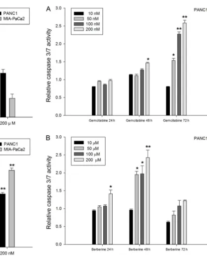
Caspase 3/7 activity after treatment
To determine whether apoptosis after treatment with berberine or gemcitabine was caspase 3/7-dependent, PANC-1 cells were treated with IC50 values of 15mM
berberine and 10 nM gemcitabine. MIA-PaCa2 cells were
Figure 3.Cell cycle after treatment of MIA-PaCa2 with berberine or gemcitabine. MIA-PaCa2 cells were treated with distilled water (control), 10mM berberine, or 7 nM gemcitabine for 72 h. The cell cycle distribution was analyzed by the ModFit LT software and depicted using histograms (A) and bar plots (B). Cell cycle was analyzed as the percentage of cells at each stage of the cell cycle after DNA staining with PI. Data from a representative experiment (from a total of at least three) are shown. *P,0.05, **P,0.01 versus control (Student’st-test).
also treated with IC50values of 10mM berberine and 7 nM
gemcitabine for 72 h. There was no change in relative caspase 3/7 activity, suggesting that the apoptosis was caspase 3/7-independent with IC50 concentrations of
berberine and gemcitabine. Figure 5A shows that when treated with 10, 50, 100, or 200mM berberine, PANC-1
and MIA-PaCa2 cells did not show significant caspase 3/7 activation compared to untreated cells. However, caspase 3/7 activity for PANC-1 cells gradually increased with higher concentrations of gemcitabine. When the gemci-tabine concentration was increased from 10 to 50 nM for a 72-h treatment period, caspase 3/7 activity for MIA-PaCa2 increased abruptly and then gradually decreased
with further increases in gemcitabine concentration (Figure 5B). To better explain this phenomenon, the relative activity was examined over time with varying concentrations of the chemicals. In the case of gemcita-bine, the relative activity with PANC-1 increased gradually with time up to 72 h (Figure 6A). With increasing concentrations of berberine, activity peaked at 48 h and decreased at 72 h (Figure 6B). The relative activity with MIA-PaCa2 was also examined. In the case of gemcita-bine, activity gradually increased up to 72 h. However, in the case of berberine, activity was highest at 24 h and decreased with time (Figure 7). Collectively, these data indicate that the apoptotic response with berberine was
Figure 4.Annexin V staining after treatment with berberine and gemcitabine.A, PANC-1 cells were treated with distilled water (control), 15mM berberine, or 10 nM gemcitabine for 72 h.B, MIA-PaCa2 cells were treated with distilled water, 10mM berberine, or 7 nM gemcitabine for 72 h.C, PANC-1 cells were treated with distilled water, 15mM berberine, or 10 nM gemcitabine for 72 h.D,
MIA-PaCa2 cells were treated with distilled water, 10mM berberine, or 7 nM gemcitabine for 72 h. All cells were stained with
FITC-conjugated annexin V in a buffer containing PI and analyzed by flow cytometry. Data are reported as means±SD from a representative experiment (from a total of at least three). *P,0.05, **P,0.01 versus control (Student’st-test).
much quicker in both cell lines, but especially in MIA-PaCa2. This result also indicates that the apoptotic response induced by caspase 3/7 activity should be assessed at many time points; a single measurement is not sufficient.
Following gemcitabine treatment (Figure 5B), pancrea-tic cancer cells did not show significant caspase 3/7 activation at a lower concentration of gemcitabine (10 nM) for 72 h. However, at higher concentrations (>50 nM) PANC-1 cells exhibited higher caspase 3/7 activation, but to a lower extent than observed for MIA-PaCa2. On the other hand, PANC-1 treated with berberine for 48 h showed higher overall caspase 3/7 activation than MIA-PaCa2 treated with berberine for 24 h (Figures 6B and 7B). These results suggest that caspase 3/7 was activated earlier by berberine than by gemcitabine, and that the mechanism for cell death was caspase 3/7-independent at a lower berberine concentration (IC50). The results in
Figures 6 and 7 also suggest that the cell death mechanism is caspase 3/7-dependent at high concentrations of both berberine (>50mM) and gemcitabine (>50 nM).
Intracellular ROS levels after treatment
We determined the effect of ROS generation induced by berberine or gemcitabine in PANC-1 and MIA-PaCa2 cells. ROS production in PANC-1 cells incubated with 10 nM gemcitabine or 10mM berberine for 72 h was nearly 4 times
that of control cells (Figure 8). ROS production in PANC-1 cells was dose dependent. For MIA-PaCa2 cells treated with 10 nM gemcitabine for 72 h, ROS production increased approximately 2.7-fold. In cells treated with 10mM berberine, ROS production increased 2.6-fold
relative to control cells (data not shown). The proportion of ROS production in MIA-PaCa2 cells was also dose dependent. Our results showed that treatment with both gemcitabine and berberine for 72 h significantly increased ROS levels in a dose-dependent manner. The phenomenon
Figure 5.Caspase 3/7 activity analyses of PANC-1 and MIA-PaCa2 cells after 72-h treatment with berberine (A) and with gemcitabine (B). Data are reported as mean activity (n>3) relative to control ± SD. *P,0.05, **P,0.01 versus control (Student’st-test).
Figure 6. A, Caspase 3/7 activities after treatment of PANC1 cells with gemcitabine (A) and with berberine (B) for 24, 48, and 72 h. Data are reported as means±SD activity (n>3) relative to control. *P,0.05, **P,0.01 versus control (Student’s t-test).
was also observed following treatment for 24 and 48 h. When treated with the same concentration for the same amount of time, the overall ROS level in PANC-1 cells was higher than that in MIA-PaCa2 (data not shown).
Discussion
The poor prognosis for pancreatic cancer underscores the need to identify new therapeutic agents and targets. Recent studies have shown that berberine exerts a variety of pharmacological effects and contributes to the inhibition of cell proliferation of a variety of cancers (11-15). Here, we tested its anticancer effects in PANC-1 and MIA-PaCa2 human pancreatic cancer cells.
Berberine significantly inhibited the proliferation and reduced the viability of PANC-1 and MIA-PaCa2 cells. These results suggest that berberine may be an effective chemotherapeutic agent for pancreatic cancer. The
inhibitory effect of berberine on pancreatic cancer cells was also due to its ability to induce cell cycle arrest. Whereas G1 arrest was induced with berberine treatment, arrest in the S phase was induced when PANC-1 cells were treated with gemcitabine for 72 h. The same results were obtained with MIA-PaCa2. Collectively, these results suggest that berberine inhibits pancreatic cancer cell proliferation by inducing G1-phase cell cycle arrest. These results are in agreement with various studies that treated other cancer cell lines with berberine. It has been reported that berberine inhibits growth by inducing G1-phase arrest in cholangiocarcinoma cells, prostate cancer cells, and lung cancer cells; berberine also inhibits cell growth by causing G2/M-phase arrest in prostate cancer cells (10,16-18).
The G1 phase can allow cells to induce repair mechanisms or apoptotic pathways. Thus, the effects of
Figure 7.A, Caspase 3/7 activity after treatment of MIA-PaCa2 cells with gemcitabine (A) and berberine (B) for 24, 48, or 72 h. Data are reported as means±SD activity (n>3) relative to control. *P,0.05, **P,0.01 versus control (Student’st-test).
Figure 8.Intracellular reactive oxygen species (ROS) levels after 72-h gemcitabine (A) and berberine (B) treatment. Data are reported as means±SD activity (n>3) relative to control. DCFH-DA: 29,79-dichlorofluorescein diacetate. **P,0.01 versus control (Student’st-test).
berberine on apoptosis induction of PANC-1 and MIA-PaCa2 cells were determined, and the results indicated that treatment of pancreatic cancer cells with berberine effectively induced apoptosis, as has been observed for breast cancer, prostate cancer, and colorectal cancer cells (10,19-21). A recent study reported that berberine efficiently suppresses cancer stem cells (22). In particular, the current results indicated that berberine had a greater apoptotic effect in PANC-1 cells than did gemcitabine, which is considered the standard treatment for pancreatic cancer. In some cancers, cells can become resistant to apoptosis and do not respond to chemotherapeutic agents (23). Thus, a variety of agents are useful, as long as they can induce apoptosis via either caspase-dependent or caspase-incaspase-dependent pathways. Upon treatment of PANC-1 and MIA-PaCa2 for 72 h at IC50,
the caspase 3/7 activities were almost the same as in control cells. This effect of berberine has also been observed in another pancreatic cancer cell line, PxPC-3
(21). Our caspase 3/7 assay results suggest that the mechanism for apoptosis was caspase 3/7-independent when berberine and gemcitabine are administered at IC50
values. However, at much higher concentrations, the relative caspase 3/7 activities increased several fold, indicating that apoptosis becomes caspase 3/7 depen-dent. Additionally, when the time courses of caspase 3/7 activities were observed for both compounds, berberine activated caspase 3/7 activity before gemcitabine. Our study results are in agreement with previous findings that ROS production is increased in various cancer cells by treatment with anticancer drugs (10,24,25).
Acknowledgments
This research was supported by the Korea Health Technology R&D Project (#B110053), Ministry of Health and Welfare and by a Korea University grant.
References
1. Jemal A, Siegel R, Ward E, Hao Y, Xu J, Thun MJ. Cancer statistics, 2009.CA Cancer J Clin2009; 59: 225-249, doi: 10.3322/caac.20006.
2. Wang SJ, Gao Y, Chen H, Kong R, Jiang HC, Pan SH, et al. Dihydroartemisinin inactivates NF-kappaB and potentiates the anti-tumor effect of gemcitabine on pancreatic cancer bothin vitroandin vivo.Cancer Lett2010; 293: 99-108, doi: 10.1016/j.canlet.2010.01.001.
3. O’Reilly EM, Abou-Alfa GK. Cytotoxic therapy for advanced pancreatic adenocarcinoma. Semin Oncol 2007; 34: 347-353, doi: 10.1053/j.seminoncol.2007.05.009.
4. Burris HA III, Moore MJ, Andersen J, Green MR, Rothenberg ML, Modiano MR, et al. Improvements in survival and clinical benefit with gemcitabine as first-line therapy for patients with advanced pancreas cancer: a randomized trial.J Clin Oncol1997; 15: 2403-2413. 5. Maitra A, Hruban RH. Pancreatic cancer.Annu Rev Pathol
2008; 3: 157-188, doi: 10.1146/annurev.pathmechdis.3. 121806.154305.
6. Bezakova L, Misik V, Malekova L, Svajdlenka E, Kostalova D. Lipoxygenase inhibition and antioxidant properties of bisbenzylisoqunoline alkaloids isolated from Mahonia aqui-folium.Pharmazie1996; 51: 758-761.
7. Misik V, Bezakova L, Malekova L, Kostalova D. Lipoxygenase inhibition and antioxidant properties of proto-berberine and aporphine alkaloids isolated from Mahonia aquifolium.Planta Med1995; 61: 372-373, doi: 10.1055/s-2006-958107.
8. Chen J, Zhao H, Wang X, Lee FS, Yang H, Zheng L. Analysis of major alkaloids inRhizoma coptidisby capillary electrophoresis-electrospray-time of flight mass spectro-metry with different background electrolytes. Electro-phoresis 2008; 29: 2135-2147, doi: 10.1002/elps.2007 00797.
9. Zhu XZ, Li XY, Liu J. Recent pharmacological studies on natural products in China.Eur J Pharmacol2004; 500: 221-230, doi: 10.1016/j.ejphar.2004.07.027.
10. Mantena SK, Sharma SD, Katiyar SK. Berberine, a natural product, induces G1-phase cell cycle arrest and caspase-3-dependent apoptosis in human prostate carcinoma cells. Mol Cancer Ther 2006; 5: 296-308, doi: 10.1158/1535-7163.MCT-05-0448.
11. Amin AH, Subbaiah TV, Abbasi KM. Berberine sulfate: antimicrobial activity, bioassay, and mode of action.Can J Microbiol1969; 15: 1067-1076, doi: 10.1139/m69-190. 12. Bova S, Padrini R, Goldman WF, Berman DM, Cargnelli G.
On the mechanism of vasodilating action of berberine: possible role of inositol lipid signaling system.J Pharmacol Exp Ther1992; 261: 318-323.
13. Akhter MH, Sabir M, Bhide NK. Anti-inflammatory effect of berberine in rats injected locally with cholera toxin.Indian J Med Res1977; 65: 133-141.
14. Lee YS, Kim WS, Kim KH, Yoon MJ, Cho HJ, Shen Y, et al. Berberine, a natural plant product, activates AMP-activated protein kinase with beneficial metabolic effects in diabetic and insulin-resistant states.Diabetes2006; 55: 2256-2264, doi: 10.2337/db06-0006.
15. Kong W, Wei J, Abidi P, Lin M, Inaba S, Li C, et al. Berberine is a novel cholesterol-lowering drug working through a unique mechanism distinct from statins. Nat Med2004; 10: 1344-1351, doi: 10.1038/nm1135.
16. He W, Wang B, Zhuang Y, Shao D, Sun K, Chen J. Berberine inhibits growth and induces G1 arrest and apoptosis in human cholangiocarcinoma QBC939 cells.J Pharmacol Sci 2012; 119: 341-348, doi: 10.1254/jphs. 12052FP.
17. Wang Y, Liu Q, Liu Z, Li B, Sun Z, Zhou H, et al. Berberine, a genotoxic alkaloid, induces ATM-Chk1 mediated G2 arrest in prostate cancer cells.Mutat Res 2012; 734: 20-29, doi: 10.1016/j.mrfmmm.2012.04.005.
18. Sung JH, Kim JB, Park SH, Park SY, Lee JK, Lee H-S, et al. Berberine decreases cell growth but increases the side population fraction of H460 lung cancer cells.J Korean Soc Appl Biol Chem2012; 55: 491-495, doi: 10.1007/s13765
-012-2119-0.
19. Kuo HP, Chuang TC, Tsai SC, Tseng HH, Hsu SC, Chen YC, et al. Berberine, an isoquinoline alkaloid, inhibits the metastatic potential of breast cancer cells via Akt pathway modulation.J Agric Food Chem2012; 60: 9649-9658, doi: 10.1021/jf302832n.
20. Tillhon M, Guaman Ortiz LM, Lombardi P, Scovassi AI. Berberine: new perspectives for old remedies. Biochem
Pharmacol 2012; 84: 1260-1267, doi: 10.1016/j.bcp.
2012.07.018.
21. Pinto-Garcia L, Efferth T, Torres A, Hoheisel JD, Youns M. Berberine inhibits cell growth and mediates caspase-inde-pendent cell death in human pancreatic cancer cells.Planta Med2010; 76: 1155-1161, doi: 10.1055/s-0030-1249931. 22. Park SH, Sung JH, Chung N. Berberine diminishes side
population and down-regulates stem cell-associated genes
in the pancreatic cancer cell lines PANC-1 and MIA PaCa-2. Mol Cell Biochem2014; 394: 209-215, doi: 10.1007/s11010-014-2096-1.
23. Pilat MJ, Kamradt JM, Pienta KJ. Hormone resistance in prostate cancer.Cancer Metastasis Rev1998; 17: 373-381, doi: 10.1023/A:1006166511344.
24. He C, Rong R, Liu J, Wan J, Zhou K, Kang JX. Effects of Coptisextract combined with chemotherapeutic agents on ROS production, multidrug resistance, and cell growth in A549 human lung cancer cells.Chin Med2012; 7: 11, doi: 10.1186/1749-8546-7-11.
25. Eom KS, Kim HJ, So HS, Park R, Kim TY. Berberine-induced apoptosis in human glioblastoma T98G cells is mediated by endoplasmic reticulum stress accompanying reactive oxygen species and mitochondrial dysfunction.Biol Pharm Bull2010; 33: 1644-1649, doi: 10.1248/bpb.33.1644.