Iranian Journal of Basic Medical Sciences
ijbms.mums.ac.ir
Effects of berberine on proliferation, cell cycle distribution and
apoptosis of human breast cancer T47D and MCF7 cell lines
Elmira Barzegar
1, Shamileh Fouladdel
1, Tahereh Komeili Movahhed
1, 2, Shekoufeh
Atashpour
1, Mohammad Hossein Ghahremani
3, Seyed Nasser Ostad
3, Ebrahim Azizi
1*
1 Molecular Research Lab, Department of Pharmacology and Toxicology, Tehran University of Medical Sciences, Tehran, Iran 2 Department of Molecular Medicine, School of Advanced Technologies in Medicine, Tehran University of Medical Sciences, Tehran, Iran 3 Department of Pharmacology and Toxicology, Faculty of Pharmacy, Tehran University of Medical Sciences, Tehran, Iran
A R T I C L E I N F O A B S T R A C T
Article type: Original article
Objective(s):Berberine, a naturally occurring isoquinoline alkaloid, has shown antitumor properties in some in vitro systems. But the effect of berberine on breast cancer has not yet been completely studied. In this study, we evaluated anticancer properties of berberine in comparison to doxorubicin.
Materials and Methods: The antiproliferative effects of berberine and doxorubicin alone and in combination were evaluated in T47D and MCF7 cell lines using MTT cytotoxicity assay. In addition, flow cytometry analysis was performed to evaluate the cell cycle alteration and apoptosis induction in these cell lines following exposure to berberine and doxorubicin alone and in combination.
Results: The IC50 of berberine was determined to be 25 µM after 48 hr of treatment in both cell lines but for doxorubicin it was 250 nM and 500 nM in T47D and MCF-7 cell lines, respectively. Co-treatment with berberine and doxorubicin increased cytotoxicity in T47D cells more significantly than in MCF-7 cells. Flow cytometry results demonstrated that berberine alone or in combination with doxorubicin induced G2/M arrest in the T47D cells, but G0/G1 arrest in the MCF-7 cells. Doxorubicin alone induced G2/M arrest in both cell lines. Furthermore, berberine and doxorubicin alone or in combination significantly induced apoptosis in both cell lines.
Conclusion: Berberine alone and in combination with doxorubicin inhibited cell proliferation, induced apoptosis and altered cell cycle distribution of breast cancer cells. Therefore, berberine showed to be a good candidate for further studies as a new anticancer drug in the treatment of human breast cancer. Article history:
Received: Aug 10, 2014 Accepted: Sep 29, 2014
Keywords: Apoptosis Berberine Breast cancer Cell cycle Cytotoxicity Doxorubicin
►
Please cite this paper as:Barzegar E, Fouladdel Sh, Komeili Movahhed T, Atashpour Sh, Ghahremani MH, Ostad SN, Azizi E. Effects of berberine on proliferation, cell cycle distribution and apoptosis of human breast cancer T47D and MCF7 cell lines. Iran J Basic Med Sci 2015; 18:334-342.
Introduction
Breast cancer is one of the most common malignancies with high cancer-related mortality among women worldwide (1, 2). Studies in Iran suggest that breast cancer affects Iranian women at least one decade earlier than the women in developed countries (3). For many years cancer research around the world has been focused on finding better therapeutic strategies and new molecular approaches to reduce the mortality (4). Although advances in the novel targeted therapies in the past decades have improved survival rate in breast cancer, there are still significant numbers of mortalities. This is mainly due to development of drug resistance in cancer cells to available anticancer compounds. A wide variety of mechanisms have been implicated in drug resistance of cancer cells including increased or decreased expression of target proteins, reduced apoptosis via alterations in the expression of Bcl2 family members and increased efflux pump activities. Thus, it is very important to
search for new therapeutic agents with less resistance potential to treat breast cancer (5).
Anthracyclines are a class of antitumor agents widely used for cancer treatment (6, 7). Doxorubicin (Dox) is a widely used anthracycline that has been associated with efficient arrest of cell division and induced apoptotic cell death by intercalation with DNA, generation of free radicals, interaction with cellular membranes, and inhibition of Topo IIA (8). Several
multi-drug resistance proteins (MRPs) have been
implicated in the elimination of Dox from the cell, including MRP1, MRP2, MRP7, multi-drug resistance 1
(MDR1), and ATP-binding cassette sub-family G
member 2 (ABCG2). Therefore, drug resistance along with side effects such as cardiotoxicity seriously limited clinical success of anthracyclines in cancer therapy (9). Cancer cell resistance to anthracyclines, intrinsic or acquired, is induced by multiple factors, such as multidrug resistant protein expression, apoptotic pathway alterations, and drug-detoxifying enzyme induction (10, 11).
Barzegar et al Anticancer properties of berberine
There has been a considerable interest in the use of phytochemicals for cancer treatment. Phytochemicals show promise as potential chemopreventive or chemotherapeutic agents against various cancers (12). Berberine (Brb) is an isoquinoline alkaloid present in the root, rhizome and stem bark of a number of
important medicinal plant species such as Berberis
aquifolium, Berberis vulgaris, Berberis aristata, Tinospora cordifolia, Rhizoma coptidis (13). Brb is currently known to have a wide range of pharmacologic effects, including anti-cancer effects, in a variety of human cancer cells (14). Brb has been reported to be able to decrease TPA-induced angiogenesis and migration factors including VEGF and FN in breast cancer cells (15). Brb also showed a decrease in side population (SP) cells in breast cancer cells that was associated with a decrease in ABCG2 expression (16). Brb showed inhibition in cell proliferation and induced apoptosis in prostate cancer cells but not in normal prostate epithelial cells (13). Brb has been reported to decrease cell proliferation in breast cancer cells that was mediated by a mitochondria and caspase-dependent apoptotic pathway (17).
Therefore, we investigated the effect of Brb and Dox alone and in combination on proliferation, apoptosis induction and cell cycle distribution of breast cancer T47D and MCF7 cell lines.
Materials and Methods
MaterialsRPMI 1640 and FBS were purchased from Biosera (UK). Pen-strep and trypsin- EDTA were purchased from Gibco (UK). MTT (3-[4, 5-dimethylthiazol-2-yl]-2, 5-diphenyl tetrazolium bromide), propidium iodide (PI), and Annexin V-FITC (Anv) were purchased from sigma (Germany). DAPI (4, 6-diamidine-2-phenylindole) and Nonidet P40 were purchased from Roche (Germany). Doxorubicin was purchased from Ebewe (Austria). Berberine was purchased from Sigma (UK).
Cell culture
MCF7 and T47D cell lines were purchased from Pasteur Institute (Iran). T47D and MCF7 cells were cultured in RPMI1640 supplemented with 10% heat-inactivated FBS, 100 U/ml penicillin and 100 mg/ml streptomycin, and incubated at 37°C in a humidified 5% CO2 incubator.
Drug preparation
Brb was initially dissolved in DMSO and diluted to different concentrations with complete cell culture medium freshly before adding to the cultured cells. Dox was diluted in complete cell culture medium freshly before adding to the cultured cells. The
sub-confluent cells were treated with different
concentrations of Brb and Dox alone or in combination and compared to control RPMI (culture
medium containing below 1% DMSO).
MTT cytotoxicity assay
Proliferation of T47D and MCF7 cells under different conditions was determined using the MTT assay. Briefly, 5000 cells per well were seeded in 96-well plates. After 48 hr, culture media was removed and the cells were treated with Brb and Dox alone or in combination at varying concentrations and time points. Then MTT solution (4 mg/ml in PBS) was
added to each well. After 3 hr incubation at 37 °C at
5% CO2, DMSO was added to each well to dissolve the formazan crystals. The absorbance of each well was read at 540 nm against 620 nm using a microplate reader (Sunrise, Tecan, Switzerland). The results were presented as a percentage to the control RPMI. Drug concentration that inhibited cell proliferation to 50% of the control RPMI (IC50) was determined from at least three independent experiments in quadruplicate format for each treatment.
Apoptosis assay
T47D and MCF7 cells were seeded into 6-well
plates at a density of 2.5×10⁵ cells/well. The cells
were exposed to IC50 of Brb and Dox alone or in combination for 48 hr and then cells were harvested, washed twice with PBS, resuspended in binding buffer, and stained with Annexin V-FITC (Anv) plus
PI for 15 min at 4 °C in dark. Then stained cells were
resuspended in binding buffer and assessed for apoptosis by Partec-PAS (Germany) flow cytometer and data was processed using FloMax software. In addition, stained cells were examined under the fluorescent microscope (Olympus IX81, Japan) using FITC (Green) and PI (Red) filters. Green (Annexin V-FITC+) cells are apoptotic and red (PI+) cells are necrotic.
Cell cycle distribution analysis
T47D and MCF7 cells were seeded into 6-well
plates at a density of 2.5×10⁵ cells/well. The cells
were exposed to IC50 of Brb and Dox alone or in
combination for 48 hr and then cells were harvested, washed twice with PBS and stained with DAPI
cocktail containing NDP40 for 30 min at 4 °C in dark.
Cell cycle distribution, based on DNA content of cells, was then determined by Partec-PAS flow cytometer, and data was processed using FloMax software.
Statistical analysis
All experiments were repeated at least three times and data presented as mean±SD. Statistical analysis was performed using GraphPad Prism version 5.00 for Windows (GraphPad Software, San Diego CA). One-way ANOVA was used with Tukey test to compare the data groups with control, and
differences were considered significant if P values
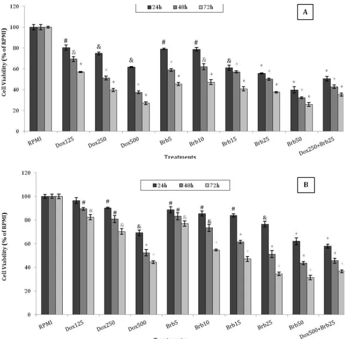
Figure 1. Cytotoxic effects of Berberine and Doxorubicin on T47D and MCF7 cells
The T47D (A) and MCF7 (B) cells were treated with different concentrations of Brb (5-50 µM) and Dox (125-500 nM). After 24 hr, 48 hr and 72 hr of treatment, cell proliferation was determined using MTT assay. Each experiment was repeated at least three times in quadruplicates for each concentration and the results are presented as mean±SD. The IC50 values were then determined and used in the subsequent experiments. # denotes P<0.05, & P<0.01, and * P<0.001 for significant difference between treatments in comparison to control RPMI.
Results
Cytotoxic effects of treatments on T47D and MCF7 cells
MTT assay was used to determine the effect of different treatments on proliferation of T47D and MCF7 cells. Cell proliferation was significantly decreased following treatment of T47D and MCF7 cells with Brb and Dox in a concentration- and
time-dependent manner (P<0.001), (Figure1 A and B). The
IC50 was observed at 25 µM concentration of Brb
after 48 hr of treatment in both cell lines. The IC50 for Dox was determined to be 250 nM and 500 nM after
48 hr treatment in T47D and MCF-7 cells, respectively. Co-treatment with Brb and Dox
significantly increased cytotoxicity in T47D
compared with MCF7 cells (P<0.05).
Apoptosis
Barzegar et al Anticancer properties of berberine
Figure 2. Apoptosis induction in T47D and MCF7 cells
The T47D (A and C) and MCF7 (B and D) cells received different treatments for 48 hr to evaluate induction of apoptosis using Annexin V-FITC (Anv) and PI double-staining by flow cytometry. Representative images of flow cytometry for apoptosis in the T47D (A) and MCF7 (B) cells are shown following treatment with Brb and Dox alone or in combination in comparison to control RPMI. The percentage of early (Q4: PI-/Anv+) and late apoptotic (Q2: PI+/Anv+) cells in T47D (C) and MCF7 (D) cells are presented for each treatment in comparison to control RPMI. Data are presented as mean±SD of three independent experiments.
# denotes P<0.05, & P<0.01, and * P<0.001 for significant difference between treatments in comparison to control RPMI
cells, the early (Q4: PI-/Anv+) and late apoptosis (Q2: PI+/Anv+) rate together for the control RPMI was 5.24%, while those of the Dox (250 nM) and Brb
(25 µM) alone were 9.69% (P<0.05) and 29.6%
(P<0.01), respectively (Figure 2-C). In MCF7 cells, the
early (Q4: PI-/Anv+) and late apoptosis (Q2: PI+/Anv+) rate together for the control RPMI was 4.13%, while those of the Dox (500 nM) and Brb (25
µM) alone were 12.74% (P<0.01) and 31.72%
(P<0.001), respectively (Figure 2-D). A significant
increase in the percentage of apoptotic cells was
observed in both T47D (41.5%, P<0.001) and MCF7
(37.8%, P<0.001) cells following co-treatment with
IC50 of Brb and Dox, in comparison to other treatments (Figure 2-C and D). In addition, stained cells with Annexin V-FITC and PI showed increase in apoptotic cells stained green (Annexin V-FITC+) following treatment with IC50 of Brb alone or in combination with Dox in comparison to Dox alone or RPMI in both T47D and MCF7 cell lines (Figure 3-A
and B). Furthermore, cells stained red (PI+) increased in cells treated with Brb alone or in combination with Dox in comparison to Dox alone or RPMI in both T47D and MCF7 cell lines (Figure 3-A and B).
Cell cycle distribution
The effect of Brb and Dox alone and in combination on cell cycle distribution of T47D and MCF7 cells was determined by flow cytometry and showed alterations in G0/G1, S, and G2/M phases of cell cycle under different treatment conditions (Figure 4-A and B). In T47D cells, 56.54% of control RPMI cells were in G0/G1 phase, 29.56% in S phase and 13.90% in G2/M phase (Figure 4-C). In MCF-7 cells, 71.41% of control RPMI cells were in G0/G1 phase, 21.71% in S phase and 6.89% in G2/M phase (Figure 4-D). Compared with control RPMI, Brb 25 µM induced cell accumulation in the G2/M phase
Figure 3. Microscopic evidence of apoptosis induction in T47D and MCF7 cells
Stained T47D (A) and MCF7 (B) cells with Annexin V-FITC and PI were examined under the microscope, which showed increase in green (Annexin V-FITC+) and to a lesser extent in red (PI+) cells following treatment of cells with Brb alone or in combination with Dox in comparison to Dox alone or control RPMI. Dual stained (PI+/Annexin V-FITC+) cells that indicate late apoptotic event are also seen at different extent in T47D and MCF7 cells.
G0/G1 (P<0.05) and S phase (P<0.001) in T47D cells.
Brb 25 µM induced G0/G1 cell accumulation in comparison to control RPMI (80.86% versus 71.41%,
P<0.001) in MCF7 cells. In comparison to control, a
significant increase in G2/M arrest was observed in
both T47D (76.69%, P<0.001) and MCF7 (32.74%,
P<0.001) cells treated with Dox at concentrations of
250 nM and 500 nM, respectively (Figure 4-C and D). The percentage of G2/M arrest following treatment of T47D cells with the combination of Brb and Dox and the pattern of cell cycle distribution was almost similar to treatment with Brb 25 µM alone (31.80% versus 33.55%). In the MCF7 cells, G0/G1 arrest was observed with the combination of Brb and Dox in comparison to control RPMI (81.42% versus 71.41%,
P<0.001), which was almost similar to Brb 25 µM
alone (81.42% versus 80.86%). Brb alone or in combination with Dox differently induced cell cycle arrest. It was G0/G1 in MCF-7 cells but G2/M in T47D cells (Figure 4-C and D).
Discussion
Results of this study showed a concentration- and time- dependent manner in cytotoxic effects of Brb on T47D and MCF7 cancer cells. In addition, Brb alone or in combination with Dox induced cell cycle arrest and apoptosis in both cancer cells.
Apoptosis contributes to cell death in tumors treated with various anticancer agents (18, 19). Despite the fact that many tumors initially respond to therapy, cells can subsequently survive and gain resistance to these treatments (20).
Anthracyclines such as Dox have been shown to be able to upregulate several pro-apoptotic and
downregulate anti-apoptotic proteins in Bcl2 family (20). In addition, anthracyclines seem to achieve
their antitumor effects by affecting several
intracellular processes, causing DNA intercalation as well as generation of free radicals. A key mechanism, however, seems to be the inhibition of the DNA damage repair enzyme topoisomerase II (TopoII) (21). Changes in Topo IIA expression levels as a major determinant of response to the Topo IIA inhibitors show that suppression of Topo IIA levels
induces resistance to Dox in vitro and in vivo (22).
Barzegar et al Anticancer properties of berberine
Figure 4. Cell cycle alterations in T47D and MCF7 cells
The T47D (A and C) and MCF7 (B and D) cells received different treatments for 48 hr and were stained with DAPI cocktail and analyzed by flow cytometry to determine cell cycle distribution pattern. Representative images of flow cytometry for cell cycle distribution of the T47D (A) and MCF7 (B) cells are shown following treatment with Brb and Dox alone or in combination in comparison to control RPMI. Alterations in the percentage of T47D (C) and MCF7 (D) cells in G0/G1, S, and G2/M phases of cell cycle are presented as the mean±SD of three independent experiments. # denotes P<0.05, & P<0.01, and * P<0.001 for significant difference between treatments in comparison to control RPMI
cells (31). In addition, treatment with Brb inhibited growth in both MDA-MB-231 and MCF7 cells (16, 32), and exhibited a significant cytotoxic effect on MCF7 cells without affecting the breast normal epithelial cells (17). It has been reported that
the highest cytotoxic concentration induced
intercalation of Brb with DNA (48). Dox, which is a commonly used drug for cancer treatment (49), has been reported to significantly inhibit the melanoma tumor growth following combination with Brb (50). In our study, results of MTT assay also showed that treatment with combination of Brb and Dox increased cytotoxicity on T47D and MCF7 cells to a greater extent than Brb and Dox alone. This suggests that Brb may be an effective chemotherapeutic agent against breast cancer.
Control of cell cycle progression in cancer cells is considered to be a potentially effective strategy for the control of tumor growth. The molecular analyses of human cancers have revealed that cell cycle regulators are frequently mutated in most common malignancies (13). It has been reported that treatment with Brb induced cell cycle arrest at G0/G1 phase in different
cancer cells including breast (32, 49), liver (43, 51), cholangiocarcinoma (52), thyroid (40), bladder (41),
neuroblastoma (53), glioblastoma (54), oral
squamous (45) and epidermal (46) cancers. In some studies on prostate cancer cells, it was reported that at low concentration, Brb induced G0/G1 arrest, and upon exposure to higher concentration of Brb, cells exhibited G2/M arrest (13, 37, 38). Similar results
have been reported in melanoma cells (55). Our in
vitro data indicated that treatment of MCF-7 cells with Brb resulted in significant G0/G1 phase arrest of cell cycle progression which indicates that one of the mechanisms by which Brb may act to inhibit the proliferation of cancer cells is inhibition of cell cycle progression. Notably, this effect was not seen in T47D cell line. Similar to Dox, treatment of T47D cells with Brb alone or in combination with Dox resulted in G2/M arrest of cell cycle progression. However, in MCF-7 cells G0/G1 arrest was observed following treatment with Brb in combination with Dox.
Similarly, in another study, treatment with
Apoptosis plays a crucial role in eliminating the mutated neoplastic and hyperproliferation neoplastic cells from the system and therefore is considered a protective mechanism against cancer progression (57). Acquired resistance toward apoptosis is a hallmark of most and perhaps all types of cancer. Apoptosis is tightly regulated by anti-apoptotic and pro-apoptotic effector molecules, including proteins of the Bcl2 family, and can be mediated by several different pathways (58). In this study, the effects of Brb alone and in combination with Dox were determined on the induction of apoptosis in both T47D and MCF7 cells. Our flow cytometry and microscopic data indicate that treatment of T47D and MCF7 cells with Brb alone and in combination with Dox resulted in significant induction of apoptosis. A significant enhancement in the percent of apoptotic cells was observed in both T47D and MCF7 cell lines following treatment with combination of Brb and Dox. Similarly, apoptosis induction was reported after combination treatment with Brb and Paclitaxel in digestive tract cancer cells (56).
Conclusion
Our findings indicate that Brb and Dox, alone and in combination, exhibit antiproliferative effects against human breast cancer T47D and MCF7 cell lines, and this effect was mediated through interference with normal cell cycle distribution and induction of apoptosis. Therefore, Brb may be used as a good candidate drug for treatment of human breast cancer. More importantly, our findings suggest that Brb combined with Dox can be a novel combination in treatment of breast cancer with greater effects and possibly less side effects. Further
investigation and in particular in vivo evaluations are
required to explore clinical applications of Brb alone or in combination with available anticancer drugs in breast and other cancers.
Acknowledgment
The results reported in this article are part of a PhD dissertation that was financially supported by Tehran University of Medical Sciences (TUMS), Tehran, Iran.
References
1. Razmkhah M, Ghaderi A. HLA Class I Allele Frequencies in Southern Iranian Women With Breast
Cancer. Iran J Basic Med Sci 2013; 16:140-143.
2. Cao H, Yang Zh, Jiang G. Expression and clinical significance of activating transcription factor 3 in human breast cancer. Iran J Basic Med Sci 2013; 16:1151-1154.
3. Karimian Fathi N, Shekari Khaniani M, Montazeri V, Mansoori Derakhshan S. Minor role of BRCA2 mutation (Exon2 and Exon11) in patients with early-onset breast cancer amongst Iranian Azeri-Turkish women. Iran J Basic Med Sci 2014; 17:108-111.
4. Tavakoli-Yaraki M, Karami-Tehrani F. Apoptosis Induced by 13-S hydroxyoctadecadienoic acid in the Breast Cancer Cell Lines, MCF-7 and MDA-MB-231. Iran
J Basic Med Sci 2013; 16: 661- 9.
5. Lynch MD, Cariati M, Purushotham AD. Breast cancer, stem cells and prospects for therapy. Breast Cancer Res 2006; 8:211.
6. Zhong L, Shen H, Huang C, Jing H, Cao D. AKR1B10 induces cell resistance to daunorubicin and idarubicin by reducing C13 ketonic group. Toxicol Appl Pharmacol 2011; 255:40- 7.
7. Moretti E, Oakman C, Di Leo A. Predicting anthra-cycline benefit: have we made any progress? Curr Opin Oncol 2009; 21:507- 15.
8. Hussein MA. Preclinical rationale, mechanisms of action, and clinical activity of anthracyclines in
myeloma. Clin Lymphoma Myeloma 2007;4: S145-1 9.
9. Brechbuhl HM, Kachadourian R, Min E, Chan D, Day BJ. Chrysin enhances Doxorubicin-induced cytotoxicity in human lung epithelial cancer cell lines: the role of glutathione. Toxicol Appl Pharmacol 2012; 258:1-9. 10. Hembruff SL, Laberge ML, Villeneuve DJ, Guo B,
Veitch Z, Cecchetto M, et al. Role of drug transporters
and drug accumulation in the temporal acquisition of drug resistance. BMC Cancer 2008; 8:318.
11. Zhong L, Shen H, Huang C, Jing H, Cao D. AKR1B10 induces cell resistance to daunorubicin and idarubicin by reducing C13 ketonic group. Toxicol Appl Pharmacol 2011; 255:40- 7.
12. Meeran SM, Katiyar S, Katiyar SK. Berberine-induced apoptosis in human prostate cancer cells is initiated by reactive oxygen species generation. Toxicol Appl Pharmacol 2008; 229:33-43.
13. Mantena SK, Sharma SD, Katiyar SK. Berberine, a natural product, induces G1-phase cell cycle arrest and caspase-3-dependent apoptosis in human prostate carcinoma cells. Mol Cancer Ther 2006; 5:296-308. 14. Peng PL, Hsieh YS, Wang CJ, Hsu JL, Chou FP. Inhibitory effect of berberine on the invasion of human lung cancer cells via decreased productions of urokinase-plasminogen activator and matrix metalloproteinase-2. Toxicol Appl Pharmacol 2006; 214:8-15.
15. Kim S, Han J, Lee SK, Choi MY, Kim J, Lee J, et al.
Berberine suppresses the TPA-induced MMP-1 and MMP-9 expressions through the inhibition of PKC-alpha in breast cancer cells. J Surg Res 2012; 176:e21-9.
16. Kim JB, Ko E, Han W, Shin I, Park SY, Noh DY. Berberine diminishes the side population and ABCG2 transporter expression in MCF-7 breast cancer cells.
Planta Med 2008; 74:1693-1700.
17. Patil JB, Kim J, Jayaprakasha GK. Berberine induces apoptosis in breast cancer cells (MCF-7) through mitochondrial-dependent pathway. Eur J Pharmacol 2010; 645:70- 8.
18. Rezaei PF, Fouladdel S, Hassani S, Yousefbeyk F,
Ghaffari SM, Amin G,et al. Induction of apoptosis and
cell cycle arrest by pericarp polyphenol-rich extract of Baneh in human colon carcinoma HT29 cells. Food Chem Toxicol 2012; 50:1054-1059.
19. Shahveisi K, Mousavi SH, Hosseini M, Khajavi Rad A,
Jalali SA, Rajaei Z, et al. The role of local renin-angiotensin
Barzegar et al Anticancer properties of berberine
20. Simstein R, Burow M, Parker A, Weldon C, Beckman B. Apoptosis, chemoresistance, and breast cancer: insights from the MCF-7 cell model system. Exp Biol Med (Maywood) 2003; 228: 995-1003.
21. Lonning PE, Knappskog S, Staalesen V, Chrisanthar R, Lillehaug JR. Breast cancer prognostication and prediction in the postgenomic era. Ann Oncol 2007; 18:1293-1306.
22. Bouchalova K, Cizkova M, Cwiertka K, Trojanec R, Hajduch M. Triple negative breast cancer--current status and prospective targeted treatment based on HER1 (EGFR), TOP2A and C-MYC gene assessment. Biomed Pap Med Fac Univ Palacky Olomouc Czech Repub 2009; 153: 13-17.
23. Kuo CL, Chi CW, Liu TY. The anti-inflammatory
potential of berberine in vitro and in vivo. Cancer Lett
2004; 203:127-137.
24. Kim S, Choi JH, Kim JB, Nam SJ, Yang JH, Kim JH, et
al. Berberine suppresses TNF-alpha-induced MMP-9
and cell invasion through inhibition of AP-1 activity in MDA-MB-231 human breast cancer. Cells Molecules 2008; 13:2975-2985.
25. Ramasamy S, Abdul Wahab N, Zainal Abidin N, Manickam S. Effect of extracts from Phyllanthus watsonii Airy Shaw on cell apoptosis in cultured human breast cancer MCF-7 cells. Exp Toxicol Pathol 2012; 65:341-349. 26. Abd El-Wahab AE, Ghareeb DA, Sarhan EE, Abu-Serie
MM, El Demellawy MA. In vitro biological assessment of
berberis vulgaris and its active constituent, berberine: antioxidants, anti-acetylcholinesterase, anti-diabetic and anticancer effects. BMC Complement Altern Med 2013; 13:218.
27. Kuo HP, Chuang TC, Tsai SC, Tseng HH, Hsu SC,
Chen YC, et al. Berberine, an isoquinoline alkaloid,
inhibits the metastatic potential of breast cancer cells via Akt pathway modulation. J Agric Food Chem 2012; 60:9649-9658.
28. Hill GM, Moriarity DM, Setzer WN. Attenuation of cytotoxic natural product DNA intercalating agents by caffeine. Sci Pharm 2011; 79:729-747.
29. Pinto-Garcia L, Efferth T, Torres A, Hoheisel JD, Youns M. Berberine inhibits cell growth and mediates caspase-independent cell death in human pancreatic cancer cells. Planta Med 2010; 76:1155-1161.
30. Tsang CM, Cheung YC, Lui VW, Yip YL, Zhang G, Lin VW, et al. Berberine suppresses tumorigenicity and growth of nasopharyngeal carcinoma cells by inhibiting STAT3 activation induced by tumor associated fibroblasts. BMC Cancer 2013; 13:619.
31. Pazhang Y, Ahmadian S, Javadifar N, Shafiezadeh M. COX-2 and survivin reduction may play a role in berberine-induced apoptosis in human ductal breast epithelial tumor cell line. Tumour Biol 2012; 33:207-214.
32. Kim JB, Lee KM, Ko E, Han W, Lee JE, Shin I, et al.
Berberine inhibits growth of the breast cancer cell lines MCF-7 and MDA-MB-231. Planta Med 2008; 74:39-42.
33. Xu LN, Lu BN, Hu MM, Xu YW, Han X, Qi Y, et al.
Mechanisms involved in the cytotoxic effects of berberine on human colon cancer HCT-8 cells. Biocell 2012; 36:113-120.
34. Chidambara Murthy KN, Jayaprakasha GK, Patil BS. The natural alkaloid berberine targets multiple pathways to induce cell death in cultured human colon cancer cells. Eur J Pharmacol 2012; 688:14-21.
35. Wu K, Yang Q, Mu Y, Zhou L, Liu Y, Zhou Q, et al.
Berberine inhibits the proliferation of colon cancer
cells by inactivating Wnt/β-catenin signaling. Int J
Oncol 2012; 41:292-298.
36. Lin JP, Yang JS, Lee JH, Hsieh WT, Chung JG. Berberine induces cell cycle arrest and apoptosis in human gastric carcinoma SNU-5 cell line. World J Gastroenterol 2006; 12:21-28.
37. Wang Y, Liu Q, Liu Z, Li B, Sun Z, Zhou H, et al.
Berberine, a genotoxic alkaloid, induces ATM-Chk1 mediated G2 arrest in prostate cancer cells. Mutat Res 2012; 734:20-29.
38. Choi MS, Oh JH, Kim SM, Jung HY, Yoo HS, Lee YM,
et al. Berberine inhibits p53-dependent cell growth through induction of apoptosis of prostate cancer cells. Int J Oncol 2009; 34:1221-1230.
39. Park KS, Kim JB, Lee SJ, Bae J. Berberine-induced growth inhibition of epithelial ovarian carcinoma cell lines. J Obstet Gynaecol Res 2012; 38:535-540.
40. Park KS, Kim JB, Bae J, Park SY, Jee HG, Lee KE, et al.
Berberine inhibited the growth of thyroid cancer cell lines 8505C and TPC1. Yonsei Med J 2012; 53:346-351.
41. Yan K, Zhang C, Feng J, Hou L, Yan L, Zhou Z, et al.
Induction of G1 cell cycle arrest and apoptosis by berberine in bladder cancer cells. Eur J Pharmacol 2011; 661:1-7.
42. Hou Q, Tang X, Liu H, Tang J, Yang Y, Jing X, et al.
Berberine induces cell death in human hepatoma cells in vitro by downregulating CD147. Cancer Sci 2011; 102:1287-1292.
43. Wang XN, Han X, Xu LN, Yin LH, Xu YW, Qi Y, et al.
Enhancement of apoptosis of human hepatocellular carcinoma SMMC-7721 cells through synergy of berberine and evodiamine. Phytomedicine 2008; 15:1062-1068. 44. Tsang CM, Lau EP, Di K, Cheung PY, Hau PM, Ching YP, et al. Berberine inhibits Rho GTPases and cell migration at low doses but induces G2 arrest and apoptosis at high doses in human cancer cells. Int J Mol Med 2009; 24:131-138.
45. Lin CC, Yang JS, Chen JT, Fan S, Yu FS, Yang JL,et al.
Berberine induces apoptosis in human HSC-3 oral cancer cells via simultaneous activation of the death
receptor-mediated and mitochondrial pathway.
Anticancer Res 2007; 27:3371-3378.
46. Mantena SK, Sharma SD, Katiyar SK. Berberine inhibits growth, induces G1 arrest and apoptosis in human epidermoid carcinoma A431 cells by regulating Cdki-Cdk-cyclin cascade, disruption of mitochondrial membrane potential and cleavage of caspase 3 and PARP. Carcinogenesis 2006; 27:2018-2027.
47. Kuo CL, Chou CC, Yung BY. Berberine complexes with DNA in the berberine-induced apoptosis in human leukemic HL-60 cells. Cancer Lett 1995; 93:193-200. 48. Letasiová S, Jantová S, Miko M, Ovádeková R, Horváthová M. Effect of berberine on proliferation, biosynthesis of macromolecules, cell cycle and induction of intercalation with DNA, dsDNA damage and apoptosis in Ehrlich ascites carcinoma cells. J Pharm Pharmacol 2006; 58:263-270.
49. Kim JB, Yu JH, Ko E, Lee KW, Song AK, Park SY, et al.
The alkaloid Berberine inhibits the growth of Anoikis-resistant MCF-7 and MDA-MB-231 breast cancer cell lines by inducing cell cycle arrest. Phytomedicine 2010; 17:436-440.
murine melanoma B16F10 cells in culture and xenograft. Phytomedicine 2014; 21:340-347.
51. Yip NK, Ho WS. Berberine induces apoptosis via the mitochondrial pathway in liver cancer cells. Oncol Rep 2013; 30:1107-1112.
52. He W, Wang B, Zhuang Y, Shao D, Sun K, Chen J. Berberine inhibits growth and induces G1 arrest and apoptosis in human cholangiocarcinoma QBC939 cells. J Pharmacol Sci 2012; 119:341-348.
53. Choi MS, Yuk DY, Oh JH, Jung HY, Han SB, Moon DC,
et al. Berberine inhibits human neuroblastoma cell growth through induction of p53-dependent apoptosis. Anticancer Res 2008; 28:3777-3784.
54. Eom KS, Hong JM, Youn MJ, So HS, Park R, Kim JM,
et al. Berberine induces G1 arrest and apoptosis in human glioblastoma T98G cells through mitochondrial/caspases
pathway. Biol Pharm Bull 2008; 31:558-562.
55. Serafim TL, Oliveira PJ, Sardao VA, Perkins E, Parke D, Holy J. Different concentrations of berberine result in distinct cellular localization patterns and cell cycle effects in a melanoma cell line. Cancer Chemother Pharmacol 2008; 6:1007-10018.
56. Lin HL, Liu TY, Wu CW, Chi CW. Berberine modulates expression of mdr1 gene product and the responses of digestive track cancer cells to Paclitaxel. Br J Cancer 1999; 81:416-422.
57. Hickman JA. Apoptosis induced by anticancer drugs. Cancer Metastasis Rev 1992; 11:121-139.