RETROPERITONEAL LYMPHADENECTOMY BY VIDEOLAPAROSCOPIC
TRANSPERITONEAL APPROACH IN PATIENTS WITH
NON-SEMINOMATOUS TESTICULAR TUMOR
M. TOBIAS-MACHADO, JOÃO P. ZAMBON, ALEXANDRE D. FERREIRA, JIMMY A.
MEDINA, ROBERTO V. JULIANO, ERIC R. WROCLAWSKI
Discipline of Urology, ABC Medicine School, Santo André, São Paulo, Brazil
ABSTRACT
Objective: The present study aims to report the preliminary experience with videolaparoscopic retroperitoneal lymphadenectomy in the treatment of patients with non-seminomatous testicular tu-mor.
Materials and Methods: Seven surgeries were performed in order to access retroperitoneal lymph nodes in patients with non-seminomatous testicular cancer. We performed the videolaparoscopic retroperitoneal lymphadenectomy (LRL) technique in 5 patients with stage I disease and laparoscopic resection of residual mass (LRRM), following chemotherapy (ChT), in 2 patients with stage II dis-ease. Initial approach was obtained through 4 trocars, using an incision in supra-umbilical midline when manual assistance was required. Surgical time was analyzed, as well as blood loss, need for analgesic drugs postoperatively, hospital stay, complications, need for blood transfusion, histopatho-logical data and tumor control in a mean follow-up of 18 months.
Results: Mean surgical time was 200 to 260 minutes in LRL and LRRM groups respectively, mean blood loss was 300 mL for the LRL group and 400 mL for the LRRM group, without need for transfusions. There was a lesion in the vena cava in the LRL group, which was managed with manual assistance and one conversion in the LRRM group, due to a 10-cm tumor mass that was adhered to the aorta. Mean hospital stay was 3 days, excluding the converted case, and the use of analgesic drugs was needed until the second postoperative day. Of the stage I patients, 2 had active disease in retroperitoneum, and underwent adjuvant ChT. The 2 residual masses were teratomas. There was no recurrence during the follow-up period.
Conclusions: Videolaparoscopic retroperitoneal lymphadenectomy is a procedure with high technical complexity and a higher potential for conversion when performed following chemotherapy.
Key words: testis; testicular neoplasms; germ cell tumors; chemotherapy; lymphadenectomy; laparoscopy
Int Braz J Urol. 2004; 30: 389-97
INTRODUCTION
Conventional retroperitoneal lymphadenec-tomy is a major surgical procedure, which confers considerable morbidity to patients with non-seminomatous testicular tumor. In patients with ad-vanced disease who are already debilitated by the ef-fects of chemotherapy, the surgical procedure will
bring an additional morbidity, impairing their quality of life (1).
there is no impairment in the oncologic control of the disease (2-12).
Pathological staging of retroperitoneal lymph nodes in patients with non-seminomatous tumors of-fers 2 advantages: patients with metastatic disease can be identified and treated, and those without metastatic disease can be closely observed (1,11,12).
In patients with metastatic testicular tumors who undergo previous ChT, there is formation of cica-tricial tissue that adheres to the great vessels, which can make tumor resection difficult following ChT (5,8). The usual chemotherapy scheme using cisplatin as basic drug provides 60 to 70% of satisfactory re-sults, while approximately 30% of patients should undergo a surgical procedure (11,12). In such cases, it is recommended that the laparoscopic surgery as a minimally invasive procedure should be performed with caution, due to the risk of vascular damage with consequent conversion to open surgery, increasing postoperative morbidity (5,7,8,10-12).
In Brazil, the use of the laparoscopic approach has been performed in a few centers, due to the rarity of the disease and the technical complexity of the pro-cedure (8). The objective of this work was to report the preliminary experience with videolaparoscopic ret-roperitoneal lymphadenectomy in the treatment of pa-tients with non-seminomatous testicular tumor.
MATERIALS AND METHODS
Selection Criteria
In the period from January 1999 to December 2002, we retrospectively studied 7 patients diagnosed with non-seminomatous testicular cancer who under-went laparoscopic retroperitoneal lymphadenectomy (Table-1). Patients did not present any contra-indica-tion for surgery and were in good general condicontra-indica-tions, with American Society of Anesthesiologists (ASA) score I and II. All patients were operated in a reference teaching institution in Brazil, by the same surgeon.
Laparoscopic retroperitoneal lymphadenec-tomy was performed in the following situations: 1) – Staging for stage I non-seminomatous testicular can-cer presenting risks factors such as vascular invasion, invasion of spermatic cord or predominance of em-bryonal carcinoma in neoplasic components. 2) –
Post-chemotherapy resection of residual masses smaller than 6 cm as measured by tomography.
The procedure was always initiated by laparoscopic transperitoneal approach. The hand-as-sisted technique was used as an alternative to avoid conversion to exclusive open surgery in those patients who presented intraoperative complications or in cases where postchemotherapy fibrosis prevented the safe dissection of the great vessels, (13).
Preoperative staging was performed using thorax and abdomen computerized tomography (CT) and tumor markers (alpha-fetoprotein, beta-subunit of human chorionic gonadotropin and lactic dehydro-genase). The employed chemotherapy scheme, if in-dicated, was 4 PEB cycles (cisplatin, etoposide and bleomycin).
Medical records were assessed in order to observe technical aspects, postoperative complication, conversion rate and postoperative outcome in rela-tion to tumor control. The clinical follow-up of pa-tients ranged from 10 to 28 months.
Five patients had their surgeries indicated for tumoral staging, with 3 cases due to the presence of a predominant embryonal component and 2 due to vas-cular invasion.
Among these, 3 patients had tumor in the right testis and 2 in the left one. Two patients underwent postchemotherapy resection of residual retroperito-neal mass.
Table 1 – General demographic data of patients who un-derwent laparoscopic retroperitoneal lymphadenectomy (LRL) for staging and laparoscopic resection of residual mass (LRRM).
Number of patients Age (years) Right-sided tumor Left-sized tumor
Tumor size on tomography Body mass index
Transperitoneal Laparoscopic
Retroperito-neal Lymphadenectomy
Surgical Technique
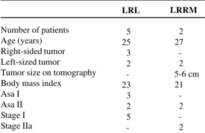
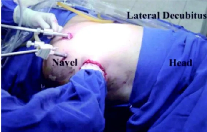
1 - Preoperative: Patients were admitted to hospital one day prior to surgery and underwent 8-hour fasting and reservation of red cells concentrate. All underwent general anesthesia with vesical catheter, nasogastric tube and antibiotic coverage during anes-thetic induction with first generation cephalosporin. Ureteral catheterization, to facilitate intraoperative identification of ureters, was not performed in any case. 2 - Positioning and installation of trocars: Af-ter positioning the patient in laAf-teral decubitus at 60 degrees, 4 trocars were placed: one 10-mm trocar in the umbilical scar, for introducing the 0-degree op-tics, two 5-mm trocars, one in the midline between the umbilical scar and the xiphoid process and the other in the midline between the pubis and the um-bilical scar. This set-up allows the port incisions to be united inside the surgical incision, in case of con-version to open surgery. A 10-mm trocar was placed 2 cm below the umbilical scar at the lateral margin of
the rectus muscle of abdomen on the side to be ap-proached (Figure-1).
3 - Dissection technique: The dissection lim-its were the same as in open surgery (7). Access to the retroperitoneal space was achieved by an inci-sion in the Toldt line, anterior and medially displac-ing the colon. For the 5 patients with stage I tumor, the modified lymphadenectomy was performed with interaortocaval dissection. For right-sided testicular tumors, the upper dissection limits were the renal hi-lum bilaterally, including the ureter at the left and extending downwards until the inferior mesenteric ar-tery. At this point, the dissection was directed to the right side, following the right aortic margin and the right common iliac artery until the crossing point of the ureter. Posterior dissection limit corresponded to the anterior spinous ligament. For tumors located in the left testis, the dissection was similar to the previ-ous one, being more economic for the contralateral side, where the inferior limit was the inferior vena cava and not the ureter.
Ultrasonic or bipolar scalpel was used for resecting the lymphatic tissue and the surgical
men was removed through entrapment in plastic pack-aging.
Hand-Assisted Retroperitoneal
Lymphadenectomy
Surgical Technique
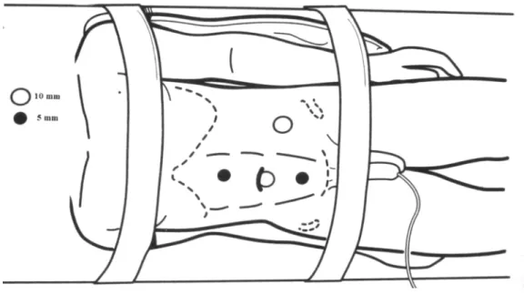
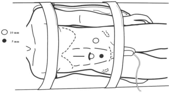
This technique was performed through a mid-line supra-umbilical incision located above the renal hilum or close to the lesion, without using manual de-vice, including whenever possible the location of one of the previously described ports (Figures-2 and 3).
In the 2 patients with post-ChT residual mass, total resection of the mass was performed.
RESULTS
In the group of patients with surgical indica-tion for staging, mean age was 25 years and mean body mass index was 23 kg/m2. Mean surgical time
was 200 minutes (160 - 360) and mean blood loss was 300 mL, with no need for transfusion.
There was one intra-operative complication with damage to the inferior vena cava, which was
resolved by vascular control, using hand-assisted ac-cess, with a 7-mm supra-umbilical midline incision, including the supra-umbilical port, without a device for pneumoperitoneum contention. After digital com-pression of the vena cava, it was possible to place 2 Doyan valves, applying a Satinski clamp and closing the lesion with 5-0 Prolene suture, externally, with no additional enlargement of the incision. In this case there was no impairment in relation to the laparoscopic access, since, once the vascular suture was completed, we were able to conclude the proce-dure through hand-assisted laparoscopic approach. Thus, we did not consider this temporary transition between open and laparoscopic techniques as a con-version to definitive open approach. There was no conversion to definitive open surgery in this group.
Figure 4 – Post-chemotherapy retroperitoneal residual mass. Surgical specimen.
Table 2 – Intraoperative data from LRL and LRRM.
Surgical time (min) Blood loss (mL) Transfusion Intraoperative complications Conversion
LRL
200 (160-360) 300
0 1 (12%)
0
LRRM
260 (240-280) 400
0 0
1
operative tomography performed 45 days before the surgery revealed a 6-cm residual mass, and for this rea-son, the patient was included in the study (Table-2).
Data relative to postoperative outcome can be observed in Table-3.
No postoperative complication was observed during the follow-up, in the 2 groups of patients.
Of the 5 stage I patients, 2 presented positive lymph nodes for non-seminomatous tumor and sub-sequently underwent adjuvant chemotherapy. In the 2 patients undergoing post-ChT resection of residual mass (Figure-4) the pathological report revealed that it was a teratoma, with no need for complementary treatment.
There was no case of local or systemic recur-rence during a mean follow-up of 18 months in both groups under study. Postoperative follow-up revealed that all patients maintained normal anterograde ejacu-lation.
COMMENTS
There is much discussion regarding the best approach to stage I non-seminomatous testicular tu-mors. While some authors advocate a careful follow-up, based on the efficacy of the chemotherapic drugs
Figure 3 – Positioning, trocars and surgical team in hand-as-sisted technique for laparoscopic retroperitoneal lymphadenec-tomy.
Table 3 – Postoperative data from LRL and LRRM.
Hospital stay (days) Analgesic use (days)
Time to oral restitution (days)
LRL
3 (1-4) 2 (1-3) 2 (1-3)
LRRM
3 (2-4) 2.5 (2-3) 2 (1-3)
used for treating this disease (12), others prefer the retroperitoneal lymphadenectomy with diagnostic and therapeutic purposes. The main argument for this management is the fact that microscopic metastases can be present in approximately 30% of cases (12-16), with 70% being free from disease without re-quiring cytotoxic chemotherapy.
accurate indication of rescue ChT in cases with an active malignant component (8-10,12,14-18).
The incidence of retroperitoneal teratoma ranges from 8 to 13% (1,8). These tumors have a proven malignant potential, grow rapidly, and can invade adjacent structures with consequent func-tional impairment to the patient. Moreover, these tumors are resistant to radio and chemotherapy (1,9). Considering this evidence, some authors advocate the performance of retroperitoneal lymphadenec-tomy even in patients with negative radiological findings (5,9,18). In cases with retroperitoneal mass larger than 1.5 cm or primary tumors up to 5 cm, regardless the response to ChT, since they present negative tumoral markers, lymphadenectomy has a precise indication (5,8-10,12). In the present study, the 2 post-ChT residual masses were teratomas and the surgery certainly benefited the patients (2,3,10,12).
Especially in laparoscopic access, where there is greater technical difficulty for lymphatic resection posterior to the great vessels, it is currently recommended to perform the modified unilateral resection or isolated resection of the post-ChT re-sidual mass. This proposal is based on the distribu-tion of retroperitoneal metastases for non-seminomatous tumors proposed by Wood et al. (11). According to these authors, the lymphatic drainage on the right side is directed to the lymph nodes lo-cated in the interaortocaval space, and to the left para-aortic and pre-aortic lymph nodes on the left side. In patients who underwent previous ChT, the sites of metastatic spread are similar to the sites of primary tumor and approximately 8% of the tumors would be outside the resection area (11,12). Previ-ously described data suggest that there is no benefit in routine retroaortic and retrocava lymphadenec-tomy. In this study, we performed retrocava dissec-tion only in our first patient (case with longer surgi-cal time). Routinely, we adopted the interaortocaval dissection regardless the side where the primary tu-mor was located, since the possibility of metastasis at this site must be taken into account. We did not observe local or systemic recurrence with the re-ported technique after a mean follow-up of 18 months.
Using the unilateral dissection below the in-ferior mesenteric artery (modified lymphadenec-tomy), approximately 85 to 90% of patients have their ejaculatory function preserved (1-3,6,9,14). In this small sample, all patients maintained preserved antegrade ejaculation. This is fundamentally impor-tant, since the main age range affected by testicular neoplasia comprises young adults in active reproduc-tive phase. In our initial patient sample, we observed surgical and outcome results very similar to other works in the literature (2,4,6,14).
As previously exposed, the videolaparoscopic technique that we employed is similar to open sur-gery. For releasing the lymphatic tissue, we preferred to use the ultra-sonic scalpel, because it reduces the risk of thermal damage to the great vessels and adja-cent organs.
Chemotherapy previously to surgery can make laparoscopic dissection difficult, due to the occurrence of local fibrosis of the retroperitoneal tissue that becomes closely adhered to vascular struc-tures, with higher conversion indexes. However, previous ChT per se do not prevent the resection of post-ChT mass by videolaparoscopic approach (5,8,10,12). Some works are contrary to resecting the post-chemotherapy residual mass by videolaparoscopic access (9). Others advocate laparoscopic surgery for masses measuring up to 5 cm, showing that this is feasible, with a higher con-version index (5,8,10,12).
Sutherland & Wright reported one case of resection of a thoracoabdominal mass successfully using thoracoscopy and hand-assisted laparoscopic access (13).
There are few works reporting the results of long-term oncologic control (19). Additionally, in series of laparoscopic surgery, patients presenting positive lymph nodes systematically receive adju-vant chemotherapy, thus it is not possible to assess surgical results separately. Anyway, the oncologic control seems to be similar to the open technique (2-12,19).
CONCLUSIONS
Videolaparoscopic retroperitoneal lym-phadenectomy is a procedure with high technical com-plexity and a higher potential for emergency conver-sion when performed after chemotherapy. In the ab-sence of laparoscopic vascular material, a manual in-cision can be sufficient to avoid conversion, maintain-ing the advantages of the minimally invasive surgery. We believe that this method is feasible for diagnosing and treating stage I and IIa non-seminomatous tumors, or when the postchemotherapy residual mass is smaller than 6 cm.
REFERENCES
1. Richie JP, Steele GS: Neoplasm of the Testis. In: Walsh PC, Retik AB, Vaughan ED, Wein JA (eds). Campbell’s Urology, ed. 8. Philadelphia, WB Saunders. 2002; pp. 2876-919.
2. Giusti G, Beltrami P, Tallarigo C, Bianchi G, Mobilio G: Unilateral laparoscopic retroperitoneal lym-phadenectomy for clinical stage I non-seminomatous testicular cancer. J Endourol. 1998; 12: 561-6. 3. Janetschek G, Hobisch A, Peschel R, Hittmair A,
Bartsch G: Laparoscopic retroperitoneal lymph node dissection for clinical stage I non-seminomatous tes-ticular carcinoma: long-term outcome. J Urol. 2000; 163: 1793-6.
4. Rassweiler JJ, Frede T, Lenz E, Seemann O, Alken P: Long-term experience with laparoscopic retroperito-neal lymph node dissection in the management of low-stage testis cancer. Eur Urol. 2000; 37: 251-60. 5. Janetschek G, Hobisch A, Hittmnair A, Holti L, Peschel
R, Bartsch G: Laparoscopic retroperitoneal lym-phadenectomy after chemotherapy for stage II B non-seminomatous testicular carcinoma. J Urol. 1999; 161: 477-81.
6. Janetschek G: Laparoscopic retroperitoneal lymph node dissection. In Sakti Das (ed). The Urologic Clin-ics of North America-Advanced Urologic Laparoscopy. Philadelphia, WB Saunders. 2001; 28: 107-14. 7. Janetschek G, Hobisch A, Holtl L, Bartsch G:
Retro-peritoneal lymphadenectomy for clinical stage I nonseminomtous testicular tumor: laparoscopy versus open surgery and impact of learning curve. J Urol. 1996; 156: 89-93.
8. Mariano MB, Tefilli MV: Laparoscopy retroperitoneal lymphadenectomy after chemotherapy for stage II B testicular tumors. Int Braz J Urol. 2001; 27: 527-34. 9. Rassweiler JJ, Seemann O, Henkel TO, Stock C, Frede
T, Alken P: Laparoscopic retroperitoneal lymph node dissection for non-seminomatous germ cell tumor: in-dications and limitations. J Urol. 1996; 156: 1108-13. 10. Rabbani F, Goldenberg SL, Gleave ME, Paterson RF, Murray N, Sullivan LD: Retroperitoneal lymphadenec-tomy for post-chemotherapy residual masses: is a modi-fied dissection and resection of residual masses suffi-cient? Br J Urol. 1998; 81: 295-99.
11. Wood DP Jr, Herr HW, Heller G, Vlamis V, Sogani PC, Motzer RJ, et al.: Distribution of retroperitoneal metastasis after chemotherapy in patients with non-seminomatous germ cell tumors. J Urol. 1992; 148: 1812-15.
12. Gelderman WA, Koops HS, Sleijfer DT, Oosterhuis JW, Oldhoff J: Treatment of retroperitoneal residual tumor after PVB chemotherapy of non-seminomatous testicular tumors. Cancer.1986; 58: 1418-21. 13. Sutherland A, Wright G: Hand-assisted laparoscopic
lymphadenectomy: a novel approach to a difficult area. ANZ J Surg. 2003; 73: 755-7.
14. Janetschek G: Laparoscopic retroperitoneal lymph node dissection. Urol Clin North Am. 2001; 28: 107-14.
15. Donohue JP, Thornhill JA, Foster RS, Rowland RG, Bihrle R: Retroperitoneal lymphadenectomy for clini-cal stage A non-seminomatous germ cell testis cancer: Review of the Indiana University experience 1965-1989. Br J Urol. 1993: 71: 326-35.
16. Atsu N, Eskicorapci S, Uner A, Ekici S, Gungen Y, Erkan I, et al.: A novel surveillance protocol for stage I non-seminomatous germ cell testicular tumors. BJU Int. 2003; 92: 32-5.
18. Bohlen D, Borner M, Sonntag RW, Fey MF, Studer UE: Long-term results following adjuvant chemo-therapy in patients with clinical stage I testicular non-seminomatous malignant germ cell tumors with high risk factors. J Urol. 1999; 161: 1148-52.
19. Bhayani SB, Ong A, Oh WK, Kantoff PW, Kavoussi LR: Laparoscopic retroperitoneal lymph node dissec-tion for clinical stage I nonseminimatous germ cell tes-ticular cancer: a long- term update. Urology. 2003; 62: 324-7.
Received: December 23, 2003 Accepted after revision: September 11, 2004
Correspondence address:
Dr. Marcos Tobias-Machado Rua Graúna 104 / 131
São Paulo, SP, 04514-000, Brazil Fax: + 55 11 288-1003
E-mail: [email protected]
EDITORIAL COMMENT
The authors describe their initial experience with transperitoneal laparoscopic retroperitoneal lym-phadenectomy in a group of 7 patients with non-seminomatous testicular tumors. Five patients pre-sented clinical stage I, and 2 patients had postchemotherapy residual masses.
Testicular cancer, though relatively rare, is the most common tumor in men aged between 15 and 35 years. It also represents the solid neoplasia with higher possibility of cure, serving as an example of an almost perfect therapeutic synergism between the different oncologic expertise fields. The dramatic improvement in survival of this population results from the combination of more accurate diagnostic techniques, availability of tumor markers, effective chemotherapy schemes and modifications in surgical techniques, which, jointly, reduced the mortality from 60% in the 70s to less than 10% in mid-90s. Thus, with the availability of effective therapeutic options, even for patients with advanced disease, efforts have been focused to reducing the morbidity, with poten-tial improvements in the current protocols.
In this setting, videolaparoscopic surgery seems to be an attractive approach, both for initial staging of non-seminomatous tumors and for selected
cases of patients with postchemotherapy residual masses.
As the authors emphasize, there is a long-lasting discussion in urologic literature concerning the best approach to stage I non-seminomatous tu-mors. While some advocate a careful follow-up, based on the efficacy of chemotherapic agents, oth-ers prefer the retroperitoneal lymphadenectomy with diagnostic and therapeutic purposes, stressing the fact that microscopic metastases can coexist in up to 30% of cases. The laparoscopic retroperitoneal lymphadenectomy appears to provide an optimal alternative in such cases, being a procedure with low morbidity that is routinely used, which allows us to be more liberal when indicating lymphadenectomy in these cases, with better acceptance by patients and clinical oncologists, thus avoiding the anxiety involved in long and consuming observation proto-cols.
retroperito-neal lymphadenectomy for residual masses. In order to reduce the procedural morbidity, we have replaced open classic lymphadenectomy for the laparoscopic approach in selected cases, as it seems to be the ten-dency of this manuscript’s authors. Despite being technically feasible following chemotherapy, we in-dicate this procedure only to single and/or unilateral
multiple residual masses measuring no more than 5 cm. Postchemotherapy laparoscopic retroperitoneal lymphadenectomy is a technically complex procedure with some potentially serious complications, and to the moment it must not be encouraged outside ser-vices with a large experience in laparoscopic retro-peritoneal surgery.