ANATOMIC ASPECTS OF EPIDIDYMIS AND TUNICA VAGINALIS IN
PATIENTS WITH TESTICULAR TORSION
LUCIANO A. FAVORITO, ANDRÉ G. CAVALCANTE, WALDEMAR S. COSTA
Urogenital Research Unit, State University of Rio de Janeiro, and Service of Urology, Souza Aguiar Municipal Hospital, Rio de Janeiro, Brazil
ABSTRACT
Objective: To analyze the morphology of epididymis and tunica vaginalis as well as their anatomical anomalies in patients with testicular torsion.
Materials and Methods: We studied 25 patients (50 testes) aged between 12 and 23 years (mean 15.6). Torsion length ranged from 2 hours to 2 days (mean 8 hours). Epididymal anatomy was classified in 6 groups: Type I - epididymis united to the testis by its head and tail; Type II - epididymis totally united to the testis; Type III - disjunction of epididymal tail; Type IV - disjunction of epididy-mal head; Type V - total disjunction between testis and epididymis, and Type VI - epididyepididy-mal atresia. The type of torsion was classified in 3 groups: Group A – intravaginal torsion; Group B – extravaginal torsion and Group C – torsion due to long mesorchium.
Results: Of the 50 analyzed testes, 40 (80%) presented bell clapper deformity (with 21 pre-senting intravaginal torsion); 8 testes (16%) had long mesorchium (4 with torsion), and only 2 (4%) presented normal anatomy in the tunica vaginalis. The most frequently found anatomical relationship between testis and epididymis was Type I - 38 cases (76%); Type II relationship was found in 6 cases (12%) and Type III relationship was found in 6 cases (12%).
Conclusions: Intravaginal torsion is the most frequent type, and torsion due to long mesor-chium is associated with cryptorchism. The most frequently found anatomical relation between testis and epididymis in the study group was Type I.
Key words: testis; epididymis; spermatic cord torsion; cryptorchidism Int Braz J Urol. 2004; 30: 420-4
INTRODUCTION
Testicular torsion is a urologic urgency. The testis will present irreversible damage if the torsion is not resolved within up to 6 hours. Testicular tor-sion can occur at any age; however, it is more fre-quent in teenagers and young adults (1). This pathol-ogy is responsible for approximately 90% of acute testicular pain in patients between 13 and 21 years old (2).
Signs and symptoms of torsion include acute scrotal pain and testicular ascent, and the testis can lie horizontally with or without inflammatory signs
(3). The diagnosis is made mainly through clinical examination, however, in doubtful cases Doppler ultrasonography of the spermatic cord and testicular scintigraphy can be used to assess testicular perfu-sion (4). Often these tests are not promptly available, thus in doubtful cases following clinical examination, when complementary exams cannot be performed, urgency scrotal exploration is the treatment of choice (3).
tu-nica is implanted too high, the testis can present ex-cessive mobility (bell clapper testis) (5). Mesorchium is the ligament that unites the testis to the epididymis (5,6). In cases of epididymal disjunction or elongated epididymis, conditions that are highly frequent in cryptorchism (7-10) the mesorchium is long and can contribute to the testicular torsion (6).
Several clinical and experimental studies on testicular torsion are described in the literature (1-4,11). Studies on the anatomic aspects of the tunica vaginalis and the association with epididymal anatomy and its anomalies in patients with testicular torsion are scarce (5,12). The objective of this work is to analyze the morphology of epididymis and tu-nica vaginalis, as well as their anatomic anomalies in patients with testicular torsion.
MATERIALS AND METHODS
In the period from May 2002 to May 2004 we studied 25 patients (50 testes and epididymis) with testicular torsion. All patients were admitted to our institution with symptoms of testicular pain. Follow-ing the clinical examination, a Doppler ultrasonogra-phy of the scrotal region was performed, in order to confirm the diagnosis. Patients were aged between
12 and 23 years (mean 15.6) and torsion duration ranged from 2 hours to 2 days (mean 8 hours).
All cases were operated by the same surgeon, who performed orchiopexy (or orchiectomy) in the twisted testis and orchiopexy in the contralateral tes-tis. During surgical exploration, the anatomy of the tunica vaginalis and the relationships between testis and epididymis were classified according to the fol-lowing system, previously described (6,12-14): Type I - epididymis united to the testis by its head and tail; Type II - epididymis totally united to the testis; Type III - disjunction of epididymal tail; Type IV – dis-junction of epididymal head; Type V - total disjunc-tion between epididymis and testis and Type VI – epididymal atresia.
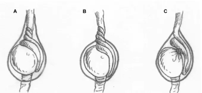
The type of testicular torsion was classified in 3 groups according to the anatomy of the tunica vaginalis and the relationship between testis and epi-didymis (Figure-1): Group A – bell clapper testicular deformity (leading to intravaginal torsion); Group B – torsion of spermatic cord (leading to extravaginal torsion) and Group C – torsion due to long mesor-chium.
We used qui-square statistical tests for con-tingency analysis of the populations under study (p < 0.05).
RESULTS
The type of testicular torsion found in the 25 patients is shown in (Table-1). Of the 25 studied pa-tients, 13 (52%) presented torsion of the right testis and 12 (48%) of the left one, with no significant dif-ference between the side of torsion. We did not find bilateral testicular torsion in any patient.
Of the 50 analyzed testes, 40 (80%) presented bell clapper deformity (with 21 presenting intravagi-nal torsion); 8 testes (16%) presented long mesor-chium (4 with torsion), and only 2 (4%) of the 50 testes under analysis, presented normal anatomy of the tunica vaginalis. These data are exposed in (Table-2). Among the 25 cases of torsion, orchiectomy was performed in 8 cases, testis fixation was performed in the remainder.
The most frequently found anatomic relation between testis and epididymis was type I - 38 cases (76%); type II relation was found in 6 cases (12%) and type III relation was found in 6 cases (12%) (Table-3).
Of the 8 cases with long mesorchium, 3 tes-tes (37.5%) were cryptorchid. One patient with tor-sion due to long mesorchium presented bilateral cryp-torchism and elongated epididymis bilaterally. In an-other case, the patient presented unilateral
cryp-Table 1 – Types of testicular torsion found during surgical exploration in the 25 patients studied.
Torsion Number of Cases (%)
Intravaginal 21 (84)
Long mesorchium 4 (16)
Total 25 (100)
Table 2 – Cases presenting bell clapper deformity of tunica vaginalis or long mesorchium in relation to the twisted and normal sides.
Anatomy of Tunica Vaginalis Twisted Testis Contralateral Testis Total
Bell clapper 21 19 40 Long mesorchium 4 4 8 Normal testis - 2 2
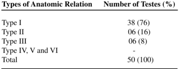
Table 3 – Incidence of types of anatomical relation be-tween testis and epididymis observed in 25 patients (50 testes). Type I - Epididymis united to the testis by its head and tail; Type II - Epididymis totally united to the testis; Type III – Disjunction of epididymal tail; Type IV – Dis-junction of epididymal head; Type V – Total disDis-junction between epididymis and testis and Type VI – Epididymal atresia.
Types of Anatomic Relation Number of Testes (%)
Type I 38 (76) Type II 06 (16) Type III 06 (8) Type IV, V and VI -Total 50 (100)
torchism with disjunction of epididymal tail. A third patient with torsion due to long mesorchium presented disjunction of the epididymal tail in the twisted tes-tis. This patient presented normal epididymal anatomy and normal layering of tunica vaginalis in the testis without torsion. Two patients presented bilateral dis-junction of epididymal tail.
COMMENTS
firmly united to the scrotum, preventing the organ to move (5,6).
Testicular torsion occurs due to anatomic anomalies of tunica vaginalis or epididymis that al-low excessive testicular mobility inside the scrotum. Due to this excessive mobility, testis can present medial rotation that ranges from 360º to 720º in its own axis, which can cause interruption of the organ’s vascularization (2).
Based on our findings, a normal anatomy of tunica vaginalis or epididymis at the side contralat-eral to the torsion is rare (2 cases - 4%), and ana-tomic anomalies occur bilaterally in the vast major-ity of cases. These findings stress the need for bilat-eral orchiopexy in cases of testicular torsion.
Bell clapper deformity (intravaginal torsion) was the most commonly found type of anomaly (80%). The relation between the presence of full cov-ering of testis and spermatic cord by tunica vaginalis (bell clapper deformity) and testicular torsion is well known. Parker & Robinson (5) in a study conducted with 40 patients found this deformity in 35% of stud-ied cases.
Cases of torsion due to long mesorchium most often occur as a consequence of anomalies of epid-idymal disjunction or elongated epididymis, condi-tions that are highly frequent in cryptorchism (5-10). Of the 8 cases with long mesorchium, 3 (37.5%) had cryptorchid testes. These findings are similar to those of Parker & Robinson (5) who found long mesorchium in 33% of studied cases.
Approximately 20% of cases of testicular tor-sion occur in patients with cryptorchism (6). Epid-idymal anomalies associated with long mesorchium are frequent in patients with cryptorchism, with an incidence ranging from (36 to 72%) (7-10) and rare in individuals with topic testes (less than 4%) (14). Due to these changes in mesorchial region, the possi-bility of testicular torsion must be considered in cryp-torchid patients presenting acute scrotal or inguinal pain.
Anatomical relations between testis and epi-didymis in patients with testicular torsion evidenced a pattern that is not different from patients without anomalies (12, 13-15). Type I and type II relations were observed in approximately 90% of cases of
tes-ticular torsion. In 2 patients with torsion with long mesorchium found in type I anatomy with elongated epididymis.
Elongated epididymis is a condition included in type I, according to the classification used in this paper (6,13-15). However it is known that patients with cryptorchism present a high index of epididy-mal anoepididy-malies (Types III, IV and V), as well as elon-gated epididymis (7-10). However, the present clas-sification includes elongated epididymis in the nor-mal group. Probably a subdivision of type I anatomic relation with elongated epididymis in a separate group will be necessary in the future.
We concluded that intravaginal torsion (bell clapper tunica vaginalis) is the most frequent type of torsion, and torsion due to long mesorchium is asso-ciated with cryptorchism. The most frequently found anatomical relation between testis and epididymis in the study group was type I (epididymis united to the testis by its head and tail).
The present research was supported by Rio de Janeiro Foundation for Research Support (FAPERJ) and National Council for Scientific and Technological Development (CNPQ).
REFERENCES
1. Cummings JM, Boullier JA, Sekhon D, Bose K: Adult testicular torsion. J Urol. 2002; 167: 2109-10. 2. Ben-Chaim J, Leibovitch I, Ramon J, Winberg D,
Goldwasser B: Etiology of acute scrotum at surgical exploration in children, adolescents and adults. Eur Urol. 1992; 21: 45-7.
3. Kass EJ, Stone KT, Cacciarelli AA, Mitchell B: Do all children with an acute scrotum require exploration? J Urol. 1993; 150: 667-9.
4. Middleton WD, Siegel BA, Melson GL, Yates CK, Andriole GL: Acute scrotal disorders: prospective com-parison of color Doppler US and testicular scintigra-phy. Radiology. 1990; 177: 177-81.
5. Parker RM, Robison JR: Anatomy and diagnosis of torsion of the testicle. J Urol. 1971; 106: 243-7. 6. Scorer CG, Farrington GH: Congenital Deformities of
7. Elder JS: Epididymal anomalies associated with hy-drocele/hernia and cryptorchidism: implications re-garding testicular descent. J Urol. 1992; 148: 624-6. 8. Gill B, Kogan S, Starr S, Reda E, Levitt S:
Signifi-cance of epididymal and ductal anomalies associated with testicular maldescent. J Urol. 1989; 142: 556-8; discussion 572.
9. Gill B, Kogan S: Cryptorchidism. Current concepts. Pediatr Clin North Amer. 1997; 44: 1211-27. 10. Marshall FF: Anomalies associated with
cryptorchid-ism. Urol Clin North Amer. 1982; 9: 339-47. 11. Noske HD, Kraus SW, Altinkilic BM, Weidner W:
Historical milestones regarding torsion of the scrotal organs. J Urol. 1998; 159:13-6.
12. Caesar RE, Kaplan GW: Incidence of the bell-clapper deformity in an autopsy series. Urology. 1994; 44: 114-6.
13. Favorito LA, Sampaio FJ: Anatomical relationships between testis and epididymis during the fetal period in humans (10-36 weeks postconception). Eur Urol. 1998; 33: 121-3.
14. Turek PJ, Ewalt DH, Snyder HM 3rd, Duckett JW: Normal epididymal anatomy in boys. J Urol. 1994; 151: 726-7.
15. Favorito LA, Sampaio FJ, Javaroni V, Cardoso LE, Costa WS: Proximal insertion of gubernaculum testis in normal human fetuses and in boys with cryptorchid-ism. J Urol. 2000; 164: 792-4.
Received: July 6, 2004 Accepted after revision: October 13, 2004
Correspondence address: Dr. Luciano Alves Favorito Unidade de Pesquisa Urogenital
Universidade do Estado do Rio de Janeiro Av. 28 de Setembro, 87, fundos, FCM, térreo Rio de Janeiro, RJ, 20551-030, Brazil Fax: + 55 21 2587-6121