Copyright © 2007 by Sociedade Brasileira de Pediatria
O
RIGINALA
RTICLEEchocardiographic abnormalities in children with
obstructive breathing disorders during sleep
Silke Anna Theresa Weber,1 Jair Cortez Montovani,2 Beatriz Matsubara,3
José Roberto Fioretto4
Abstract
Objectives:To assess cardiac morphology and function by means of echocardiograms of children with obstructed breathing while asleep.
Methods:The study enrolled 40 children of both sexes, aged from 3 to 11 years; 30 of them had obstructed breathing during sleep (group I) and 10 children were healthy controls (group II). The two groups were similar in terms of sex, age, weight and height. The 40 children underwent echocardiogram, viewing all four chambers during systole and diastole, paying special attention to the right ventricle (RV). These data were compared by means of Student’s t test (p < 0.05).
Results:In group I, increased diameter and area of the right ventricle were observed during both systole and diastole. There was less variation in RV area between systole and diastole. Reduced left ventricle (LV) diastolic diameter was also observed, together with reduced ejection fraction and reduced contraction.
Conclusions:The morphological and functional cardiac abnormalities observed in the RV and LV suggest that, in children, obstructed breathing during sleep can lead to cardiovascular repercussions. These abnormalities may expose these children to increased anesthetic and surgical risks.
J Pediatr (Rio J). 2007;83(6):518-522:Obstructive sleep apnea, child, echocardiography.
Introduction
The clinical presentation of upper airway (UA) obstruc-tion during sleep varies in intensity, from night time rhonchi to the obstructive sleep apnea-hypopnea syndrome (OSAHS).1
The obstructive sleep apnea-hypopnea syndrome is char-acterized by repeated episodes of UA obstruction, associated with intermittent hypoxia and hypercapnia.1,2Prevalence among children varied from 0.7% to 3% in a selection epide-miological studies.3-5Peak incidence is observed in preschool children, and age at which the most common obstruction of the upper airways east due to hypertrophic palatine and pha-ryngeal tonsils. The most often described symptoms are:
rhonchi, respiratory pauses, difficulty breathing, agitated sleep and nocturnal diaphoresis.2,6There may be severe clini-cal consequences of OSAHS, such ascor pulmonale,7-9in addi-tion to the negative impact on the child’s quality of life, such as retarded pondero-statural growth and facial and thoracic skeletal abnormalities,10nocturnal enuresis and behavior dis-orders, learning problems and other cognitive function deficits.11
In adults, OSAHS is considered an independent cause of systemic arterial hypertension and of cardiovascular dis-ease.12A few studies have included cardiovascular assess-ments of children. A reduction was observed in right ventricle contraction in children with UA obstructions by ventriculogra-phy, with improvements in some after adenotonsillectomy.13
1. Doutora. Professora, Departamento de Oftalmologia, Otorrinolaringologia e Cirurgia de Cabeça e Pescoço, Faculdade de Medicina de Botucatu (FMB), Univer-sidade Estadual de São Paulo (UNESP), Botucatu, SP, Brazil.
2. Professor livre-docente, Departamento de Oftalmologia, Otorrinolaringologia e Cirurgia de Cabeça e Pescoço, FMB, UNESP, Botucatu, SP, Brazil. 3. Professor livre-docente, Departamento de Clínica Médica, Cardiologia, FMB, UNESP, Botucatu, SP, Brazil.
4. Professor livre-docente, Departamento de Pediatria, Cardiopediatria, FMB, UNESP, Botucatu, SP, Brazil.
No conflicts of interest declared concerning the publication of this article.
Suggested citation:Weber SA, Montovani JC, Matsubara B, Fioretto JR. Echocardiographic abnormalities in children with obstructive breathing disorders during sleep. J Pediatr (Rio J). 2007;83(6):518-522.
Manuscript received Apr 12 2007, accepted for publication Aug 08 2007.
doi 10.2223/JPED.1720
Other studies have observed reduced left ventricle filling on echocardiogram, even reaching a collapsed state during apnea,14and also increased RV area,15with normalization after treatment of obstructive respiratory events, or CPAP (continuous positive air pressure)14or surgery.15 Adenoton-sillectomy also resulted in improved pulmonary flow param-eters and RV area in children with pulmonary hypertension, related to the VAS obstruction.16The severity of obstructive respiratory events during sleep, expressed by the more elevated rates of apnea and hypopnea, suggest that they are directly correlated with LV abnormalities.17
There is sufficient pathophysiologic evidence to allow for the suspicion that children with UA obstruction during sleep exhibit structural and functional cardiac abnormalities, nota-bly of the right ventricle.
The objective of this study was to assess cardiac morphol-ogy and function in children with obstructive respiratory dis-orders due to hypertrophic tonsils, by means of echocardiogram, and to compare them with healthy children.
Methods
This was a controlled cross-sectional study. The study investigated 40 children of both sexes, aged from three to 11 years, 30 of whom were patients being treated at the Sleep Disorders Clinic at the Otorhinolaryngology Department of FMB – UNESP, with a diagnosis of hypertrophic palatine and/or pharyngeal tonsils and clinical manifestations of obstructive respiratory disorders during sleep, such as rhonchi, respira-tory pauses, agitated sleep, breathing through the mouth, and others, and with an indication for tonsil surgery (group I). Chil-dren were excluded if they were known to have heart dis-ease, asthma or other lung conditions, neurological disease or a BMI > 30 kg/m2. The remaining 10 children were healthy controls (group II), selected from among the siblings of the patients treated at the Otorhinolaryngology Department and who agreed to take part in the study. The children in group II did not exhibit any respiratory complaints during sleep such as rhonchi, respiratory pauses, breathing through the mouth or any of the other exclusion criteria for group I. Data were collected on all of the children relating to sex, age, weight, height and body mass index (BMI) and BMI percentiles were calculated.
Children in group I who had clinical obstructive respira-tory conditions underwent complete otorhinolaryngological examination, including rigid and/or flexible nasofibroscopy to determine the degree of hypertrophy of the tonsils.18
All 40 children underwent echocardiogram, in M-mode, bidimensional and Doppler, with HP Sonos 2000 equipment, equipped with a multifrequency ultrasonic transducer at 3.0 and 2.7 MHz and a system for recording images on VHS and a video printer.
All examinations were carried out with the child awake, in a dimly lit room, with temperature controlled and always by
the same echocardiographic specialist. During examination, the child lay in left lateral decubitus, with the left arm lightly flexed over the head. The images were produced according to Canadian Society of Echocardiography recommendations.19
The morphological measurements taken were: diam-eters of the left atrium, aorta, right atrium, and systolic and diastolic diameters of the right and left ventricle and systolic and diastolic areas of both ventricles. All variables were nor-malized for each child’s BMI, since there was variation between the children in terms of age and anthropometric data.
The change in area and diameter were calculated in order to assess right ventricle function. Flow measurements were also taken for the tricuspid and pulmonary valves.
The ejection fraction and contraction of the left ventricle were calculated and flow measured at the mitral and aortic valves.
Sample size was calculated based on the assumption that the mean variation in RV area between group 1 and group 2 would be 20%, returning a figure of 10 individuals required in each group.
For each variable the mean and standard deviation were calculated from group I and group II. The results of the two groups were compared by means of Student’sttest for inde-pendent samples with normal distribution. In order to com-pare sexes across groups, the chi-square test was used. The level of significance adopted was 5% (p < 0.05).
This study received a favorable hearing from the Research Ethics Committee at the Botucatu Medical Faculty, UNESP, in 2004. Parents or guardians signed a free and informed con-sent form. The study was carried out between January and December, 2005.
Results
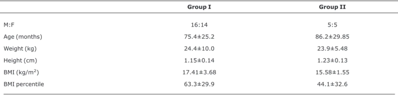
Thirty children with obstructive respiratory disturbances during sleep were studied, 16 of whom were males, and with a mean age of 75.4 months ± 25.2, where 17 children were aged 5 to 8 years. Group I and the control group were homog-enous in terms of sex, age, weight, height, BMI and BMI per-centiles (Table 1).
The area and systolic diameter of the right ventricle were enlarged, when compared with data from the control group, although only the difference in area attained significance.
Right ventricle function exhibited significantly reduced variation in area between systole and diastole (RVva). The variation in diameter was also reduced, although without sig-nificance (Table 2).
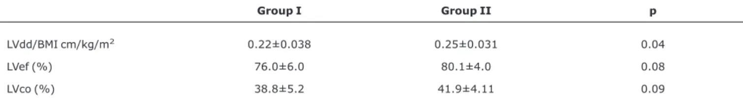
group (Table 3), although without significance. Flow measure-ments taken at the mitral, tricuspid, pulmonary and aortic valves did not yield abnormal findings in any of the groups.
Discussion
Hypertrophy of the lymphoid tissues of the pharynx is most accentuated during the preschool years, with predomi-nance among males,3,4and an increased incidence rate of obstructive symptoms and surgical tonsil interventions is observed in this age group.20In our study, despite the wide age range, there was also a predominance of preschool chil-dren, in group I, representing the target population of OSAHS in children.
Children with obstructive respiratory disorders during sleep are exposed to repeated hypopnea and apnea. During apnea, there is a progressive reduction in oxygen levels and an increase in CO2. Hypercapnia and hypoxemia provoke res-piratory acidosis and consequent vasoconstriction of the pul-monary artery. In addition to increased pulpul-monary resistance, there is also increased venous return to the right-hand car-diac chambers, which is facilitated both by the decubitus hori-zontal position during sleep and by intrathoracic pressure which becomes more negative due to respiratory effort against the obstructed area. This group of changes can lead to an enlarged right atrium and ventricle and compromise ejection during systole.12 ,21,22Concomitantly, hypoxemia
and broken sleep elevate circulating catecholamine levels, increasing peripheral resistance and leading to a reduction in left ventricle ejection. According to Steiner & Strauner,21the right-hand heart is more sensitive to small pressure varia-tions caused by pulmonary vasoconstriction, as a response to hypoxemia, than is the left ventricle.
The echocardiogram used in this study focuses on the RV and included measurements that are not routinely carried out, such as systolic and diastolic diameter, in addition to area. Right ventricle area and diameter were observed to be increased, during systole, even when measurements were adjusted for the BMI of each child. This enlargement of the RV has also been observed in other studies,13-17although not with adjustment for the weight and height of each child.
The variation between systolic and diastolic RV area and diameter was reduced, which suggests increased preload on the RV and more difficult pulmonary flow, although no increase in pulmonary vascular pressure was observed. Other studies have observed elevated pulmonary pressure, some with pulmonary hypertension,16or acute right heart failure andcor pulmonale,7as severe complications of OSAHS. In these children,c or pulmonalewith progression to acute pul-monary edema demands special care during preoperative and postoperative care for adenotonsillectomy.23Kalra et al.24 demonstrated RV and LV cardiac abnormalities similar to those
Table 1- Characteristics of the patients studied. Numerical variables are expressed as means and standard deviations*
Group I Group II
M:F 16:14 5:5
Age (months) 75.4±25.2 86.2±29.85
Weight (kg) 24.4±10.0 23.9±5.48
Height (cm) 1.15±0.14 1.23±0.13
BMI (kg/m2) 17.41±3.68 15.58±1.55
BMI percentile 63.3±29.9 44.1±32.6
BMI = body mass index; F = female; M = male.
* There were no significant differences between the groups.
Table 2- Morphological and functional right ventricle variables, by echocardiogram, for group I (DRO) and group II (controls)
Group I Group II p
RVsa/BMI cm2/kg/m2 0.37±0.099 0.31±0.065 0.05
RVsd/BMI cm/kg/m2 0.17±0.046 0.15±0.029 0.33
RVva (%) 0.35±0.06 0.50±0.05 0.04
RVvd (%) 0.27±0.02 0.33±0.04 0.09
observed in our study in children with respiratory complica-tions after adenotonsillectomy due to OSAHS, suggesting that prior identification of these cardiac abnormalities may be of aid in preventing these complications.
In the left ventricle a significant reduction in diastolic diameter was found. This finding may be either the response to right-side cardiac problems, resulting in reduced preload, or it may be a response to peripheral vasoconstriction, due to increased catecholamines in circulation. Similar LV findings have been observed in other studies.12,15,17Shiomi et al.14 demonstrated severe functional compromise to the LV by means of continuous echocardiogram. Abnormalities were only observed during sleeping obstructive respiratory epi-sodes, demonstrating that they may go unnoticed during an examination performed during the day with the child awake. This fact, and also the fact that the ECG and chest X ray very often do not detect abnormalities,25makes prior diagnosis of the cardiovascular repercussion in children with OSAHS very difficult.
In our study, children awaiting adenotonsillectomy for obstruction of the UA during sleep already exhibited altered cardiac morphology and function. Unfortunately we are unable to classify the severity of UA obstruction in these children in order to correlate the degree of echocardiographic findings with the number of obstructive respiratory events or of oxy-hemoglobin desaturation episodes. At our service, neither polysomnography nor cardiorespiratory monitoring are car-ried out as routine for adenotonsillectomy indicated due to UA obstruction in order to quantify sleeping obstructive res-piratory events and confirm the diagnosis of OSAHS.
One issue, which might be taken seriously, is that we com-pared the results obtained from group I with those from group II, since we did not pair the groups for the variables sex, age, weight or height, which could result in scientific imprecision. Despite this limitation, we do not believe that this has affected our data, since the groups were homogenous for these vari-ables and since some data proved highly asymmetrical within the groups, particularly right ventricle area and systolic diameters.
These results demonstrate to us that children with clinical manifestations and OSAHS exhibit morphological and func-tional abnormalities in right and left ventricles. Pediatricians
and otorhinolaryngologists who see mouth-breathing chil-dren with histories of rhonchi and respiratory pauses need to have a knowledge of the progression of simple UA obstruc-tion to OSAHS and of its possible severe complicaobstruc-tions. The data obtained here provide evidence of the importance of functional assessment in children with suspected severely obstructed upper airways.
References
1. Section on Pediatric Pneumology, Subcommittee on Obstructive Sleep Apnea Syndrome. American Academy of Pediatrics.Clinical practice guideline: diagnosis and management of childhood obstructive sleep apnea syndrome. Pediatrics. 2002;109:704-12.
2. Erler T, Paditz E.Obstructive sleep apnea syndrome in children: a state-of-the-art review.Treat Respir Med. 2004;3:107-22.
3. Anuntaseree W, Rookkapan K, Kuasirikul S, Thongsuksai P.
Snoring and obstructive sleep apnea in Thai school-age children: prevalence and predisposing factors.Pediatr Pulmonol. 2001; 32:222-7.
4. Gislason T, Benediktsdottir B.Snoring, apneic episode, and nocturnal hypoxemia among children 6 month to 6 years old. An epidemiologis study of lower limit of prevalence.Chest. 1995; 107:963-6.
5. Brunetti L, Rana S, Lospalluti ML, Pietrafesa A, Francavilla R, Fanelli M, et al.Prevalence of obstructive sleep apnea in a cohort of 1,207 children of southern Italy.Chest. 2001;120:1930-5.
6. Balbani AP, Weber SA, Montovani JC.Atualização em síndrome da apnéia obstrutiva do sono na infância. Rev Bras Otorrinolaringol. 2005;71:74-80.
7. Menashe VD, Farrehi C, Miller M. Hypoventilation and cor pulmonale due to chronic upper airway obstruction. J Pediatr. 1965;67:198-203.
8. Levy AM, Tabakin BS, Hanson JS, Narkewicz RM.Hypertrophied adenoids causing pulmonary hypertension and severe congestive heart failure.N Engl J Med. 1967;277:506-11.
9. Goodman RS, Goodman M, Gootman N, Cohen H.Cardiac and pulmonary failure secondary to adenotonsillar hypertrophy.
Laryngoscope. 1976;86:1367-74.
10. Di Francesco RC, Passerotii G, Paulucci B, Miniti A.Respiração oral na criança: repercussões diferentes de acordo com o diagnóstico.Rev Bras Otorrinolaringol. 2004;70:665-70.
Table 3- Morphological and functional left ventricle variables, by echocardiogram, for group I (DRO) and group II (controls)
Group I Group II p
LVdd/BMI cm/kg/m2 0.22±0.038 0.25±0.031 0.04
LVef (%) 76.0±6.0 80.1±4.0 0.08
LVco (%) 38.8±5.2 41.9±4.11 0.09
11. Weber SA, Lima Neto A, Ternes FJ, Montovani J.Distúrbio de hiperatividade e déficit de atenção na síndrome de apnéia obstrutiva do sono: há melhora com o tratamento cirúrgico?Rev Bras Otorrinolaringol. 2006;72:124-129.
12. Phillips B.Sleep-disordered breathing and cardiovascular disease.Sleep Med Rev. 2005;9:131-40.
13. Tal A, Leiberman A, Margulis G, Sofer S.Ventricular dysfunction in children with obstructive sleep apnea: radionuclide assessment.Pediatr Pulmonol. 1988;4:139-43.
14. Shiomi T, Guilleminault C, Stoohs R, Schnittger I.Obstructed breathing in children during sleep monitored by echocardiography.Acta Paediatr. 1993;82:863-71.
15. Görür K, Döven O, Uenal M, Akkus N, Özcan C.Preoperative and postoperative cardiac and clinical findings of patients with adenotonsillar hypertrophy. Int J Pediatr Otorhinolaryngol. 2001; 59:41-6.
16. Miman MC, Kirazli T, Ozyurek R.Doppler echocardiography in adenotonsillar hypertrophy.Int J Pediatr Otorhinolaryngol. 2000; 54:21-6.
17. Amin RS, Kimball TR, Bean JA, Jeffries JL, Willging JP, Cotton RT, et al.Left ventricular hypertrophy and abnormal ventricular geometry in children and adolescents with obstructive sleep apnea.Am J Respir Crit Care Med. 2002;165:1395-9.
18. Cassano P, Gelardi M, Cassano M, Fiorella ML, Fiorella R.
Adenoid tissue rhinopharyngeal obstruction grading based on fiberendoscopic findings: a novel approach to therapeutic management.Int J Pediatr Otorhinolaryngol. 2003;67:1303-9. 19. Rakowski H, Appleton C, Chan KL, Dumesnil JG, Honos G, Jue J, et al. Canadian consensus recommendations for the measurements and reporting of diastolic dysfunction by echocardiography: from the Investigators of Consensus on Diastolic Dysfunction by Echocardiography. J Am Soc Echocardiogr. 1996;9:736-60.
20. Suen JS, Arnold JE, Brooks LJ.Adenotonsillectomy for treatment of obstructive sleep apnea in children.Arch Otolaryngol Head Neck Surg. 1995;121:525-30.
21. Steiner S, Strauer BE. Funktionelle Dynamik des rechten Ventrikels und des Lungenkreislaufes bei obstruktiver Schlafapnoe.Therapeutische Konsequenzen. Internist (Berl). 2004;45:1101-7.
22. Verrier RL, Harper RM, Hobson JA. Cardiovascular physiology: central and autonomic regulation. In: Kryger MH, Roth T, Dement WC, editors. Principles and practice of sleep medicine. 3rd ed. Philadelphia: WB Saunders; 2000. p. 179-91.
23. McColley AS, April MM, Carroll JL, Naclerio RM, Loughlin GM.
Respiratory compromise after adenotonsillectomy in children with obstructive sleep apnea.Arch Otolaryngol Head Neck Surg. 1992;118:940-3.
24. Kalra M, Kimball T, Daniels SR, LeMasters G, Willging PJ, Rutter M, et al.Structural cardiac changes as a predictor of respiratory complications after adenotonsillectomy for obstructive breathing during sleep in children.Sleep Med. 2005; 6:241-5.
25. James AL, Runciman M, Burton MJ, Freeland AP.Investigation of cardiac function in children with suspected obstructive sleep apnea. J Otolaryngol. 2003; 32:151-4.
Correspondence: Silke Anna Theresa Weber
Faculdade de Medicina de Botucatu – UNESP Depto. OFT/ORL/CCP
Distrito de Rubião Júnior, s/nº CEP 18618-970 – Botucatu, SP – Brazil Tel.: +55 (14) 3811.6256