Saliva as a diagnostic luid in sports medicine:
potential and limitations
Saliva como fluido diagnóstico para utilização na medicina esportiva:
potencialidades e limitações
Lázaro Alessandro Soares Nunes1; Denise Vaz de Macedo2
First submission on 21/12/12; last submission on 08/02/13; accepted for publication on 02/04/13; published on 20/08/13
1. Doctor in Functional and Molecular Biology (Biochemistry area) by Universidade Estadual de Campinas (UNICAMP)-Biology Institute; associate researcher at the Exercise Biochemistry Laboratory (Laboratório de Bioquímica do Exercício [LABEX]) from UNICAMP-Biology Institute; professor of Biomedical Sciences at Faculdade Integrada Metropolitana de Campinas (METROCAMP).
2. Doctor in Biochemistry by UNICAMP; associate professor at LABEX Biochemistry Department from UNICAMP-Biology Institute.
ABSTRACT
The use of saliva in the diagnosis of pathologies and/or monitoring of athletes in competitions or trainings is an attractive alternative due to the fact that samples are easily obtained and it is mostly a less invasive method in comparison with venous blood collection. The saliva is a hypotonic luid in relation to plasma, containing compounds produced in the salivary glands (immunoglobulin A [IgA] and α-amylase) as well as compounds diffused in the plasma (water, electrolytes, proteins, metabolites and hormones). It plays a pivotal role in the protection of oral mucosa against microbes and in food digestion. Its production and composition depend on the sympathetic and parasympathetic nervous system activity, whose antagonistic action may result in different saliva volumes with distinct ionic and protein proiles. The aim of this review was to present a critical analysis of the potential and limitations of saliva as a diagnostic tool in sports medicine. Although there are studies that have deployed it to monitor athletes in training and doping, the standardization of some pre-analytical variables are still required, among which the following ones are worth mentioning: the accurate choice of collection system, which allows the easy quantiication of volume with adequate sample recovery; well-deined collection schedules in accordance with the circadian variations of the analyte; prevention of sample contamination with blood from oral mucosa lesions. Another key point for its application in sports is the establishment of reference intervals for analytes quantiied in the saliva, collected from a population that comprises healthy individual that exercise regularly and systematically, with physical activity progression.
Key words: oral luid; saliva collection; preanalytical variation; exercise; reference values.
INTRODUCTION
The need to collect venous blood samples at different moments of the season represents a drawback to laboratory monitoring, insofar as the procedure is uncomfortable for many athletes. A previous article from our laboratory demonstrated that ingerprick may be an interesting alternative to venipuncture(44).
This technique is less invasive and less stressful, enabling the monitoring of athletes during and after physical tests, training or competition, without major changes in routine. Furthermore, the currently available automated pieces of equipment allow the use of increasingly lower sample volumes, enabling the analysis of
blood samples in microtubes(44). However, some individuals have
reported some discomfort with the procedure.
The interest in less invasive biological luids instead of blood has grown exponentially in recent years(21). In this context, the
saliva is a biological luid that has some distinct advantages: it is a non-invasive procedure, which requires relatively simple instructions for collection. Moreover, it does not pose major risks during collection, allowing a safer management(30).
Some studies have assessed the saliva in the diagnosis and
monitoring of renal disease(8), metabolic disorders(56), detection
monitoring(32). There are also some studies proposing the use of
saliva in the monitoring of athletes in exercise situations and doping(12, 42, 55, 59).
We believe, however, that the standardization of major pre-analytical variables is still required, of which the following may be highlighted: the accurate choice of collection system, which allows to quantify the volume easily, with good sample recovery; well deined collection schedules in accordance with possible circadian variations of the analyte; the prevention of sample contamination with blood from oral mucosa lesions. The objective of this review was to conduct an analysis of the potentialities and limitations of the use of saliva as diagnostic luid in sports medicine.
PRODUCTION OF SALIVA
The luid present in the oral cavity originates mainly from three salivary glands: parotid, submandibular and sublingual. Additionally, other minor glands and gingival crevicular luid may contribute, albeit in small volume, to the formation of what is designated as “oral luid” or “whole saliva”(27).
Each salivary gland secretes a characteristic type of saliva, with different ionic(29) and protein(15) concentrations. Adults
normally produce 0.5 to 1.5 l of saliva daily(11). The production
of saliva varies if the individual is not provided with stimulation (submandibular = 65%, parotid gland = 20%, sublingual = 5% and minor glands = 10%) in comparison with artiicial stimulation when the parotid gland contributes to over 50% of total salivary secretions(27). Under normal conditions, the unstimulated saliva
FIGURE – Transport mechanism of plasma components into the salivary gland
(a) entry of components by simple iltration; (b) entry of liposoluble compounds by passive diffusion; (c) entry of components by active transport; (d) active pumping of Na+ ions and concomitant entry of H
2O; (e) component produced and secreted by the salivary glands; (f) pumping of Na
+ ions into the blood, producing hypotonic luid.
Adapted from Wong, D. T. (2006)(60).
H
2O electrolytes
Capillaries
Capillaries
Blood
Saliva
Blood
Liposoluble
hormones
d b
f
c
a
k+
e
Na
-presents a secretion rate of approximately 0.1 ml/min, reaching a peak of 7 ml/min when artiicially stimulated(27). During and
after high intensity exercise (above the anaerobic threshold), the secretion of saliva decreases, mainly due to adrenergic action, dehydration or evaporation(11).
The salivary glands comprise acinar cells, duct cells and myoepithelial cells irrigated by capillary networks (Figure). In acinar cells, the primary saliva is secreted as an isotonic luid in relation to plasma(2). Depending on the original gland, this
secretion may be serous (parotid), mucous (minor glands) or mixed (sublingual and submandibular)(27). The various acini are
connected by intercalated ducts and the secreted saliva is drained to the oral cavity through striated and excretory ducts.
The low of plasma components into the saliva may involve several processes (Figure): (a) ultrailtration through gap junctions between secretory cells. In this case, only molecules with a molecular weight below 1.9 kDa can be transported (ions, water, and some hormones); (b) selective transport through passive diffusion of lipophilic molecules (steroid hormones); (c) active transport through ion channels; (d) active pumping of Na+ ions
with concomitant entrance of H2O.
Other elements such as bacteria, epithelial cells, erythrocytes, leukocytes, food debris or contamination from gingival crevicular luid (due to gingival inlammation) may also be present in saliva. This fact cannot be neglected, since it may cause several interferences in analytical assays(2, 10, 30).
FUNCTIONS AND REGULATION
The components present in the saliva play several vital roles in the oral mucosa immunity (immunoglobulin A [IgA], mucins and cystatins), in the protection of teeth against the action of microorganisms (lysozyme, lactoferrin, histamine)(43), in food
digestion (alpha-amylase and protease)(33) and in the buffering
of acidic substances (bicarbonate, phosphate and proteins)(10).
Several physiologic or pathologic factors may alter the production of saliva quantitatively and qualitatively, for instance, mastication, psychological factors, medication, age, oral hygiene(2) and physical
activity(1, 11).
The low and composition of saliva are regulated mainly by the activity of the autonomic nervous system: the serous glands are controlled by the sympathetic nervous system and seromucous glands by both sympathetic and parasympathetic systems. α or
β-adrenergic stimulation (neural/pharmacological) may modify the quantity, viscosity, protein and ionic concentrations of the
saliva(2, 10). α-adrenergic stimulation causes calcium inlux
into the secretory cells, which results in liquid with high protein concentration. Due to the low presence of mucins, this type of saliva presents low volume and viscosity(2), whereas β-adrenergic
stimulation results in luid with high protein content, high viscosity, low volume and foamy appearance(10).
The mucous glands receive only cholinergic stimulation (parasympathetic). This type of stimulation results in aqueous saliva, with high volume, rich in electrolytes, low viscosity and low protein concentration(2). Thus, it is important to consider the
inluence of the circadian cycle on some saliva constituents when interpreting results(45).
CONSTITUENTS OF THE SALIVA
Proteins and enzymes
Mucins are virtually secreted by all salivary glands. They play a fundamental role in their rheological properties such as viscosity and elasticity. Many other proteins have been identiied and studied through comprehensive analytical techniques such as proteomics(26) or speciic methods such as capillary
electrophoresis, mass spectrometry, radioimmunoassay (RIA), enzyme immunoassay (ELISA) and liquid chromatography(14, 15, 25).
Among these proteins, we highlight the following ones: proline-rich protein (PRP), albumin, immunoglobulins, lysozyme, lactoferrin, lactoperoxidase, histatins and α-amylase(26). Apart
from the action of the autonomic nervous system(40), some
diseases such as cystic ibrosis, diabetes(51), cancer(28) and epilepsy
seem to alter the salivary protein proile(2). Recent investigations
into the proteome of saliva from individuals with type II diabetes demonstrated signiicant rises in the expression of proteins related to the metabolism and immune system in comparison with saliva from non-diabetic subjects(51). These studies may provide
information on the development of new tests for early diagnosis of cancer, diabetes and other pathologies(28).
Exercising may also alter saliva protein content. Among the most abundant salivary proteins in the sports area are the
salivary α-amylase (AAS) and IgA, which play a key role in oral mucosa immunity(4, 33). IgA is synthesized by B lymphocytes in the
periphery of the secretory epithelium, transported through the cell membrane of the salivary cells and then secreted in the saliva(4).
The salivary IgA is widely studied as a marker of immune system function, predominating in mucosa secretions(18). Its
function is to provide the irst line of defense, acting against the adhesion and penetration of microorganisms in the mucosa(4). Furthermore, it disrupts the replication of pathogens
by intracellular neutralization, binding to antigens in order to facilitate its removal from the epithelium(4). Individuals who have
IgA deiciency and low salivary low commonly present a high incidence of respiratory infections in the upper tract(16).
The results yielded by studies that assessed the effects of exercise on IgA concentrations are contradictory. Some studies reported a decrease after high intensity exercise sessions(42), others
did not report any change(36) or increase(5). When subjects are at
rest, there is no difference between IgA levels in the saliva samples from athletes or sedentary individuals, with exception of those who are under intensive training(18). The lack of standardization
in collection, analysis and expression of the results yielded in these studies encumbers the comparison among them(4).
SAA corresponds to approximately 40% to 50% of the total protein produced by the glands and released in the saliva(45). It is
synthesized chiely by the parotid gland (80%) and is involved in the digestion of carbohydrates and oral mucosa immunity(33). Its
release is conditioned to the activation of the autonomic nervous system through the action of α and β-adrenergic mediators(41).
More recently, SAA has been used as an indicator of physical(34)
and psychological stress(9, 41), inasmuch as its activity in saliva
apparently correlates with serum levels of epinephrine and cortisol(9). Studies have shown that SAA activity increases in
response to stressful conditions, such as exercise, heat, cold, and behavioral tests(9, 20).
It is worth noting that the peak of this enzyme is reached between 15 and 20 minutes before the rise in salivary cortisol concentration. This pattern is due to the response time of the sympathetic nervous system (SNS), which is faster when compared with the response of the pituitary-adrenal axis. α-amylase is synthesized and secreted directly from the salivary gland into the oral cavity, whereas cortisol needs to be released by the adrenal gland, go through the circulation and then diffuse passively until reaching the saliva. This may be one of the reasons why some studies that evaluated stress did not reveal much correlation between α-amylase and salivary cortisol(20).
Another strong SNS activator able to alter the concentrations
of α-amylase in saliva is physical exercise. Several studies have demonstrated increases in the concentration of salivary α-amylase during and after exercise in comparison with basal values(11,13). In
this context, Calvo et al.(7) proposed the use of SAA as an indicator
of anaerobic threshold in incremental exercise on treadmill. The enzyme α-amylase is produced by the salivary gland along with other components of the saliva, therefore all factors that stimulate salivation may inluence α-amylase secretion. Additionally, the production of SAA follows the variation of the circadian cycle, hence its lower levels in the early morning with peak values between 4 and 5 PM(39, 45). This implies that the
inluence of diet and circadian variation during the day should be contemplated when SAA is used in longitudinal studies(39, 45),
always following the same sample collection schedule.
Inorganic compounds
The saliva contains mainly water and strong and weak ions (Na+, K+, Mg+, Ca2+, Cl-, bicarbonate and phosphate), which may
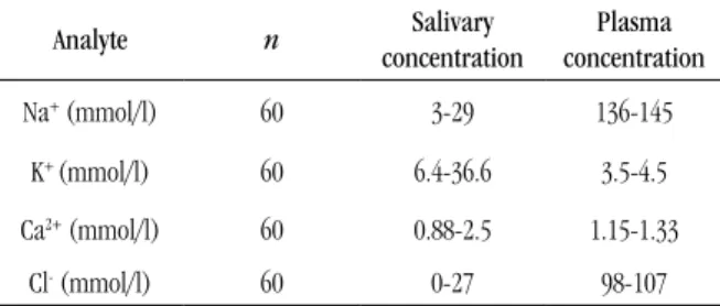
function as buffering agents. The electrolyte concentrations in the saliva may be quantiied by lame photometry and ion-selective electrode. Table 1 shows reference intervals for some electrolytes in the saliva compared with plasma(52).
K+ concentration in saliva is well above the values observed in
plasma. Conversely, the concentration of Na+ and Cl- in this luid is lower
in relation to plasma(2). As a result of this difference in concentrations
when analyzing potassium through ion selective electrode (ISE), the dilution of saliva samples is recommended. On the other hand, sample
dilution is not required to measure Na+ and Cl-. Colorimetric methods
or lame photometry are preferable, insofar as salivary concentrations are below the detection range reported by ISE(52).
Physical exercise of varying intensity and duration may change saliva ionic concentration, especially Na+ and K+(11). The
salivary concentrations of Na+, K+ and Cl- appear to be linked
with the lactate anaerobic threshold during incremental test on ergometric cycle(12). These changes may be credited to sympathetic
stimulation, which induces changes in salivary low, reabsorption and secretion of electrolytes in the secretory cells(11).
Non-protein organic compounds
Some non-protein organic compounds may be found in low concentrations in the saliva. They fulill different functions and mostly originate from the plasma by ultrailtration. They may be occasionally present due to contamination with blood from lesions of the oral mucosa. Thus, negligible amounts of bilirubin, creatinine, glucose, cholesterol and triglycerides may be detected in the saliva from healthy individuals(52).
The salivary concentrations of urea (2.9 to 6.8 mmol/l)(52) and
uric acid (0.07-32 mmol/l)(45) correspond to metabolic changes
resulting from pathologies such as renal dysfunction and gout. In individuals with renal involvement, salivary concentrations of urea may rise dramatically (from 6.1 to 29.6 mmol/l), following plasma levels(8).
Uric acid is the major antioxidant present in the saliva and contributes to approximately 70% of the total antioxidant capacity(38). Its quantiication has been deployed in monitoring
the effects of hemodialysis(6) and in the assessment of patients with
gout(47). Acute exercise promotes increases in salivary uric acid
levels(19, 47) and total antioxidant capacity when compared with
baseline(19). Nevertheless, Youssef et al.(61) demonstrated that the
total salivary antioxidant capacity of triathletes fell signiicantly by the end of the season when the values were compared with the beginning of the training period. Other important organic
TABLE 1 – Reference interval (2.5 to 97.5 percentile) for
electrolytes measured in saliva collected by passive method and plasma
Analyte n Salivary
concentration
Plasma concentration
Na+ (mmol/l) 60 3-29 136-145
K+ (mmol/l) 60 6.4-36.6 3.5-4.5
Ca2+ (mmol/l) 60 0.88-2.5 1.15-1.33
Cl- (mmol/l) 60 0-27 98-107
molecules such as ascorbic acid and vitamin E make up the salivary antioxidant defense system(38).
Automated spectrophotometric techniques that evaluate non-enzymatic salivary antioxidant capacity may be useful for monitoring athletes due to its non-invasive nature. However, speciic salivary reference values are required, insofar as there is no signiicant correlation between the antioxidant capacity in saliva and plasma(45).
Another important compound to be applied in sports is lactate. Some studies analyzed the salivary lactate response to exercise in an attempt to determine the intensity of training or assess the correlation between blood and salivary lactate(11, 46). No change
was observed in salivary lactate during aerobic exercise compared with rest period.
HORMONES
Most hormones present in the plasma may be measured in the saliva due to a connection between plasma and salivary glands(10).
The primary entry route of steroid hormones and other small and neutral molecules in the saliva is through passive diffusion. The salivary glands are intensively irrigated by capillary networks and many of the liposoluble blood components easily pass through the capillary walls into the salivary glands(21).
Serum proteins and globulins that bind to hormones are large molecules that do not pass through the cell membranes of salivary glands. Accordingly, only unbound hormones (free fraction) present in plasma may diffuse into saliva. Thus, unconjugated steroid hormones (such as cortisol, estriol, progesterone and testosterone) diffuse predominantly intracellularly, do not inluence salivary low and present very similar concentrations of free plasma fraction(58).
On the other hand, electrically charged steroids or those conjugated with proteins are able to pass, albeit in small quantities, through the gap junctions between salivary gland cells. As this process is very slow, these hormones tend to have their values strongly affected by salivary low, with much lower concentrations in relation to plasma(58). It is particularly worth mentioning that these analyzes
may encounter interference due to contamination with blood. Small
amounts of blood originated from oral and gingival lesions may
cause false rises in the concentrations of salivary analytes(54).
Cortisol is a hormone produced by the adrenal glands and released into the circulation. It has important metabolic functions, such as controlling blood pressure, cardiovascular function and immune system(53). In peripheral tissues, cortisol
stimulates lipolysis, increases protein degradation and lowers protein synthesis in muscle cells, resulting in higher release of amino acids and lipids in the circulation(31). The secretion
of this hormone undergoes circadian variation, with higher concentrations in the morning and lower concentrations in the
afternoon(39). Some studies reveal that the free fraction in plasma
cortisol correlates with salivary concentrations(35, 58), enabling the
use of this marker in the assessment of responses to physical and psychological stress(53).
Physical activity is able to raise dramatically the concentration of salivary cortisol in athletes subjected to different forms of exercise(3). In addition to evaluating the adaptive responses to
training, cortisol may be an interesting biomarker to investigate the levels of tolerance or intolerance to training when performed in conjunction with testosterone measurement and subsequent calculation of testosterone: cortisol ratio(57).
Despite the convenience of the use of salivary cortisol in the assessment of athletes and patients with pathologies that alter the secretion of corticosteroids, caution is required in interpreting data obtained with saliva samples. Few studies establish reference values for salivary cortisol considering circadian variation and in accordance with the protocol proposed by the International Federation of Clinical Chemistry (IFCC)(48).
Other synthetic or natural androgenic hormones that are administered illegally with the aim to improve performance may diffuse passively into the saliva(58). Although it is possible to
quantify testosterone, epitestosterone, tetrahydrogestrinone and other hormones used by athletes for illicit purposes, the World Anti Doping Agency (WADA) provides no information on the use of saliva in the detection and control of doping(21).
METHODS FOR SALIVA COLLECTION
Saliva can be easily collected through passive drool directly into plastic tubes (unstimulated saliva). The passive collection is the most recommended method, inasmuch as most analytes may be quantiied without hindrances. However, the supplied volume is low in this case(10). Neither teeth brushing nor food and liquid
ingestion (except water) are recommended for at least 30 minutes prior to collection. Mouth cleaning with water (preferably distilled) helps to eliminate residues that may hamper the analysis(10).
The stimulated saliva may be collected through gustatory stimulation, mastication or the use of citric acid(30). There are some
and recovery of the collected liquid. Among the most common commercial systems that use solid bases are: Oral Salimetrics Swab (Salimetrics® LLC) Salivette® and Cortisol® Salivette (Sarstedt,
Newton, NC) and Orapette (Trinity Biotech, Dublin, Ireland)(22).
None of the collection systems mentioned above allow direct and accurate volume quantiication, which is a common drawback. Furthermore, saliva and analyte recovery may vary considerably in each method, mainly owing to the fact that some analytes may adhere to the cotton, causing false lower values(14, 22, 37).
Another currently available collection system is the Saliva Collection System (SCS)® (Greiner Bio-One, GmgH,
Kremsmuenster, Austria), which uses a buffered base of citric acid for saliva collection. This collection system is more elaborate and presents a solution for oral cavity cleaning and standardization of collection procedures. After applying a cleaning solution, the individual places the extracting solution in their mouth for 2 minutes and then spits out all the solution into a beaker. The collected saliva (saliva + extraction solution) is subsequently transferred via vacuum system to graduated transport tubes, which permit to quantify the total volume of collected solution(22, 45).
This system of saliva collection has been recently tested using routine laboratory methods and has demonstrated reliable and reproducible results for measurement of calcium, magnesium(49),
α-amylase, uric acid(45), IgA, cortisol and therapeutic drugs(22).
Nonetheless, certain analytes are prone to interference due to pH and additives present in the extraction solution(45). Compared with
other commercially available systems, SCS requires further care during the collection so that there is no exchange of washing and extraction solutions. Moreover, it is highly costly(22).
Pre-analytical and analytical interferences in the
analysis of saliva samples
One of the most important pre-analytical interferences to be considered is the need to quantify time and volume of collected
saliva in order to estimate the saliva secretion rate. The saliva low rate through the salivary ducts determines, for instance, the concentration of electrolytes. The increase in salivary low through artiicial stimulation may change the ionic composition, raising concentrations of Na+, Cl- and bicarbonate and reducing
salivary K+(2). Some salivary components such as IgA and
dehydroepiandrosterone-sulfate (DHEA-S) are dependent on salivary low, requiring correction according to volume and collection time(2, 58).
The use of citric acid and chewing gum as stimulation and materials with cotton or polyester may inaccurately augment salivary testosterone levels. The use of collection devices with cotton is not recommended for the analysis of hormones due to the possibility of interference in the performance of immunoassays(21).
Cotton may bind to cortisol and dehydroepiandrosterone (DHEA) and cause a false decrease(17, 23).
Besides the choice of a suitable collection system, the collection time should always be standardized and observed. Hormones and other saliva constituents may present major circadian variations, which may require a pre-established collection schedule.
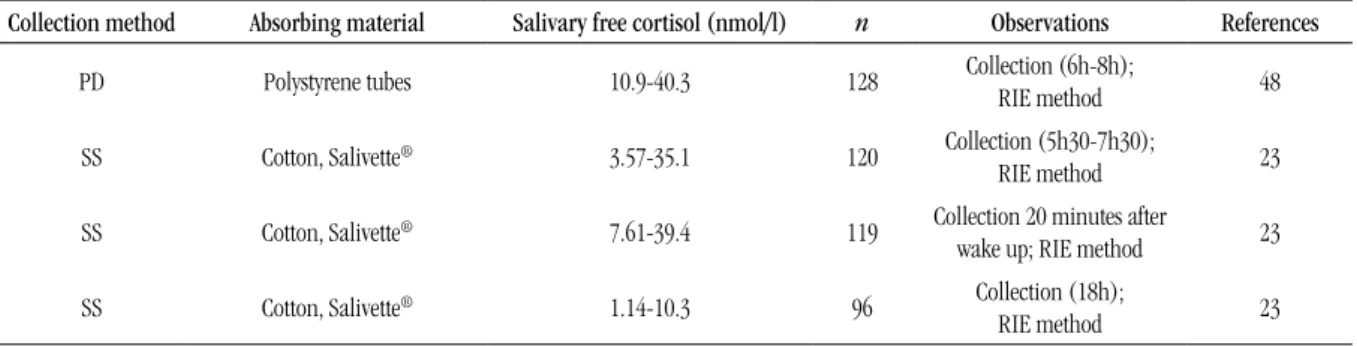
Table 2 shows that the reference interval for salivary
cortisol may vary with the method or time of collection. The intake of alcohol, caffeine and protein-rich diets may alter cortisol levels(24).
Saliva samples should be refrigerated (4ºC) if they are processed within 3 to 6 hours after collection. When the sample is stored for long periods, the temperature should be kept at -80ºC to prevent bacterial growth(24). The salivary cortisol is stable at 5ºC
for three months and up to one year when stored within -20ºC to -80ºC. Repeated freezing and thawing cycles do not affect cortisol concentrations(24). The addition of sodium azide in saliva
may inhibit bacterial growth and preserve the sample, though immunoassays using horseradish peroxidase may be subject to interference(10).
TABLE 2 –Reference intervals for salivary free cortisol at different collection periods
Collection method Absorbing material Salivary free cortisol (nmol/l) n Observations References
PD Polystyrene tubes 10.9-40.3 128 Collection (6h-8h); RIE method 48
SS Cotton, Salivette® 3.57-35.1 120 Collection (5h30-7h30);
RIE method 23
SS Cotton, Salivette® 7.61-39.4 119 Collection 20 minutes after
wake up; RIE method 23
SS Cotton, Salivette® 1.14-10.3 96 Collection (18h);
RIE method 23
Reference range (2.5 to 97.5 percentile).
CONCLUSION
The use of saliva in laboratory testing has great potential, although it is still required to standardize some preanalytical variables, such as the collection system, taking into account the analyte to be quantiied, the collection schedules, direct quantiication of volume, sample recovery and prevention of sample contamination with blood from oral mucosa lesions.
The lack of correlation between some analytes present in saliva and blood does not preclude the possibility of using this luid in laboratories, though it requires the establishment of speciic reference intervals for these analyses through traditional methods, with minor modiications in the techniques to maximize sensitivity.
RESUMO
A utilização de saliva como alternativa para o diagnóstico de patologias e/ou monitoramento de atletas em competições ou treinos é muito atrativa devido à facilidade de obtenção da amostra e, principalmente, pela natureza menos invasiva que a coleta de sangue venoso. A saliva é um luído hipotônico em relação ao plasma; contém compostos produzidos localmente nas glândulas salivares (imunoglobulina A [IgA] e α-amilase), além de compostos difundidos do plasma (água, eletrólitos, proteínas, metabólitos e hormônios). A saliva desempenha funções importantes na proteção da mucosa oral contra microrganismos e na digestão dos alimentos. Sua produção e sua composição são dependentes da atividade do sistema nervoso autônomo simpático e parassimpático, cuja ação antagônica pode resultar em diferentes volumes de saliva com peris proteico e iônico distintos. O objetivo da presente revisão é apresentar uma análise crítica das potencialidades e limitações da utilização da saliva como ferramenta diagnóstica para a medicina esportiva. Embora existam estudos que a utilizam para o monitoramento de atletas em situações de exercício e doping, ainda é necessário padronizar algumas variáveis pré-analíticas, como a escolha correta do melhor sistema de coleta, que permite quantiicar facilmente o volume, com boa recuperação de amostra; os horários de coleta bem deinidos, de acordo com as possíveis variações circadianas do analito; e a contaminação da saliva com sangue proveniente de lesões da mucosa oral, que tem de ser evitada. Outro ponto fundamental para aplicação no esporte é o estabelecimento de valores de referência para analitos quantiicados na saliva, obtidos de uma população composta de sujeitos saudáveis e exercitados de forma constante e sistematizada, com progressão de cargas de esforço.
Unitermos: luido oral; coleta de saliva; variáveis pré-analíticas; exercício; valores de referência.
REFERENCES
1. ALLGROVE, J. E. et al. Effects of exercise intensity on salivary antimicrobial proteins and markers of stress in active men. J Sports Sci,
v. 26, n. 6, p. 653-61, 2008.
2. APS, J. K.; MARTENS, L.C. Review: the physiology of saliva and transfer of drugs into saliva. Forensic Sci Int, v. 150, p. 119-31, 2005.
3. AUBETS, J.; SEGURA, J. Salivary cortisol as a marker of competition related stress. Science & Sports, v. 10, p. 149-54, 1995.
4. BISHOP, N.C.; GLEESON, M. Acute and chronic effects of exercise on markers of mucosa immunity. Front Biosci, v. 14, p. 4444-56, 2009. 5. BLANNIN, P. J. et al. The effect of exercising to exhaustion at different intensities on saliva immunoglobulin A, protein and electrolyte secretion.
Int J Sports Med, v. 19, p. 546-52, 1998.
6. BLICHARZ, T. M. et al. Use of colorimetric test strips for monitoring
the effect of hemodialysis on salivary nitrite and uric acid in patients
with end-stage renal disease: a proof of principle. Clin Chem, v. 54, n. 9,
p. 1473-80, 2008.
7. CALVO, F. et al. Anaerobic threshold determination with analysis of
salivary amylase. Can J Appl Physiol, v. 22, p. 553-61, 1997.
8. CARDOSO, E. M. L. et al. Assessment of salivary urea as a less invasive alternative to serum determinations. Scand J Clin Lab Invest, v. 69, n. 3, p. 330-4, 2009.
9. CHATTERTON, R. T. et al. Salivary alpha-amylase as a measure of endogenous adrenergic activity. Clin Physiol, v. 16, n. 4, p. 433-8, 1996. 10. CHIAPPIN, S. et al. Saliva specimen: a new laboratory tool diagnostic and basic investigation. Clin Chim Acta, v. 383, p. 30-40, 2007. 11. CHICHARRO, J. L. et al. Saliva composition and exercise. Sports Med,
v. 26, n.1, p. 17-27, 1998.
14. DeCARO, J. A. Methodological considerations in the use of salivary a-amylase as a stress marker in ield research. Am J Hum Biol, v. 20,
p. 617-9, 2008.
15. DENNY, P. et al. The proteome of human parotid and submandibular sublingual gland salivas collected as the ductal secretions. J Proteome Res, v. 7, p. 1994-2006, 2008.
16. FOX, P. C. Xerostomia: evaluation of a symptom with increasing signiicance. J Am Dent Assoc, v. 110, n. 4, p. 519-25, 1985.
17. GALLAGHER, P. et al. Assessing cortisol and dehydroepiandrosterone (DHEA) in saliva: effects of collection method. J Psychopharmacol, v. 20,
n. 5, p. 643-9, 2006.
18. GLEESON, M.; PYNE, D. B. Exercise effects on mucosa immunity.
Immunol Cell Biol, v.78, p. 536-44, 2000.
19. GONZALEZ, D.; MARQUINA, R.; RONDÓN, N. Effects of aerobic exercise on uric acid, total antioxidant activity, oxidative stress, and nitric oxide in human saliva. Res Sports Med, v. 16, p.128-37, 2008.
20. GRANGER, D. A. et al. Salivary α-amylase in biobehavioral research.
Ann NY Acad Sci, v. 1098, p. 122-44, 2007.
21. GRÖSCHL, M. Current status of salivary hormone analysis. Clin Chem, v. 54, n. 11, p. 1759-69, 2008.
22. GRÖSCHL, M. et al. Evaluation of saliva collection devices for the analysis of steroids, peptides and therapeutic drugs. J Pharma Biomed Anal, v. 47, p. 478-86, 2008.
23. HANSEN, A. M. et al. Evaluation of a radioimmunoassay and establishment of a reference interval for salivary cortisol in healthy subjects in Denmark. Scand J Clin Lab Invest, v. 63, p. 303-10, 2003. 24. HANSEN, A. M.; GARDE, A. H.; PERSSON, R. Sources of biological and methodological variation in salivary cortisol and their impact on measurement among healthy adults: a review. Scand J Clin Lab Invest,
v. 68, n. 6, p. 448-58, 2008.
25. HU, S.et al. Large scale identiication of proteins in human salivary proteome by liquid chromatography/mass spectrometry and two-dimensional gel electrophoresis mass spectrometry. Proteomics, v. 5, p. 1714-28, 2005.
26. HUANG, C. M. Comparative proteomic analysis of human whole saliva. Arch Oral Biol, v. 49, n.12, p. 951-62, 2004.
27. HUMPHREY, S. P.; WILLIAMSON, R.T. A review of saliva: normal composition, low, and function. J Prosthet Dent, v. 85, n. 2, p.162-9, 2001.
28. JOU, Y. J. et al. Proteomic identiication of salivary transferrin as a biomarker for early detection of oral cancer. Anal Chim Acta, v. 681, p. 41-8, 2010.
29. KALK, W. W. I. et al. Sialometry and sialochemistry: a non-invasive
approach for diagnosing Sjögren’s syndrome. Ann Rheum Dis, v. 61, p. 137-44, 2002.
30. KAUFMAN, E.; LAMSTER, I. B. The diagnostic application of saliva.
Crit Rev Oral Biol Med, v. 13, n. 2, p. 197-202, 2002.
31. KRAEMER, W. J.; RATAMESS, N. A. Hormonal responses and adaptations to resistance exercise and training. Sports Med, v. 35, n. 4, p. 339-61, 2005. 32. LANGMAN, L. J. The use of oral luid for therapeutic drug management.
Ann NY Acad Sci, v. 1098, p.145-66, 2007.
33. LEVINE, M. J. Salivary macromolecules. A structure/function synopsis.
Ann N Y Acad Sci, v. 20, n. 694, p. 11-6, 1993.
34. LI, T. L.; GLEESON, M. The effect of single and repeated bouts of prolonged cycling and circadian variation on saliva low rate, immunoglobulin A and-amylase responses. J Sports Sci, v. 22, p. 1015-24,
2004.
35. LIPPI, G. et al. Measurement of morning saliva cortisol in athletes.
Clin Biochem, v. 42, p. 904-6, 2009.
36. McDOWELL, S. L. et al. The effect of exercise intensity and duration on salivary immunoglobulin A. Eur J Appl Physiol, v. 63, p.108-11, 1991. 37. MICHISHIGE, F. et al. Effect of saliva collection method on the concentration of protein components in saliva. J Med Invest, v. 53,
p.140-6, 2006.
38. NAGLER, R. M. et al. Characterization of the differentiated antioxidant
proile of human saliva. Free Radic Biol Med, v. 32, n. 3, p. 268-77, 2002. 39. NATER, U. M. et al. Determinants of the diurnal course of salivary alpha-amylase. Psychoneuroendocrinology, v. 32, p. 392-401, 2007.
40. NATER, U. M. et al. Human salivary alpha-amylase reactivity in a psychosocial stress paradigm. Int J Psychophysiol, v. 55, n. 3, p. 333-42,
2005.
41. NATER, U. M.; ROHLEDER, N. Salivary alpha-amylase as a non-invasive biomarker for the sympathetic nervous system: current state of research. Psychoneuroendocrinology, v. 34, p. 486-96, 2009.
42. NIEMAN, D. C. et al. Change in salivary IgA following a competitive
marathon race. Int J Sports Med, v. 23, p. 69-75, 2002.
43. NIEUW AMERONGEN, A.V.; LIGTTENBERG, A. J.; VEERMAN, E. C. Implications for diagnostics in the biochemistry and physiology of saliva.
Ann NY Acad Sci, v. 1098, p. 1-6, 2007.
44. NUNES L. A. S. et al. Adequacies of skin puncture for evaluating
biochemical and hematological parameters in athletes. Clin J Sport Med, v.16, n. 5, p. 418-21, 2006.
45. NUNES L. A. S. et al. Reference intervals for saliva analytes collected by a standardized method in a physically active population. Clin Biochem, v. 44, p. 1440-4, 2011.
46. OHKUWA, T. et al. Salivary and blood lactate after supramaximal exercise in sprinters and long distance runners. Scand J Med Sci Sports, v. 5, p. 285-90, 1995.
47. OWEN-SMITH, B.; QUINEY, J.; READ, J. Salivary urate in gout, exercise and diurnal variation. Lancet, v. 351, n. 27, p. 1932, 1998.
48. PATEL, R. S. et al. Production of gender-speciic morning salivary cortisol reference intervals using internationally accepted procedures.
Clin Chem Lab Med, v. 42, n. 12, p. 1424-9, 2004.
49. RAGGAM, R. B. et al. Evaluation of a novel standardized system for collection and quantiication of oral luid. Clin Chem Lab Med, v. 46,
n. 92, p. 287-91, 2008.
50. RAGGAM, R. B. et al. Reliable detection and quantiication of viral
nucleic acids in oral luid: liquid phase-based sample collection in conjunction with automated and standardized molecular assays. J Med
Virol, v. 80, p. 1684-8, 2008.
51. RAO P. V. et al. Proteomic identiication of salivary biomarkers of type-2 diabetes. J Proteome Res, v. 8, n. 1, p. 239-45, 2009.
52. REHAK, N. N.; CECCO, S. A.; CSAKO, G. Biochemical composition and electrolyte balance of “unstimulated” whole human saliva. Clin Chem
MAILING ADDRESS
Lázaro Alessandro Soares Nunes
Rua Monteiro Lobato, s/n; Instituto de Biologia; Departamento de Bioquímica, sala 12; Barão Geraldo; CEP: 13083-970; Campinas-SP; Brazil; e-mail: [email protected].
.
53. SAPOLSKY, R. M.; ROMERO, L. M.; MUNCK, A. U. How do glucocorticoids inluence stress responses? Integrating permissive, suppressive, stimulatory, and preparative actions. Endocr Rev, v. 21, p. 55-89, 2000.
54. SCHWARTZ, E. B.; GRANGER, D. A. Transferrin enzyme immunoassay for quantitative monitoring of blood contamination in saliva. Clin Chem,
v. 50, n. 3, p. 546-55, 2004.
55. STRANO-ROSSI, S. et al. Analysis of stimulants in oral luid and urine by gas chromatography-mass spectrometry II: pseudoephedrine. J Anal
Toxicol, v. 34, n. 4, p. 210-5, 2010.
56. TRILCK, M. et al. Salivary cortisol measurement - a reliable method for diagnosis of cushing´s syndrome. Exp Clin Endocrinol Diabetes, v. 113, p. 225-30, 2005.
57. URHAUSEN, A.; KINDERMANN, W. Diagnosis of overtraining. What tools do we have? Sports Med, v. 32, n. 2, p. 95-102, 2002.
58. VINING, R. F.; McGINLEY, R. A.; SYMONS, R. G. Hormones in saliva: mode of entry and consequent implications for clinical interpretation.
Clin Chem, v. 29, n. 10, p. 1752-6, 1983.
59. WALSH, N. P. et al. Salivary IgA response to prolonged exercise in a cold environment in trained cyclists. Med Sci Sports Exerc, v. 34, p.1632-7, 2002.
60. WONG, D. T. Salivary diagnostics powered by nanotechnologies, proteomics and genomics. JADA, v. 137, p. 313-21, 2006.