Greater resistance of the rabbit antropyloric region
to experimental acute gastric ischemia
Maior resistência da região antropilórica de coelhos
à isquemia gástrica aguda experimental
Maria Angélica B. Magalhães1; Andy Petroianu2; Juliano A. Figueiredo3; Luiz R. Alberti4; Alfredo J. A. Barbosa5
First submission on 13/05/13; last submission on 17/06/13; accepted for publication on 17/06/13; published on 20/08/13 1. Assistant professor at Universidade José do Rosário Vellano-Medical School (Unifenas), Belo Horizonte campus. 2. Professor of Surgery at Universidade Federal de Minas Gerais-Medical School (UFMG).
3. Associate professor at Faculdade de Saúde e Ecologia Humana de Minas Gerais (FASEH). 4. Associate professor at UFMG-Medical School.
5. Professor of Pathology at UFMG-Medical School.
ABStrACt
Introduction: Gastric ischemia represents an important medical challenge in pathology and surgical practice. Objective: The purpose of this study was to assess the effects of acute gastric ischemia on different regions of the stomach. Method: Rabbit stomachs were subjected to devascularization of the greater and lesser curvatures for 3, 6 and 12 hours. After these periods, the stomachs were removed for macro and microscopic analysis. Results: Hemorrhagic necrosis was more marked in the gastric fundus and body. In contrast, the antropylorus remained preserved in 100% of the rabbits after 3 hours of ischemia (group I), and in 80% of the rabbits after 6 and 12 hours of ischemia (groups II and III). Necrosis of the gastric body and fundus mucosa were observed in all animals after 6 and 12 hours of ischemia. Conclusion: We concluded that this experimental model of acute gastric ischemia was effective in producing hemorrhagic necrosis of the gastric fundus and body in rabbits even within a short period of time. Furthermore, the antropyloric region was preserved in most animals.
Key words:experimental acute gastric ischemia; gastric devascularization; stomach; gastric infarction; rabbit.
introDuCtion
Gastric ischemia may occur due to reduced or complete obstruction of the arterial or venous blood low to the stomach mainly due to thromboembolism, phlebitis and gastric volvulus(9, 19).
Non-obstructive factors such as sepsis, congestive heart failure, side effects of digitalis and alpha-adrenergic agents may provoke a reduction of cardiac output, tissue hypoperfusion and gastric ischemia(9, 18). Moreover, the stomach is an organ commonly
subjected to a large number of surgical procedures which are prone
to cause gastric ischemia. Among these surgeries, some are worth mentioning, namely surgical correction of portal hypertension, reversion of gastric volvulus, subtotal gastrectomies to treat gastric neoplasias, esophago-gastroplasties to reconstruct the digestive tube after esophagectomies, and gastroplasties of obese patients(6, 11, 17, 20).
The most severe complication of gastric ischemia is necrosis of the stomach and its life threatening complication.
few studies dealing with acute gastric ischemia in the various regions of the stomach are available. The purpose of the present investigation was to study the effects of acute ischemia due to simultaneous arterial and venous obstruction on the wall of the fundus, body and antrum of the stomach.
MEthoD
This study was performed on ifteen White New Zealand male rabbits weighing 2.500 to 3.000 g. The experiment was carried out according to the ethical norms for animal experimentation and was approved by the Ethics Committee for Animal Experimentation of the Federal University of Minas Gerais, Belo Horizonte, Brazil.
The animals were anaesthetized with an injection into the gluteal region of 2% xylazine at 10 mg/kg in combination with 10% ketamine at 60 mg/kg. When necessary, a quarter of the initial anesthetic dose was additionally applied(4, 5). The same
surgical technique was deployed for all animals. After laparotomy, the entire gastric vasculature, including veins and arteries, was ligated and sectioned along the greater and lesser curvature of the stomach, with the organ ixed in place only through the esophagus and duodenum. Afterwards, laparorrhaphy was performed.
The animals were divided into three groups of ive animals each according to time of observation after surgery: 3 hours (group I), 6 hours (group II) and 12 hours (group III). After the predetermined observation periods, the animals were euthanized with an overdose of anesthetic followed by an intravenous injection of 10% potassium chloride. Subsequently, the stomachs were removed en bloc for macro- and microscopic study.
The removed stomachs were opened along the greater curvature and washed with running water. Changes compatible with hemorrhagic necrosis of the fundus, body and antrum were recorded and fragments of these areas and of pre-established areas
of each gastric region were collected, ixed in 4% formaldehyde, processed for parafin embedding, and stained with hematoxylin and eosin (HE) for histological study.
rESuLtS
Macroscopic examination of the stomach of all animals in the three groups showed localized or diffuse changes indicative of hemorrhagic necrosis (Figures 1 and 2). The gastric fundus and
body were the most affected regions in all rabbits. In most animals the antral region was preserved, presenting only mild congestive changes of the mucosa in one animal each in groups II and III. All animals of groups I, II and III displayed macroscopic lesions indicative of hemorrhagic necrosis of various extension in the body and fundus and of greater intensity in groups II and III (6 and 12 hours) compared to group I (3 hours) (Table 1).
tABLE 1 – Macroscopic changes indicative of localized or diffuse hemorrhagic necrosis of the antrum,
body and fundus of rabbit stomachs submitted to gastric ischemia for 3, 6 and 12 hours (groups I, II and III, respectively)
Groups
Antrum Body Fundus
Localized n (%)
Diffuse n (%)
Localized n (%)
Diffuse n (%)
Localized n (%)
Diffuse n (%) Group I
n = 5 - - 2 (40) 3 (60) 2 (40) 3 (60)
Group II
n = 5 - - 1 (20) 4 (80) - 5 (100)
Group III
n = 5 - - - 5 (100) - 5 (100)
Microscopic changes in stomach tissues became more conspicuous with increasing time of gastric ischemia. Edema and vessel congestion were observed in the mucosa, submucosa and muscle layers of the three gastric regions in all animals. Areas of mucosal necrosis of the body and fundus occurred in all animals after three hours of gastric ischemia and only one animal in this group exhibited necrosis of the muscle layer in the region of the gastric fundus (Table 2 and Figures 3 and 4).
Necrosis of the mucosa of the antropyloric region was detected in only one (20%) rabbit each in groups II and III, and the muscle layer of this region was preserved in all animals of the three groups. Hemorrhagic necrosis of the mucosa of the gastric body and fundus was observed in all animals of groups II
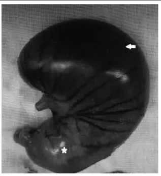
figurE 2 – Macroscopic aspect of the opened stomach of the same rabbit of Figure 1 after 6 hours of gastric ischemia: presence of diffuse hemorrhagic necrosis in the body and fundus regions (arrow), whereas the antral region (*) is preserved
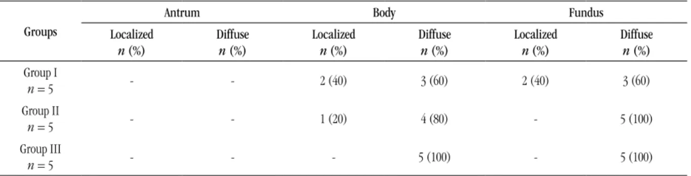
figurE 3 – Macroscopic aspect of the closed stomach of the rabbit after 12 hours of gastric ischemia. The outer surface of the stomach exhibits diffuse hemorrhagic necrosis of the body and fundus regions (arrow), whereas the antropyloric region (*) is preserved
(6 hours) and III (12 hours). Two animals (40%) developed necrosis of the muscle layer in the gastric body and fundus after 6 hours of ischemia (group II). After 12 hours of ischemia (group III), necrosis of the muscle layer was noticed in the gastric body of three animals (60%) and in the fundus of four animals (80%) (Table 2).
DiSCuSSion
Gastric ischemia is commonly followed by poor prognosis. Despite the rich vascular arcade of the stomach, complications of ischemic origin affecting this organ have been frequent in humans, due in part to the increasing number of abdominal operations that interfere with the vascularization of this organ. The symptoms of
tABLE 2 – Microscopic changes of the rabbit stomach indicative of hemorrhagic necrosis in the mucosal and muscle
layers of the antrum, body and fundus regions after 3, 6 and 12 hours of gastric ischemia (groups I, II and III, respectively)
Groups
Antrum Body Fundus
Mucosal n (%)
Muscle n (%)
Mucosal n (%)
Muscle n (%)
Mucosal n (%)
Muscle n (%) Group I
n = 5 - - 5 (100) - 5 (100) 1 (20)
Group II
n = 5 1 (20) - 5 (100) 2 (40) 5 (100) 2 (40)
Group III
gastric ischemia are usually nonspeciic, ranging from local pain to an intense acute abdomen(12). Even without a precise diagnosis,
surgical treatment must be rapid, otherwise mortality may be above 80%(7). Resection of necrotic segment is the appropriate
treatment and total gastrectomy may be sometimes required(12).
The effects of gastric devascularization on the vitality of the stomach have been evaluated in few experimental studies. In dogs, complete devascularization of the stomach wall may result in gangrene of the organ and cause death of the animals(3).
In humans, partial stomach devascularization is a frequent procedure. Ligation of the right and left gastric veins and those of the right and posterior upper wall of the stomach are the treatments adopted to reduce the hypertension of gastric varices(2).
Acute hemorrhagic lesions of the gastroduodenal mucosa refractory to clinical treatment represent other indications for local devascularization of the stomach(14, 15, 21). Venous obstruction alone
may have the same ischemic effects as arterial obstruction on the gastric wall. Venous thrombosis, phlebitis and coagulopathies are predisposing factors of ischemia(13).
In the present study, devascularization was performed in the entire wall of the stomach, including all veins and arteries. The yielded results showed that the model of gastric ischemia was effective in all animals, as conirmed by macro- and microscopic examination, which revealed varied degrees of necrosis of the gastric wall. Gastric ischemia, even when induced for a relatively
short period of time, may cause severe and irreversible injuries to one or more different tissues of the stomach wall.
As observed, the gastric fundus and body were the regions more sensitive to ischemia, whereas the antrum was preserved in practically all animals. Since all the vessels of the greater and lesser curvatures were sectioned, the most likely explanation for the preservation of the antropyloric region is based on two possibilities. The irst refers to the rich vascular anastomosis present in the gastroduodenal interface. These anastomoses derive from small branches of the gastroduodenal artery, which derive from the hepatic artery and are largely responsible for the formation of the vascular plexuses present in the submucosa of the more distal regions of the stomach. Submucosal microvessels originating from the duodenum probably provide supplementary blood irrigation to the gastric antrum, leading to a greater resistance to ischemia. This vascular distribution is well known to occur in humans and is closely similar to that of rabbits(1, 10, 16). In addition, the anatomical
variations of each individual may contribute to the maintenance of blood irrigation in speciic regions of the stomach(8). Thus, it
is possible that the results obtained with this experimental model may be worth to be applied to humans. Secondly, the preservation of the gastric antrum vitality in the animals studied herein may be explained by the relatively short time of induced ischemia (3 to 12 hours). As this region of the stomach appears to beneit from a larger number of vascular anastomoses, it may resist to general gastric ischemia for a longer period of time. This possibility is supported by the macro- and microscopic indings, which revealed lesions of greater intensity in the upper gastric regions even when the time of ischemia was increased to 12 hours.
In conclusion, the experimental model of acute ischemia of the stomach proposed herein, based on ligation and section of all gastric vessels, was effective in promoting hemorrhagic necrosis of the gastric body and fundus within a short period of time. On the other hand, the antropylorus was signiicantly more resistant to ischemia, showing no signiicant changes in most animals, probably owing to the presence of rich microvascular anastomosis in the gastroduodenal junction.
ACknowLEDgEMEntS
This study received inancial support from Fundação de Amparo à Pesquisa do Estado de Minas Gerais (FAPEMIG) and the National Council for Scientiic and Technological Development (Conselho Nacional de Desenvolvimento Cientíico e Tecnológico [CNPq]), Brazil. The authors thank Mrs. Fernanda Césari Barros for technical assistance.
rESuMo
Introdução: Isquemia gástrica representa um importante desaio médico nas áreas de Patologia e Cirurgia. O objetivo do presente
trabalho foi avaliar os efeitos da isquemia gástrica aguda nas diferentes regiões do estômago. Método: Estômagos de coelhos foram
submetidos à completa desvascularização das curvaturas maior e menor durante 3, 6 e 12 horas. Após esses períodos de tempo,
os órgãos foram removidos para análise macro e microscópica. Resultados: Necrose hemorrágica foi mais evidente nas regiões
de corpo e fundo. Por outro lado, a região antropilórica manteve-se preservada em 100% dos coelhos após 3 horas de isquemia (grupo I), e em 80% dos coelhos após 6 e 12 horas de isquemia (grupos II e III). Necrose da camada mucosa do corpo e do fundo
gástricos foi observada em todos os animais após 6 e 12 horas de isquemia. Conclusão: Conclui-se que esse modelo de isquemia
gástrica aguda foi eicaz para produzir necrose hemorrágica do corpo e fundo de coelhos mesmo por um curto período de tempo.
Além disso, a região antropilórica manteve-se preservada na maioria dos animais.
Unitermos: isquemia gástrica experimental; desvascularização gástrica; estômago; infarto gástrico; coelho.
rEfErEnCES
1. ABIDU-FIGUEIREDO, M. et al. Celiac artery in New Zealand rabbit:
anatomical study of its origin and arrangement for experimental research and surgical practice. Pesq Vet Bras, v. 28, p. 237-40, 2008.
2. BERMAN, J. K.; HULL, J. E. Hepatic splenic and left gastric arterial ligations in advanced portal cirrhosis. Arch Surg, v. 65, p. 37-53, 1952.
3. BERNHEIM, B. M. Partial and total devascularization of the stomach. Ann Surg, v. 96, p. 179-83, 1932.
4. FLECKNELL, P. A. Anaesthesia of animals for biomedical research. Br J Anaesth, v. 71, p. 885-94, 1993.
5. FONSECA, N. M. et al. Anestesia em coelhos. Acta Cir Bras, v. 11, p. 82-104, 1996.
6. HARRISON, B. J.; GLANGES, E.; SPARKMAN, R. S. Gastric istula following splenectomy: it’s cause and prevention. Ann Surg, v. 185, p. 210-3, 1977.
7. KOYAZOUNDA, A. et al. Necrose gastric par dilatation gastrique aigue. J Chir, v. 122, p. 403-7, 1985.
8. MALINOVSKY, L. et al. Vascular anastomosis among abdominal organs in laboratory animals. G Chir, v. 18, p. 602-4 1997.
9. MCKINSEY, J. F.; GEWERTZ, B. L. Acute mesenteric ischemia. Surg Clin North Am, v. 77, p. 307-18, 1997.
10. MOSKALEWSKI, S. et al. Venous outlow system in rabbit gastric mucosa. Folia Morphol, v. 63, p. 151-7, 2004.
11. PETROIANU, A. et al. Tensão de ruptura dos órgãos que constituem o tubo digestório com e sem o uso de corticoide em camundongos. Rev Col Bras Cir, v. 27, p. 69-72, 1999.
12. REEVE, T. et al. Near-total gastric necrosis caused by acute gastric dilatation. South Med J, v. 81, n. 4, p. 515-7, 1988.
13. RIBEIRO, M. E; YOSHIDA, W. B. Lesões intestinais decorrentes de isquemia e reperfusão: isiopatologia e modelos experimentais. J Vasc Bras, v. 4, p. 183-94, 2005.
14. RICHARDSON, J. D.; AUST, J. B. Gastric devascularization: a useful salvage procedure for massive hemorrhagic gastritis. Ann Surg, v. 185, p. 649-55, 1977.
15. RITTENHOUSE, M.; McFEE, A. S.; AUST, J. B. Gastric devascularization: an alternate approach to the surgical treatment of the massive diffuse hemorrhage from gastritis. South Med, v. 69, 892-9, 1976.
16. ROTTENBERG, N. et al. The vascular terminal network device in the
anterior stomach wall. Morphol Embryol, v. 35, p. 155-8, 1989. 17. SCHEIN, M.; SAADIA, R. Postoperative gastric ischemia. Brit J Surg, v. 76, p. 844-8, 1989.
18. STONEY, R. J.; CUNNINGHAM, C. G. Acute mesenteric ischemia.
Surgery, v.114, p. 489-90, 1993.
19. TANNER, N. C. Chronic and recurrent volvulus of the stomach. Am J Surg, v. 115, p. 105-9, 1968.
20. TRAPIELLO NETO, V. et al. Alterações da mucosa gástrica na
gastrojejunostomia isoperistáltica e anisoperistáltica em rato. Arq Gastroenterol, v.36, p. 94-8, 1999.
21. UDASSIN, B. et al. Gastric devascularization. An emergency treatment for hemorrhagic gastritis in neonate. J Ped Surg, v. 18, p. 579-80, 1983.
MAiLing ADDrESS
Alfredo J. A. Barbosa