J Bras Pneumol. 2007;33(4):484-486
Case Report
* Study carried out in the Pavilhão Pereira Filho da Santa Casa de Porto Alegre; Postgraduate Program of Medicine/Pulmonology of the Universidade Federal do Rio Grande do Sul – UFRGS, Federal Universtiy of Rio Grande do Sul; Department of Thoracic Surgery of the Fundação Faculdade Federal de Ciências Médicas de Porto Alegre – FFFCMPA, Federal Foundation School of Medical Sciences of Porto Alegre – Porto Alegre (RS) Brazil.
1. Masters in Pulmonology from the Universidade Federal do Rio Grande do Sul – UFRGS, Federal University of Rio Grande do Sul) – Porto Alegre (RS) Brazil. 2. Thoracic Surgeon at the Ernesto Dornelles Hospital, Porto Alegre (RS) Brazil.
3. Medical Student at the Fundação Faculdade Federal de Ciências Médicas de Porto Alegre – FFFCMPA, Federal Foundation School of Medical Sciences of Porto Alegre – Porto Alegre (RS) Brazil.
4. Assistant Professor of Thoracic Surgery in the Department of Surgery of the Fundação Faculdade Federal de Ciências Médicas de Porto Alegre – FFFCMPA, Federal Foundation School of Medical Sciences of Porto Alegre – Porto Alegre (RS) Brazil.
5. Pulmonologist at the Moinhos de Vento Hospital, Porto Alegre (RS) Brazil.
6. Pathologist in the Department of Pathology of the Santa Casa de Porto Alegre, Porto Alegre (RS) Brazil.
Correspondence to: Pablo Gerardo Sánchez. Santa Casa de Porto Alegre, Pavilhão Pereira Filho, Avenida Prof. Annes Dias, 285, 1º andar, Centro, CEP 90020-090, Porto Alegre, RS, Brasil.
Tel 55 51 3225-6831. E-mail: [email protected]
Submitted: 24 May 2006. Accepted, after review: 10 August 2006.
Endobronchial inflammatory pseudotumor: a case report*
Pablo Gerardo Sanchez1, Gabriel Ribeiro Madke2, Eduardo Sperb Pilla2, Rafael Foergnes3,José Carlos Felicetti4, Enio do Valle5, Geraldo Geyer6
Abstract
Inflammatory pseudotumor of the lung is a rare form of benign neoplasia and is generally characterized by a solitary pulmonary nodule. The endobronchial presentation is uncommon. Conservative surgery remains the treatment of choice, and surgeons should always strive to achieve tumor-free margins due to the possibility of local recidivism. This article reports the case of a 36-year-old male patient with recurrent attacks of wheezing and cough. The patient underwent successful bronchoplasty for the resection of an endobronchial inflammatory pseudotumor.
Keywords: Granuloma, plasma cell; Coin lesion, pulmonary; Surgery.
Introduction
Originally described as a plasma cell granuloma,(1)
inflammatory pseudotumor of the lung has been referred to by a series of names, such as fibrous histiocytoma or fibrox-anthoma, which demonstrates a limited understanding of its etiopathogenesis. It primarily affects young patients
and has no predisposition for race or gender.(2) Although its
origin is unknown, one-third of patients report a history of
lung infection.(3) Its most common presentation is a solitary
pulmonary nodule, and the endobronchial presentation is uncommon.
Case report
Endobronchial inflammatory pseudotumor: a case report
J Bras Pneumol. 2007;33(4):484-486
485
between the proximal part of the intermediate bronchus and the middle lobe bronchus. The postop-erative evolution was favorable and uneventful. The patient was discharged on postoperative day 5. The tumor originated in the apical segmental bronchus of the right lower lobe (Figure 2). The anatomical and pathological diagnosis was that of inflamma-tory fibrohistiocytic pseudotumor (Figure 3), which was confirmed through immunohistochemistry. there was no evidence of local, regional, or remote
impairment, the patient underwent a right lateral thoracotomy for resection of the lower lobe. Due to the extent of the endobronchial involvement, with impairment of the intermediate bronchus but without invasion of the middle lobe bronchus, we decided to perform a lower lobectomy with partial resection of the intermediate bronchus and recon-struction, including the creation of an anastomosis
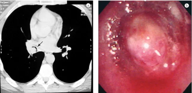
Figure 1 - Radiological and endoscopic aspects: a) Computed tomography scan of the chest showing nearly complete obstruction of the lumen of the intermediate bronchus (arrow); and b) Endoscopic view of the tumor.
Figure 2 - Excised section: longitudinal opening of the right lower lobe bronchus showing the tumor origin in the upper segment (arrow). Distal accumulation of secretion.
Figure 3 - Photomicrograph showing an area in which there is a proliferative, fusocellular storiform pattern, next to an area presenting an inflammatory pattern.
486 Sanchez PG, Madke GR, Pilla ES, Foergnes R, Felicetti JC, Valle E, Geyer G
J Bras Pneumol. 2007;33(4):484-486
lobe. Enucleation of nodules is not recommended,
due to the possibility of recurrence.(5) Radiotherapy
and corticosteroid treatment can be considered in patients with recidivism, in patients who underwent incomplete resection, or in patients considered unfit
to undergo pulmonary resection.(7,8) The prognosis
seems to be excellent when complete surgical
resec-tion is performed.(1,5)
In conclusion, inflammatory pseudotumor of the lung is a rare, locally invasive, and generally asymptomatic form of benign neoplasia, except in its endobronchial presentation, whose clinical and radiological aspects suggest other types of neoplasia. The preoperative tests are rarely diagnostic of the tumor, and complete resection is necessary for adequate treatment and a definitive diagnosis.
References
1. Cerfolio RJ, Allen MS, Nascimento AG, Deschamps C, Trastek VF, Miller DL et al. Inflammatory pseudotumors of the lung. Ann Thorac Surg. 1999;67(4):933-6.
2. Bahadori M, Liebow AA. Plasma cell granulomas of the lung. Cancer. 1973;31(1):191-208.
3. Alexiou C, Obuszko Z, Beggs D, Morgan WE. Inflammatory pseudotumors of the lung. Ann Thorac Surg. 1998;66(3):948-50.
4. Chen D, Ryan G, Edwards M. Bronchial sleeve resection for a patient with an inflammatory pseudotumor. ANZ J Surg. 2001;71(3):187-9.
5. Copin MC, Gosselin BH, Ribet ME. Plasma cell granuloma of the lung: difficulties in the diagnosis and prognosis. Ann Thorac Surg. 1996;61(5):1477-82.
6. Maeda M, Matsuzaki Y, Edagawa M, Shimizu T, Onitsuka T, Kataoka H. Successful treatment of a bronchial inflammatory pseudotumor by bronchoplasty in an 8-year-old boy: report of a case. Surg Today. 2000;30(5):465-8.
7. Shirakusa T, Kusano T, Motonaga R, Eimoto T. Plasma cell granuloma of the lung --resection and steroid therapy. Thorac Cardiovasc Surg. 1987;35(3):185-8.
8. Imperato JP, Folkman J, Sagerman RH, Cassady JR. Treatment of plasma cell granuloma of the lung with radiation therapy. A report of two cases and a review of the literature. Cancer. 1986;57(11):2127-9.
Discussion
Inflammatory pseudotumor accounts for less
than 1% of all lung tumors.(4) The mean age at
onset is 40 years, and 15% of the patients are under
10 years of age.(5) Although most patients are
asymp-tomatic, they can present cough, fever, hemoptysis, dyspnea, and chronic bronchitis. This type of tumor usually manifests as a solitary pulmonary nodule or as a mass with well-defined borders, and it is often confused with malignant pulmonary neoplasia. The endobronchial presentation is uncommon, accounting for less than 12% of the cases, and constitutes the most symptomatic form, which frequently leads to an early diagnosis. Although the cytological examination of the sputum and of the bronchial lavage fluid cannot be used to confirm the diagnosis, it should be performed in order to rule out active infections, such as tuberculosis, and other types of neoplasia.
Histological examination reveals local prolifera-tion of mature plasma cells and of reticuloendothelial cells in a granulation tissue stroma with no signs of mitosis or cellular atypia. The differential diagnosis includes sclerotic hemangioma, pseudolymphoma, malignant proliferation of plasma cells, and infec-tious granulomas.
Conservative surgery remains the treatment of
choice,(1) although the locally aggressive behavior
of the tumor occasionally requires the use of more radical procedures, such as pneumonectomy. Other
reports in the literature(4,6) report favorable results