Bronchodilator effect on maximal breath-hold
time in patients with obstructive lung disease*
Efeito do broncodilatador no tempo de apneia voluntária máxima em pacientes com distúrbios ventilatórios obstrutivos
Raqueli Biscayno Viecili, Paulo Roberto Stefani Sanches, Denise Rossato Silva, Danton Pereira da Silva, André Frota Muller, Sergio Saldanha Menna Barreto
Abstract
Objective: To identify the role of bronchodilators in the maximal breath-hold time in patients with obstructive lung disease (OLD). Methods: We conducted a case-control study including patients with OLD and a control group. Spirometric tests were performed prior to and after the use of a bronchodilator, as were breath-hold tests, using an electronic microprocessor and a pneumotachograph as a flow transducer. Respiratory flow curves were displayed in real time on a portable computer. The maximal breath-hold times at end-inspiratory volume and at end-expiratory volume (BHTmaxVEI and BHTmaxVEE, respectively) were determined from the acquired signal. Results: A total of 35 patients with OLD and 16 controls were included. Prior to the use of a bronchodilator, the BHTmaxVEI was significantly lower in the OLD group than in the control group (22.27 ± 11.81 s vs. 31.45 ± 15.73 s; p = 0.025), although there was no significant difference between the two groups in terms of the post-bronchodilator values (24.94 ± 12.89 s vs. 31.67 ± 17.53 s). In contrast, BHTmaxVEE values were significantly lower in the OLD group than in the control group, in the pre- and post-bronchodilator tests (16.88 ± 6.58 s vs. 22.09 ± 7.95 s; p = 0.017; and 21.22 ± 9.37 s vs. 28.53 ± 12.46 s; p = 0.024, respectively). Conclusions: Our results provide additional evidence of the clinical usefulness of the breath-hold test in the assessment of pulmonary function and add to the existing knowledge regarding the role of the bronchodilator in this test.
Keywords: Respiratory function tests; Pulmonary disease, chronic obstructive; Bronchodilator agents; Apnea.
Resumo
Objetivo: Identificar o papel do broncodilatador no tempo de apneia voluntária máxima em pacientes com distúrbios ventilatórios obstrutivos (DVOs). Métodos: Estudo caso-controle incluindo pacientes com DVOs e grupo controle. Foram realizadas espirometrias antes e após o uso de broncodilatador, assim como testes de apneia respiratória, utilizando-se um microprocessador eletrônico e um pneumotacógrafo como transdutor de fluxo. As curvas de fluxo respiratório foram exibidas em tempo real em um computador portátil, e os tempos de apneia voluntária inspiratória e expiratória máximos (TAVIM e TAVEM, respectivamente) foram determinados a partir do sinal adquirido. Resultados: Um total de 35 pacientes com DVOs e 16 controles foram incluídos no estudo. O TAVIM sem o uso de broncodilatador foi significativamente menor no grupo DVO que no grupo controle (22,27 ± 11,81 s vs. 31,45 ± 15,73; p = 0,025), mas essa diferença não foi significativa após o uso de broncodilatador (24,94 ± 12,89 s vs. 31,67 ± 17,53 s). Os valores de TAVEM foram significativamente menores no grupo DVO que no grupo controle antes (16,88 ± 6,58 s vs. 22,09 ± 7,95 s; p = 0,017) e após o uso de broncodilatador (21,22 ± 9,37 s vs. 28,53 ± 12,46 s; p = 0,024). Conclusões: Estes resultados fornecem uma evidência adicional da utilidade clínica do teste de apneia na avaliação da função pulmonar e do papel do broncodilatador nesse teste.
Descritores: Testes de função respiratória; Doença pulmonar obstrutiva crônica; Broncodilatadores; Apneia.
* Study carried out under the auspices of the Graduate Program in Medical Sciences, Department of Pulmonology, Hospital de Clínicas de Porto Alegre – HCPA, Porto Alegre Hospital de Clínicas – Universidade Federal do Rio Grande do Sul – UFRGS, Federal University of Rio Grande do Sul – Porto Alegre, Brazil.
Correspondence to: Raqueli Biscayno Viecili. Rua Dona Firmina, 414, casa 8, São José, CEP 91520-210, Porto Alegre, RS, Brasil. Tel. 55 51 3086-0761. E-mail: [email protected]
Financial support: None.
maneuvers, under similar conditions, on HR. Those two experiments were early indicators of the close relationship of the cardiorespiratory mechanism in health and disease.(4,5)
Apnea is an unstable state, changes occurring in numerous interrelated variables. The breath-hold test is simple and rapid. It consists of determining how long individuals can hold their breath. Maximal breath-hold time (BHTmax) varies from individual to individual and depends on chemical and nonchemical stimuli.(6,7)
It has been demonstrated that BHT is reduced by something that increases the response of diaphragmatic afferents (a tonic activity of the diaphragm and, possibly, arterial hypoxia and hypercapnia) or that increases central respiratory rhythm (arterial hypoxia or hypercapnia, decreased lung volume, or increased metabolic rate).(8)
The breath-hold test was tested in certain clinical settings and proved to be extremely useful clinically.(8-10) The test can be used both
as a screening test, raising the suspicion of OLD, and as a lung function parameter, similar to FEV1 and FVC.
A potential clinical and functional respiratory test, BHT can complement clinical examination, raising the suspicion of pathophysiological abnormalities, such as lung hyperinflation.
The objective of the present study was to evaluate the effect of bronchodilator use on the BHTmax in patients with OLD.
Methods
This was a prospective case-control study. The participants were over 18 years of age and had been referred to the Pulmonary Physiology Clinic of the Pulmonology Department of the Hospital de Clínicas de Porto Alegre (HCPA, Porto Alegre Hospital de Clínicas), in Porto Alegre, Brazil, for spirometry. Patients with OLD constituted the study group, whereas those with normal spirometry results constituted the control group.
The participants were interviewed, data having been collected with a standardized questionnaire comprising questions regarding demographic data, smoking habit, and comorbidities. The study protocol was approved by the HCPA Research Ethics Committee, and all of the participants gave written informed consent.
Introduction
The World Health Organization estimates that COPD kills more than 2.75 million people each year. Every 10 seconds, one person dies from COPD. According to the World Health Organization, COPD is, together with AIDS/HIV, the fourth leading cause of death worldwide, after heart diseases, cerebrovascular diseases, and pneumonia. In Brazil, approximately 40,000 people die from COPD each year, and approximately 7 million Brazilians have the disease. For the Brazilian Unified Health Care System, COPD is the respiratory disease that has the highest cost.(1)
Patients with COPD present with various functional changes, the most typical finding being a persistent reduction in forced expiratory flow. Clinical trials examining the reversibility of obstructive lung disease (OLD) in COPD are usually based on expiratory flow measurements and their variation after the use of inhaled bronchodilators, FEV1 being the parameter that is most commonly used in order to characterize airflow limitation in OLD.(2) It is convenient to
think of FEV1 as the mean flow during the first second of FVC. In addition, FEV1 is used in order to determine the degree of airflow obstruction (mild, moderate, or severe) in COPD patients, as well as being used in the functional follow-up of such patients.(3)
Other measurements, taken when tracing a volume-time curve for FVC, are also useful in grading OLD. The transition from functionally normal airways to mildly obstructed airways is generally gradual.(2,3)
When patients with airflow obstruction are tested after the use of placebo or without active medication, limits of variation in functional parameters are established. A statistically significant response (i.e., beyond random variability) is characterized by such limits being exceeded after bronchodilator use. Increases of 0.2 L or more in FEV1 and 0.35 L or more in FVC are widely used in order to characterize that response.(2,3)
BHTmaxVEE, and BHT) were automatically quantified (in seconds) from selected segments of the flow curve.
The system is heated in order to prevent condensation within the capillary tubes, which can result in reading errors. In the capillary tubes there is a stainless steel screen that is heated and provides resistance; a mouthpiece with a disposable filter was attached to the device in order to avert the impact of particulate matter and aid in creating a laminar flow.(3,12)
The statistical analysis was performed with the Statistical Package for the Social Sciences, version 18.0 (SPSS Inc., Chicago, IL, USA). The data were expressed as frequencies, means ± SD, or medians (interquartile ranges). Pearson’s correlation coefficient was used in order to determine the correlations among the variables BHTmaxVEI, BHTmaxVEE, and FEV1/FVC ratio prior to and after bronchodilator use. In order to identify correlations (minimum r = 0.60) among those variables, with a power of 80% and a significance of 5%, 19 patients were required. The level of significance was set at p < 0.05 for all analyses.
Results
Between May and November of 2010, 51 individuals (35 patients with OLD and 16 controls) were included in the present study. In the OLD group, the physician who requested spirometry diagnosed the following underlying diseases: asthma, in 17 (48.6%); COPD, in 11 (31.4%); dyspnea, in 3 (8.57%); pulmonary nodules, in 2 (5.71%); and preoperative period of vascular surgery, in 2 (5.71%). Of the patients with OLD, 6 (17.14%) were classified as having incipient OLD, 13 (37.14%) were classified as having mild OLD, 10 (28.57%) were classified as having moderate OLD, and 6 (17.14%) were classified as having severe OLD.(17) Table 1
shows the general characteristics of the study population.
The mean age of OLD group patients was 57.4 ± 13.1 years, whereas that of control group patients was 44.6 ± 16.8 years (p = 0.05). The prevalence of smoking was higher in the OLD group than in the control group (62.9% vs. 31.3%; p = 0.004). Pre-bronchodilator BHTmaxVEI was shorter in the OLD group than in the control group (22.27 ± 11.81 s vs. 31.45 ± 15.73 s; p = 0.025). There were no statistically significant Patients who presented with severe coronary
artery disease, cardiac arrhythmias, acute myocardial infarction, traumatic brain injury, glaucoma, hemoptysis, unstable angina, retinal detachment, hypertension, or pulmonary edema were excluded from the present study, as were pregnant women.(10)
Pulmonary function was evaluated with a MasterScreen Body spirometer (Jaeger, Würzburg, Germany), in accordance with the American Thoracic Society/European Respiratory Society guidelines(10,11) and previously published
reference values.(12-16)
In order to determine the BHTmax at end-inspiratory volume (BHTmaxVEI), we instructed the participants to inhale deeply three times (with a mouthpiece and a nose clip); at the end of the third maximal inspiratory maneuver, the participants were instructed to hold their breath for as long as they could. In order to determine the BHTmax at end-expiratory volume (BHTmaxVEE), we instructed the participants to inhale and exhale deeply (under the same conditions as those described above) three times and then hold their breath for as long as they could. Each maneuver was performed three times.
Spirometry was performed prior to and after the use of a bronchodilator in order to determine the effect of the bronchodilator on the BHTmaxVEI and BHTmaxVEE.
The respiratory flow and apneas were monitored by a pneumotachograph (Hans Rudolph, Kansas City, MO, USA). The pneumotachograph was originally described by Fleisch in 1925; since then, the device has undergone various modifications in an attempt to improve the original concept. The pneumotachograph can measure the respiratory flow and is usually used in order to measure gas flow rates in ICU monitors, respirators, and ventilators.(3,12) The pneumotachograph design
Discussion
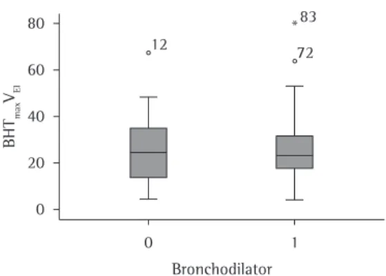
The objective of the present case-control study was to evaluate the effect of bronchodilator use on the BHTmax in patients with OLD and in normal individuals. We demonstrated that pre-bronchodilator BHTmaxVEI and BHTmaxVEE values were lower in the OLD group than in the control group. However, after bronchodilator use, only BHTmaxVEE values were significantly lower in the OLD group. In addition, after bronchodilator use, neither BHTmaxVEI nor BHTmaxVEE correlated significantly with any of the pulmonary function parameters (Figures 1 and 2).
The breath-hold test is one of many methods used in order to induce a sensation of dyspnea and provide information regarding the onset of and resistance to the sensation of dyspnea. In conscious individuals, immediately after the onset of apnea in functional residual capacity, there is a period of 20-30 s in which no particular respiratory sensation is experienced. That period differences between the two groups in terms of
the post-bronchodilator BHTmaxVEI.
Pre-bronchodilator BHTmaxVEE values were significantly lower in the OLD group than in the control group (16.88 ± 6.58 s vs. 22.09 ± 7.95 s; p = 0.017), as were post-bronchodilator BHTmaxVEE values (21.22 ± 9.37 s vs. 28.53 ± 12.46 s; p = 0,024).
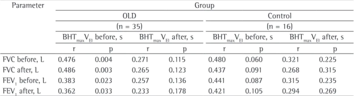
In the OLD group, we found significant, positive bivariate correlations between pre-bronchodilator BHTmaxVEI and the following: pre-bronchodilator FEV1 in L (r = 0.383; p = 0.023); post-bronchodilator FEV1 in L (r = 0.362; p = 0.033); pre-bronchodilator FVC in L (r = 0.476; p = 0.004); and post-bronchodilator FVC in L (r = 0.486; p = 0.004). Post-bronchodilator BHTmaxVEI did not correlate significantly with any of the pulmonary function parameters in the OLD group. Neither pre- nor post-bronchodilator BHTmaxVEE correlated significantly with any of the pulmonary function parameters in either group (Table 2).
Table 1 - General characteristics of the study population.a
Parameter Group p
OLD Control
(n = 35) (n = 16)
Age, years 57.40 ±13.13 44.56 ± 16.79 0.05
FVC before, L 2.81 ± 0.93 3.51 ± 0.81 0.012
FVC after, L 3.04 ± 0.99 3.53 ± 0.74 0.088
FEV1 before, L 1.74 ± 0.83 3.01 ± 0.71 < 0.0001
FEV1 after, L 1.95 ± 0.88 3.12 ± 0.66 < 0.0001
BHTmaxVEI before, s 22.27 ± 11.81 31.46 ± 15.73 0.025 BHTmaxVEI after, s 24.94 ± 12.89 31.67 ± 17.54 0.130
BHTmaxVEE before, s 16.88 ± 6.58 22.09 ± 7.95 0.017
BHTmaxVEE after, s 21.22 ± 9.37 28.54 ± 12.47 0.024
OLD: obstructive lung disease; before: before bronchodilator use; after: after bronchodilator use; BHTmaxVEI: maximal breath-hold time at end-inspiratory volume; and BHTmaxVEE: maximal breath-hold time at end-expiratory volume.
aValues expressed as mean ± SD.
Table 2 - Correlations between the maximal breath-hold time at end-inspiratory volume and spirometric parameters.*
Parameter Group
OLD Control
(n = 35) (n = 16)
BHTmaxVEI before, s BHTmaxVEI after, s BHTmaxVEI before, s BHTmaxVEI after, s
r p r p r p r p
FVC before, L 0.476 0.004 0.271 0.115 0.480 0.060 0.321 0.225
FVC after, L 0.486 0.003 0.265 0.123 0.437 0.091 0.268 0.315
FEV1 before, L 0.383 0.023 0.257 0.136 0.441 0.087 0.315 0.235 FEV1 after, L 0.362 0.033 0.233 0.178 0.421 0.105 0.294 0.269
pulmonary abnormalities, even in cases in which spirometry results are normal.(2)
In the present study, pre- and post-bronchodilator BHTmaxVEE values were significantly lower in the OLD group than in the control group. Nevertheless, in both groups, BHTmaxVEE values were found to improve significantly after bronchodilator use. Although a previous study suggested that the breath-hold test was useless as a pulmonary function test,(19)
various studies have shown that the breath-hold test can play a role in the evaluation of dyspnea and that bronchodilator use increases BHTmaxVEE values.(8-10,20-22)
We found no significant differences between the OLD group and the control group in terms of post-bronchodilator BHTmaxVEI. In the OLD group, bronchodilator use increased the BHTmaxVEI, the difference between the two groups having become less than significant. In a previous study, in which the BHTmaxVEI was sequentially measured in the postoperative period, the authors reported that the test was easy to perform, was well accepted by patients, and was an interesting tool to be used in the clinical follow-up of patients in the postoperative period, having aided in detecting complications during that period.(22-26) However, that study did
not evaluate the role of bronchodilator use in the BHTmaxVEI.
Our study has some limitations. First, the investigation was conducted in a single center. Second, our study sample was small. Despite those limitations, our results provide additional evidence of the effect of bronchodilator use on the BHTmaxVEI and BHTmaxVEE.
In conclusion, we found that pre- and post-bronchodilator BHTmaxVEI and BHTmaxVEE values were lower in the OLD group than in the control group However, after bronchodilator use, only BHTmaxVEE values were significantly lower in the OLD group. The breath-hold test can aid in recognizing severe pulmonary changes, as well as in promoting a more effective behavioral intervention. Post-bronchodilator BHTmaxVEI and BHTmaxVEE values should be interpreted with caution, given that they can be significantly different from pre-bronchodilator values. Further studies are needed in order to confirm the usefulness of the breath-hold test and the effect of bronchodilator use, particularly as a screening tool for the evaluation of dyspnea. ends with the onset of dyspnea and is followed
by a progressive increase in the intensity of dyspnea until apnea is interrupted. By measuring the apnea period, we can obtain information regarding the threshold of the sensation of dyspnea. Therefore, the measurement of the entire apnea period provides a yardstick of the behavior of the tolerance limit for the sensation of dyspnea.(17)
The breath-hold test has previously been used in patients with panic disorder; in addition, the test has been used in combination with the Borg scale and the FEV1/FVC ratio in order to evaluate the perception of dyspnea in asthma patients, having detected a low perception of dyspnea in those patients.(18) A recent study(2)
demonstrated another possible contribution of the breath-hold test. In individuals who smoke, are obese, or both, the breath-hold test reveals
Figure 1 - Maximal breath-hold time at end-inspiratory volume (BHTmaxVEI) prior to bronchodilator use (0) and after bronchodilator use (1).
14. Crapo RO, Morris AH, Clayton PD, Nixon CR. Lung volumes in healthy nonsmoking adults. Bull Eur Physiopathol Respir. 1982;18(3):419-25.
15. Nishino T. Pathophysiology of dyspnea evaluated by breath-holding test: studies of furosemide treatment. Respir Physiol Neurobiol. 2009;167(1):20-5.
16. Masdrakis VG, Markianos M, Vaidakis N, Papakostas YG, Oulis P. Caffeine challenge and breath-holding duration in patients with panic disorder. Prog Neuropsychopharmacol Biol Psychiatry. 2009;33(1):41-4. 17. Tavares FM, Silva LC, Rubin AS. Measuring forced
expiratory volume in one second alone is not an accurate method of assessing response to bronchodilators in chronic obstructive pulmonary disease. J Bras Pneumol. 2005;31(5):407-14.
18. Inoue H, Yamauchi K, Kobayashi H, Shikanai T, Nakamura Y, Satoh J, et al. A new breath-holding test may noninvasively reveal early lung abnormalities caused by smoking and/or obesity. Chest. 2009;136(2):545-53. 19. Crapo RO, Morris AH, Gardner RM. Reference
spirometric values using techniques and equipment that meet ATS recommendations. Am Rev Respir Dis. 1981;123(6):659-64.
20. Barreto SS, Gottschall CA. Comparação entre provas espirométricas e testes clínico-funcionais pulmonares. J Pneumol. 1978;4(1):21-9.
21. Miller MR, Hankinson J, Brusasco V, Burgos F, Casaburi R, Coates A, et al. Standardisation of spirometry. Eur Respir J. 2005;26(2):319-38.
22. Dales RE, Spitzer WO, Tousignant P, Schechter M, Suissa S. Clinical interpretation of airway response to a bronchodilator. Epidemiologic considerations. Am Rev Respir Dis. 1988;138(2):317-20.
23. Rodrigues Jr R, Pereira CA. Resposta a broncodilatador na espirometria: que parâmetros e valores são clinicamente relevantes em doenças obstrutivas? J Pneumol. 2001;27(1):35-47.
24. Pereira CA, Rebello CB, Diccini S, Sato T. Resposta a broncodilatador em doenças obstrutivas -- asma vs DPOC. J Pneumol. 1996;22(Suppl 1):50.
25. Gaensler EA, Rayl DF, Donnelly DM. The breath holding test in pulmonary insufficiency; evaluation of 1,000 studies. Surg Gynecol Obstet. 1951;92(1):81-90. 26. Courtney R, Cohen M. Investigating the claims of
Konstantin Buteyko, M.D., Ph.D.: the relationship of breath holding time to end tidal CO2 and other proposed measures of dysfunctional breathing. J Altern Complement Med. 2008;14(2):115-23.
References
1. Global Initiative for Chronic Obstructive Lung Disease [homepage on the Internet]. Bethesda: Global Initiative for Chronic Obstructive Lung Disease. [cited 2007 Oct 22]. Global Strategy for the Diagnosis, Management and Prevention of COPD 2006. [Adobe Acrobat document, 100p]. Available from: http://www.goldcopd.org/ uploads/users/files/GOLDReport2006_0122.pdf 2. Light RW, Conrad SA, George RB. The one best test for
evaluating the effects of bronchodilator therapy. Chest. 1977;72(4):512-6.
3. Pereira CA. Espirometria. J Pneumol. 2002;28(Suppl 3):S1-S82.
4. Sourk RL, Nugent KM. Bronchodilator testing: confidence intervals derived from placebo inhalations. Am Rev Respir Dis. 1983;128(1):153-7.
5. McMechan FH. The diagnostic and prognostic value of breath-holding test. Cal State J Med. 1922;20(11):377-80.
6. Tweeddale PM, Alexander F, McHardy GJ. Short term variability in FEV1 and bronchodilator responsiveness in patients with obstructive ventilatory defects. Thorax. 1987;42(7):487-90.
7. Mitrouska I, Tsoumakidou M, Prinianakis G, Milic-Emili J, Siafakas NM. Effect of voluntary respiratory efforts on breath-holding time. Respir Physiol Neurobiol. 2007;157(2-3):290-4.
8. Parkes MJ. Breath-holding and its breakpoint. Exp Physiol. 2006;91(1):1-15.
9. Nannini LJ, Zaietta GA, Guerrera AJ, Varela JA, Fernández OM, Flores DM. Breath-holding test in subjects with near-fatal asthma. A new index for dyspnea perception. Respir Med. 2007;101(2):246-53.
10. Pinheiro CT, Barreto SS, Gottschall CA. Determinação do tempo de apnéia inspiratória máximo TAIM no pós-operatório. J Pneumol. 1994;20(1):16-23.
11. Macintyre N, Crapo RO, Viegi G, Johnson DC, van der Grinten CP, Brusasco V, et al. Standardisation of the single-breath determination of carbon monoxide uptake in the lung. Eur Respir J. 2005;26(4):720-35.
12. Wanger J, Clausen JL, Coates A, Pedersen OF, Brusasco V, Burgos F, et al. Standardisation of the measurement of lung volumes. Eur Respir J. 2005;26(3):511-22. 13. Crapo RO, Morris AH. Standardized single breath normal
About the authors
Raqueli Biscayno Viecili
Visiting Researcher. Graduate Program in Medical Sciences, Department of Pulmonology, Hospital de Clínicas de Porto Alegre – HCPA, Porto Alegre Hospital de Clínicas – Universidade Federal do Rio Grande do Sul – UFRGS, Federal University of Rio Grande do Sul – Porto Alegre, Brazil.
Paulo Roberto Stefani Sanches
Biomedical Engineer. Hospital de Clínicas de Porto Alegre – HCPA, Porto Alegre Hospital de Clínicas – Universidade Federal do Rio Grande do Sul – UFRGS, Federal University of Rio Grande do Sul – Porto Alegre, Brazil.
Denise Rossato Silva
Professor. Graduate Program in Pulmonology, Universidade Federal do Rio Grande do Sul – UFRGS, Federal University of Rio Grande do Sul – Porto Alegre, Brazil.
Danton Pereira da Silva
Biomedical Engineer. Hospital de Clínicas de Porto Alegre – HCPA, Porto Alegre Hospital de Clínicas – Universidade Federal do Rio Grande do Sul – UFRGS, Federal University of Rio Grande do Sul – Porto Alegre, Brazil.
André Frota Muller
Biomedical Engineer. Hospital de Clínicas de Porto Alegre – HCPA, Porto Alegre Hospital de Clínicas – Universidade Federal do Rio Grande do Sul – UFRGS, Federal University of Rio Grande do Sul – Porto Alegre, Brazil.
Sergio Saldanha Menna Barreto