D I A B E T E S & M E T A B O L I S M J O U R N A L
his is an Open Access article distributed under the terms of the Creative Commons At-tribution Non-Commercial License (http://creativecommons.org/licenses/by-nc/3.0/) which permits unrestricted non-commercial use, distribution, and reproduction in any medium, provided the original work is properly cited.
Copyright © 2011 Korean Diabetes Association http://e-dmj.org
Diabetes Metab J 2011;35:8-11
New Perspectives on Diabetic Vascular Complications:
he Loss of Endogenous Protective Factors Induced by
Hyperglycemia
In-Kyung Jeong1
, George L. King2
1Department of Endocrinology and Metabolism, Kyung Hee University School of Medicine, Seoul, Korea 2Section on Vascular Cell Biology, Joslin Diabetes Center, Harvard University, Boston, MA, USA
Diabetic vascular complications are among the leading causes of morbidity and mortality in diabetic patients. In the past, many studies have focused on the mechanisms of hyperglycemia-induced chronic vascular complications via the formation of toxic metabolites such as oxidative stress, advanced glycosylated end products, persistent activation of protein kinase C, and increased sorbitol concentrations. However, vascular complications result from imbalances caused by increases in systemic toxic metabo-lites, such as those that occur under conditions of hyperglycemia and dyslipidemia, and by reductions in endogenous protective factors such as insulin, vascular endothelial growth factor, and platelet derived growth factor. his review outlines some of the evidence supporting the importance of enhancing endogenous regenerative factors.
Keywords: Diabetic vascular complication; Hyperglycemia; Platelet derived growth factor; Vascular endothelial growth factor
Corresponding author: George L. King
Section on Vascular Cell Biology, Joslin Diabetes Center One Joslin Place, Boston, MA 02215, USA
E-mail: [email protected]
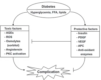
The vascular complications of diabetes mellitus affect many organ systems, including the retina, kidney, nerve, and cardio-vascular system. hese serious complications are the leading causes of mortality and morbidity in diabetic patients. Diabetic vascular complications result from imbalances caused by increases in the toxic efects of systemic metabolic abnormalities such as hyperglycemia, dyslipidemia, and hyper-tension, and reductions in the regenerative efects of endoge-nous protective factors such as insulin, vascular endothelial growth factor (VEGF), platelet derived growth factor (PDGF), nitric oxide (NO), and antioxidant enzymes (Fig. 1). In the past, many studies on the mechanisms of diabetic complica-tions have focused on the mechanisms by which hyperglyce-mia might lead to the chronic vascular complications via the formation of toxic metabolites such as oxidants and advanced
glycosylated products. hese mechanisms include increases in oxidative stress, persistent activation of protein kinase C (PKC) and other signaling pathways, increased sorbitol concentra-tions through the aldose reductase pathway, the elevated for-mation of advanced glycosylation end products, and increased lux through the hexosamine pathway [1]. However, few stud-ies have evaluated the importance of endogenous protective factors or the inhibitory efects of hyperglycemia in neutraliz-ing these protective factors durneutraliz-ing the initiation and progres-sion of diabetic complications. his review outlines some of the evidence supporting the importance of preventing and de-laying the progression of diabetic complications. Further, the lack of success in inding efective clinical therapeutics to neu-tralize the toxic efects of hyperglycemia could be due to the need for enhancing protective factors.
Review
he loss of endogenous protective factors induced by hyperglycemia
9
Diabetes Metab J 2011;35:8-11 http://e-dmj.org
Hyperglycemia-induced cellular apoptosis is a common pa-thology of many diabetic complications such as for retinal peri-cytes, renal podoperi-cytes, and vascular endothelial cells. In the case of diabetic retinopathy, accelerated pericyte apoptosis is one of the earliest and most speciic indings of diabetic com-plications. Enge et al. [2] reported that PDGF-B or PDGF receptor-β knockout mice exhibited pericyte apoptosis and retinal microvascular abnormalities similar to the early stages of diabetic retinopathy, indicating that PDGF-B is a very im-portant survival factor for retinal pericytes. However, the level of PDGF-B expression was observed to be elevated in diabetic state compared with that in non-diabetic animal [3]. hus, it was not clear whether pericyte loss results from PDGF-B abun-dance or PDGF-B resistance. Recently, Geraldes et al. [4] clear-ly demonstrated that hypergclear-lycemia induced a persistent acti-vation of PKC-δ, which leads to PDGF resistance in the retina. Under normal glucose condition, PDGF-B stimulated DNA synthesis, inhibited cellular apoptosis, and increased p-AKT and p-ERK activation in retinal pericytes. However, PDGF-in-duced activation of p-AKT and p-ERK signaling was blunted by hyperglycemic levels, which was restored in cells expressing dominant negative PKC-δ or in PKC-δ knockout mice. Geral-des et al. also identiied SHP-1, a tyrosine kinase, as a cellular target of PKC-δ and p38α that inhibited the survival action of tyrosine kinase growth factor receptors such as PDGFR-β, by
an NF-KB-independent pathway. Therefore, hyperglycemia induces activation of PKC-δ - p38α MAPK – SHP-1, which leads to the inhibition of PDGF–related survival actions that cause pericyte apoptosis in diabetic retinopathy. his study has identiied SHP-1 as a new potential therapeutic target, to be in-hibited as a treatment for diabetic vascular complications. Another survival factor is VEGF, which is one of the most important endogenous angiogenic polypeptides that respond to hypoxia under normal physiological conditions. he expres-sion of VEGF is increased in the retina, causing diabetic pro-liferative retinopathy; however, its abnormally low expression in the myocardium and peripheral lesions causes poor collat-eral vessel formation in periphcollat-eral tissues. During angiogene-sis, VEGF interacts with several other angiogenic factors, play-ing an important role in cell proliferation, diferentiation, mi-gration, cell survival, NO production, release of other growth factors, and sympathetic innervation. VEGF may have actions in non-endothelial cells. VEGF is also produced by renal podo-cytes. Podocytes and fenestrated endothelial cells are two ma-jor cells of the glomerular iltration barrier. Mesangial cells are located between the capillary loops and provide structural support. he apoptosis of podocytes is an early event of dia-betic nephropathy and a likely cause of proteinuria. Sison et al. [5] reported that VEGF-A released by podocytes was required for glomerular endothelial cell migration, diferentiation, and survival through cell-speciic gene targeting. he tyrosine ki-nase receptors for VEGF (VEGFR-1 and VEGFR-2) were ex-pressed by glomerular endothelial cells. heir podocyte-specif-ic, heterozygous, conditional knock out of VEGF-A mice showed a loss of endothelial cell fenestrations, endothelial cell necrosis, loss of podocyte foot process, and a marked loss of mesangial cells. his suggests that the paracrine efects of VEGF are crucial for glomerulus development and maintaining the function of the glomerular iltration barrier.
Microalbuminuria is an early detectable clinical abnormali-ty in diabetic glomerulopathy. Metabolic pathways activated by hyperglycemia, glycated proteins, hemodynamic factors, and oxidative stress are key players in the pathogenesis of dia-betic nephropathy. A variety of growth factors and cytokines are then induced through complex signal transduction path-ways. Podocyte-derived VEGF is an angiogenic factor whose expression is increased in animal models of diabetic kidney disease [6]. Controversies exist on the efect of the diabetes-induced enhanced expression of VEGF in the renal glomeruli. Some have suggested that VEGFs act in a novel autocrine
sig-Diabetes
Hyperglycemia, FFA, lipids
Toxic factors - AGEs - ROS - Osmolytes (sorbitol) - Angiotensin - PKC activation
Protective factors - Insulin - PDGF - VEGF - APC - Anti-oxidant enzymes
Complication
Jeong I-K, et al.
10 Diabetes Metab J 2011;35:8-11 http://e-dmj.org
naling mode to induce the podocytopathy of diabetes, espe-cially the genesis of albuminuria. hus, treatment with anti-VEGF antibodies may attenuate glomerular basement mem-brane thickening and progression of diabetic nephropathy. However, recently Eremina et al. [7] reported proteinuria, hy-pertension, and renal failure in several patients treated with anti-VEGF agent, bevacizumab. Pathologic indings from kid-ney biopsies were prominent endothelial swelling, red cell fragments, mesangiolysis, and thrombi in capillary loops which were similar to those in podocyte-speciic VEGF-A knockout mice. he study also suggested that anti-VEGF therapy inhib-its the VEGF action of podocytes and results in endothelial damage and thrombotic microangiopathy.
An alternative explanation is that the increased VEGF ex-pression in the kidney is a compensatory action to protect the glomeruli from the toxic efects of hyperglycemia. his means that the increase of VEGF is a compensatory response to over-come ischemic or other stressful injury which can induce VEGF resistance in the renal glomeruli by diabetes.
In contrast with diabetic microvascular complications, VEGF expression is reduced in diabetic heart and macrovasculature. Ventricular tissues from muscle insulin receptor knockout mice, which lack insulin receptors in the myocardium, have significant reductions in insulin but not IGF-I signaling, VEGF expression, and vascular density before and ater isch-emia versus controls [8]. his suggests that impaired insulin signaling has decreased VEGF expression and aggravated ischemic injury. hus, VEGF is an important angiogenic and survival cytokine in protecting micro- and macro-vascular tis-sues from conditions such as hyperglycemia.
Insulin is another important survival factor for vascular en-dothelial cells. Insulin has been shown to have both anti-ath-erosclerotic and pro-athanti-ath-erosclerotic actions in vascular cells. he anti-atherosclerotic actions of insulin include decreased endothelial cell apoptosis and reactive oxygen species, and in-creased NO by activation of endothelial NO synthase and heme oxygenase-1. In contrast, insulin has also been reported to have pro-atherosclerotic actions such as increasing endo-thelin-1 and plasminogen activator inhibitor-1, and enhanc-ing the migration and growth of vascular smooth muscle cells. In diabetes, hyperglycemia can selectively inhibit insulin’s an-ti-atherosclerotic actions, while hyperinsulinemia, as observed in insulin-resistant type 2 diabetes mellitus, can enhance insu-lin’s pro-atherosclerotic efects to accelerate the prevalence of atherosclerotic diseases [9]. Rask-Madsen et al. [10] recently
reported that loss of insulin signaling in endothelial cells ac-celerated atherosclerosis in apolipoprotein E-null mice. Dou-ble knockout mice with a deletion of the endothelial insulin receptor and apolipoprotein E gene (EIRAKO) were created using the Cre/loxP system to achieve tissue-speciic deletion of these genes. Despite retaining systemic insulin sensitivity, glucose tolerance, plasma lipids, and blood pressure were not different between the EIRAKO and control mice; however, more severe atherosclerotic lesions, impaired vasodilatation, and increased VCAM-1 expression were observed in the EI-RAKO mice vs. the apolipoprotein E-null mice. herefore, im-proving insulin sensitivity in the endothelium may decrease the risk for atherosclerosis in insulin resistance and diabetes states.
Clinical evidences are also available in support of the impor-tance of endogenous protective factors. Recently, we reported that a signiicant number of type 1 diabetes mellitus patients with a duration of diabetes of 50 years or longer do not have signiicant retinopathy and nephropathy (Joslin Medalist Study) [11]. his study demonstrated a lack of association between degree of hyperglycemia and prevalence of microvascular complications, although as a group, glycemic control in the study participants is very good, with a mean HbA1c of 7.3%. These results suggest that unknown endogenous protective factors are greatly reducing microvascular complications in this group of type 1 diabetic subjects of extremely long dura-tion. If the protective factors could be found from the study of these patients, we may be able to prevent or delay microvascu-lar complications in all diabetic patients.
In summary, new therapeutic approaches for diabetic vas-cular complications need to focus on decreasing the toxic fac-tors of diabetes, but also need to focus on increasing the action of protective factors.
REFERENCES
1. Brownlee M. Biochemistry and molecular cell biology of dia-betic complications. Nature 2001;414:813-20.
2. Enge M, Bjarnegard M, Gerhardt H, Gustafsson E, Kalen M, Asker N, Hammes HP, Shani M, Fassler R, Betsholtz C. Endo-thelium-specific platelet-derived growth factor-B ablation mimics diabetic retinopathy. EMBO J 2002;21:4307-16. 3. Yokota T, Ma RC, Park JY, Isshiki K, Sotiropoulos KB,
endothe-he loss of endogenous protective factors induced by hyperglycemia
11
Diabetes Metab J 2011;35:8-11 http://e-dmj.org
lin-1 in the retina of diabetic rats and cultured retinal capillary pericytes. Diabetes 2003;52:838-45.
4. Geraldes P, Hiraoka-Yamamoto J, Matsumoto M, Clermont A, Leitges M, Marette A, Aiello LP, Kern TS, King GL. Activation of PKC-delta and SHP-1 by hyperglycemia causes vascular cell apoptosis and diabetic retinopathy. Nat Med 2009;15:1298-306. 5. Sison K, Eremina V, Baelde H, Min W, Hirashima M, Fantus
IG, Quaggin SE. Glomerular structure and function require paracrine, not autocrine, VEGF-VEGFR-2 signaling. J Am Soc Nephrol 2010;21:1691-701.
6. Ziyadeh FN. Different roles for TGF-beta and VEGF in the pathogenesis of the cardinal features of diabetic nephropathy. Diabetes Res Clin Pract 2008;82 Suppl 1:S38-41.
7. Eremina V, Jeferson JA, Kowalewska J, Hochster H, Haas M, Weisstuch J, Richardson C, Kopp JB, Kabir MG, Backx PH, Gerber HP, Ferrara N, Barisoni L, Alpers CE, Quaggin SE. VEGF inhibition and renal thrombotic microangiopathy. N Engl J Med 2008;358:1129-36.
8. He Z, Opland DM, Way KJ, Ueki K, Bodyak N, Kang PM, Izu-mo S, Kulkarni RN, Wang B, Liao R, Kahn CR, King GL.
Reg-ulation of vascular endothelial growth factor expression and vascularization in the myocardium by insulin receptor and PI3K/Akt pathways in insulin resistance and ischemia. Arte-rioscler hromb Vasc Biol 2006;26:787-93.
9. Rask-Madsen C, King GL. Mechanisms of disease: endothelial dysfunction in insulin resistance and diabetes. Nat Clin Pract Endocrinol Metab 2007;3:46-56.
10. Rask-Madsen C, Li Q, Freund B, Feather D, Abramov R, Wu IH, Chen K, Yamamoto-Hiraoka J, Goldenbogen J, Sotiropou-los KB, Clermont A, Geraldes P, Dall’Osso C, Wagers AJ, Huang PL, Rekhter M, Scalia R, Kahn CR, King GL. Loss of insulin signaling in vascular endothelial cells accelerates ath-erosclerosis in apolipoprotein E null mice. Cell Metab 2010;11: 379-89.