Dedifferentiation of the Epithelium and Imbalance in Sex
Steroid Signaling
Ida Bjo¨rkgren1,2, Lauri Saastamoinen1, Anton Krutskikh3, Ilpo Huhtaniemi3, Matti Poutanen1,4, Petra Sipila¨1,4*
1Department of Physiology, Institute of Biomedicine, University of Turku, Turku, Finland,2Turku Graduate School of Biomedical Sciences, Turku, Finland,3Institute of Reproductive and Developmental Biology, Imperial College London, Hammersmith Campus, London, United Kingdom,4Turku Center for Disease Modeling, (TCDM), University of Turku, Turku, Finland
Abstract
Background:The postnatal development of the epididymis is a complex process that results in a highly differentiated epithelium, divided into several segments. Recent studies indicate a role for RNA interference (RNAi) in the development of the epididymis, however, the actual requirement for RNAi has remained elusive. Here, we present the first evidence of a direct need for RNAi in the differentiation of the epididymal epithelium.
Methodology/Principal Findings:By utilizing the Cre-LoxP system we have generated a conditional knock-out of Dicer1 in
the two most proximal segments of the mouse epididymis. Recombination ofDicer1, catalyzed byDefb41iCre/wt, took place
before puberty, starting from 12 days postpartum. Shortly thereafter, downregulation of the expression of two genes specific for the most proximal epididymis (lipocalin 8 and cystatin 8) was observed. Following this, segment development continued until week 5 at which age the epithelium started to regress back to an undifferentiated state. The dedifferentiated epithelium also showed an increase in estrogen receptor 1 expression while the expression of androgen receptor and its target genes; glutathione peroxidase 5, lipocalin 5 and cysteine-rich secretory protein 1 was downregulated, indicating imbalanced sex steroid signaling.
Conclusions/Significance:At the time of the final epididymal development, Dicer1 acts as a regulator of signaling pathways essential for maintaining epithelial cell differentiation.
Citation:Bjo¨rkgren I, Saastamoinen L, Krutskikh A, Huhtaniemi I, Poutanen M, et al. (2012) Dicer1 Ablation in the Mouse Epididymis Causes Dedifferentiation of the Epithelium and Imbalance in Sex Steroid Signaling. PLoS ONE 7(6): e38457. doi:10.1371/journal.pone.0038457
Editor:Joel R. Drevet, Clermont-Ferrand Univ., France
ReceivedJanuary 20, 2012;AcceptedMay 5, 2012;PublishedJune 6, 2012
Copyright:ß2012 Bjo¨rkgren et al. This is an open-access article distributed under the terms of the Creative Commons Attribution License, which permits unrestricted use, distribution, and reproduction in any medium, provided the original author and source are credited.
Funding:This work was supported by research grants from The Academy of Finland (project number 138561, http://www.aka.fi), and Sigrid Juse´lius Foundation (http://www.sigridjuselius.fi/foundation). IB is supported by the Turku Doctoral Programme of Biomedical Sciences (http://www.tubs.utu.fi). The funders had no role in study design, data collection and analysis, decision to publish, or preparation of the manuscript.
Competing Interests:The authors have declared that no competing interests exist.
* E-mail: petra.sipila@utu.fi
Introduction
After development in the testis, the spermatozoa travel through the epididymis where they mature, gaining motility and the ability to fertilize the oocyte. Despite consisting of a single long duct, the epididymis is a complex organ, highly convoluted and divided into several anatomical and functional segments, i.e. initial segment (IS), caput (CAP), corpus (COR) and cauda (CAU). Each segment synthesizes and secretes a specific set of proteins, thus creating the unique luminal environment needed for the sperm maturation process [1–3]. The most proximal segments (IS and CAP) have been proven essential for sperm maturation as the disruption of their development or function often leads to male infertility [4–7]. The epididymis develops from the mesonephric tubules and the proximal Wolffian duct (WD) [8]. During the embryonic stage, and before epithelial differentiation, mesenchymal androgen receptor (AR), along with inhibin beta A (Inhba), facilitates the elongation and convolution/coiling of the tubule [9–11]. Devel-opment of the epididymis continues after birth with differentiation
of the epithelial cells into principal, basal and narrow/clear cells [2,2,12,12,13]. At the onset of spermatogenesis, the epididymal epithelium develops segment-specific gene expression [14]. From studies with genetically modified mice, it has become evident that leucine-rich repeat-containing G protein-coupled receptor 4 (LGR4, also known as GPR48) is needed for the postnatal epididymal coiling and the differentiation of IS [15,16]. In addition, the proto-oncogene Ros1 (ROS1, also known as c-ros) is necessary for the formation of IS [5]. Recent studies with epididymal AR knock-out mice have revealed that androgen signaling is required for the formation of IS and differentiation of principal and basal cells [6,7,10]. However, there are still many unresolved issues regarding the regulatory pathways responsible for differentiation of the epididymal segments and their specific gene expression.
cleavage [17]. One class of small non-coding RNAs is the microRNAs (miRNAs) which are initially produced as longer precursors that need to be processed by the RNaseIII enzyme Dicer1 to become fully functional [18]. The,22 nt-long mature
miRNAs then control protein expression and modulate diverse cellular events such as differentiation, proliferation, apoptosis and cell metabolism [19,20]. Consequently, Dicer1 deficient mice die already on embryonic day 7.5 owing to complete loss of pluripotent stem cells [21]. To study the effect of RNAi in the development and function of specific organs, Dicer1 conditional knock-out (Dicer1cKO) mice can be used. The need for Dicer1 in the differentiation of several cell types, including sensory epithelial cells, T cells and pancreaticb-cells, has been shown inDicer1cKO mice by crossingDicer1fl/flmice with those expressing Crein the cells of interest [12,22–24]. Furthermore, a requirement for Dicer1 in the patterning of the liver and colon was also confirmed with Dicer1 cKO mice. Hepatocyte specific Dicer1 ablation compro-mises region-specific protein expression while Dicer1 depletion in the developing colon leads to a disorganized epithelium [25,26].
Previous studies on human and rat epididymides have shown that miRNAs are differentially expressed at juvenile and adult stages [27,28], indicating a role for miRNAs in the postnatal development of the epididymis. To study the function of RNAi in the developing epididymis we generated epididymis-specificDicer1 cKO mice by crossing Dicer1fl/fl mice withDefb41iCre mice. This resulted in the elimination of Dicer1 expression from the pre-pubertal epididymis. Our results demonstrate the importance of Dicer1 in sex steroid signaling and in maintenance of the differentiated state of the epididymal epithelium.
Results
Generation ofDicer1fl/fl; Defb41iCre/wt Mice
Quantitative RT-PCR of the mouse epididymis showed a continuous expression of Dicer1 from birth into adulthood (Figure 1C). As the full knock-out ofDicer1is embryonically lethal [21], we generated aDicer1cKO mouse line by crossingDicer1fl/fl mice [29] with a mouse line expressing iCreunder the Defensin beta 41 (Defb41) promoter. TheDicer1flallele consists of two loxP sites flanking exon 24, which contains a major part of the second RNaseIII domain (Figure 1A). The heterozygousDefb41iCremouse line did not show any phenotypic defects or fertility problems and expressediCrein the epithelium of the most proximal part of the epididymis, IS and CAP. Recombination ofDicer1was observed in 12 day-oldDicer1fl/fl; Defb41iCre/wtmouse IS and CAP by genomic PCR (Figure 1B) and qRT-PCR studies revealed a significant reduction in Dicer1 expression levels at the age of 2 months (Figure 1D).
Morphology of the Epididymis and Fertility ofDicer1fl/fl; Defb41iCre/wt Males
Macroscopic evaluation of 2 month-oldDicer1fl/fl; Defb41iCre/wt mice epididymides revealed an underdeveloped IS and, in addition, the mice frequently presented with enlarged efferent ducts (Figure 2A, B). The IS of control mice can be clearly visualized owing to the endogenousb-galactosidase activity in the segment. The much smaller IS of Dicer1fl/fl; Defb41iCre/wt mice could not be distinguished from CAP with X-gal staining (Figure 2A). Furthermore, the intense vasculature typical of WT IS was missing from theDicer1 cKO IS. Histological evaluation showed a division of the epididymis into different segments (Figure 2B) but the epithelial cell layer of both IS and CAP was disorganized (Figure 3H).Dicer1fl/flmice have a similar phenotype to WT mice epididymides and were used as controls throughout
the study.Dicer1cKO epididymides were significantly smaller than those of control mice (30.461.5 mg, control: 35.460.7 mg, P#0.01). No significant difference in the weight of 6 month-old Dicer1 cKO and control mice epididymides was observed. However, the epithelial cell layer of the 6 month-old Dicer1fl/fl; Defb41iCre/wtmouse was further disturbed, with neoplastic changes in the efferent ducts causing their progressive obstruction (Figure S1). Even though sperm were detected in the CAU, 2- to 3-month-oldDicer1fl/fl; Defb41iCre/wt male mice failed to produce offspring when mated with WT females (Table 1). The number of sperm was reduced in Dicer1fl/fl; Defb41iCre/wt mouse epididymides as histological staining showed some tubular cross sections with no sperm. At 6 months of age the number of tubular cross sections without sperm was further increased due to the obstruction of the Dicer1fl/fl; Defb41iCre/wt mouse efferent ducts. The testis of the 6 month-oldDicer1fl/fl; Defb41iCre/wtmouse also displayed disruption of the seminiferous epithelium owing to fluid back-pressure (data not shown). Further morphological analyses revealed that the number of sperm with angulated tails was not significantly increased in 2 month-old Dicer1fl/fl; Defb41iCre/wt mouse CAU (22.963.4% of all sperm, control: 16.361.2%).
Effects ofDicer1 cKO on Epididymal Cell Differentiation
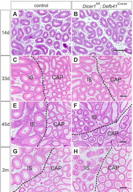
At the age of one month, the IS of both control andDicer1fl/fl; Defb41iCre/wt mice was distinguishable from the CAP by its high, columnar-shaped epithelial cells (Figure 3C, D). However, the height of the epithelium ofDicer1cKO IS was reduced below that of control mouse IS. In the 45 day-old mouse, the epithelial cells of theDicer1cKO IS had regressed to an undifferentiated state with a disorganized epithelial cell pattern (Figure 3E, F), resembling those of the epididymis of 14 day-old mice (Figure 3A, B). Furthermore, the height of the epithelial cells of IS was significantly reduced (average cell height in control IS: 45mm, in Dicer1 cKO IS: 28mm, P#0.0001) in 2 month-old Dicer1fl/fl; Defb41iCre/wt mice. However, the phenotype of theDicer1 cKO IS varied between individuals from a severely disorganized epithelial cell layer to a thin epithelial cell layer resembling that of COR (Figure S2) in some individuals.
To study further the effect of Dicer1ablation on epithelial cell differentiation, the presence of different epithelial cell types of the epididymal epithelium was analyzed by immunohistochemistry. Phalloidin conjugated to TRITC was used to stain the F-actin of principal cells, and antibodies against vacuolar H+
-ATPase (V-ATPase) and Keratin 5 (Krt5) were used to stain clear/narrow cells and basal cells, respectively. The results indicated that all cell types were present in theDicer1 cKO epididymis (Figure 4). The epithelial cell types were found in similar numbers and locations in theDicer1cKO epithelium as in control animals. However,a-actin staining revealed an increase in muscle cell layer thickness (Figure 4G, H). TheDicer1fl/fl; Defb41iCre/wt mice displayed three layers of muscle cells surrounding the epididymal duct, whereas control mice typically had one.
mice, from 21 days postpartum onward (Figure S3A, B). However, the expression levels ofRos1did not differ significantly from that of control mice (Figure S3C).
Cell Proliferation and Apoptosis
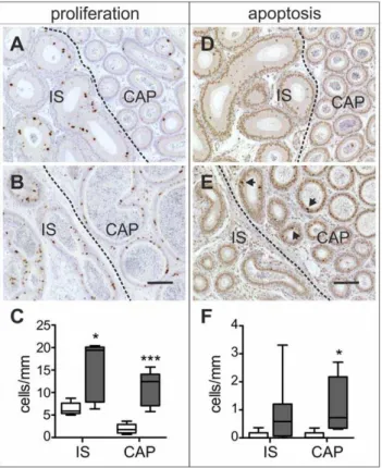
Immunohistochemical studies showed a marked increase in cell proliferation throughout the proximal epididymis of 2 month-old Dicer1fl/fl; Defb41iCre/wt mice (Figure 5B). The number of Ki-67 positive cells was on average 2 times higher in Dicer1 cKO IS (15.263.0 cells/mm, control: 6.460.6 cells/mm, P#0.05) and almost 6 times higher inDicer1 cKO CAP (11.161.7 cells/mm, control: 1.960.5 cells/mm, P#0.001) compared with those of control mice epididymides (Figure 5C). Although the epithelium was highly proliferative, it did not lead to a marked increase in the size of the Dicer1fl/fl; Defb41iCre/wt mouse epididymides. On the contrary, at the age of 2 months theDicer1 cKO epididymis was significantly smaller than that of the control mouse. Furthermore, the number of apoptotic cells was increased inDicer1cKO IS and CAP (Dicer1cKO IS: 0.8960.39 cells/mm, control IS: 0.0760.07 cells/mm; Dicer1cKO CAP: 1.1260.34 cells/mm, control CAP: 0.0760.07 cells/mm,P#0.05) (Figure 5D–F). The difference seen in IS was not statistically significant owing to high variation between the individual mice.
Expression of Sex Steroid and Fibroblast Growth Factor Receptors in theDicer1cKO IS and CAP
To study whether the observed epididymal phenotype is caused by changes in sex steroid signaling, the expression ofAr,estrogen
receptor 1 (Esr1, also known asERa) and estrogen receptor 2 (Esr2, also known asERb) was analysed by qRT-PCR and immunohis-tochemistry in 2 month-oldDicer1cKO and control epididymides. The qRT-PCR results showed a significantly weaker expression of ArinDicer1cKO IS and CAP (Figure 6E), and downregulation of AR target gene expression, cysteine-rich secretory protein 1 (Crisp1), glutathione peroxidase 5 (Gpx5) and lipocalin 5 (Lcn5), was also observed (Figure 6F). Esr2 showed a similar reduction in mRNA expression levels asAr, while the expression ofEsr1did not significantly differ betweenDicer1cKO and control IS and CAP (Figure 6E). However, immunohistological staining displayed an altered ESR1 expression pattern in Dicer1 cKO IS. ESR1 was foremost expressed in the narrow cells of control mouse IS, whereas the Dicer1 cKO IS showed equal expression of the receptor in almost all epithelial cells (Figure 6A, B). Furthermore, immunohistochemical analyses clearly showed weaker AR staining in a number of theDicer1cKO epididymides analysed (Figure 6D). Nevertheless, the AR findings were variable and someDicer1cKO epididymides had an AR staining equal to that of the controls. Furthermore, the relative expression values of fibroblast growth factor receptors 1–4 (Fgfr1–4)were not statistically different when comparing qRT-PCR results from Dicer1fl/fl; Defb41iCre/wt mice with control mouse IS and CAP (data not shown).
Discussion
At birth, the epididymis consists of a long convoluted duct lacking both cell type and segment specific markers. In the mouse Figure 1. The epididymis specificDicer1knock-out.(A) Schematic diagram of theDicer1locus with loxP sites flanking exon 24 (e24). Arrows indicate the location of genotyping primers used for analyzing the deletion of e24. (B) Genomic PCR of 12 day-old mice showing the intact locus (Dicer1fl) and the recombinant locus with e24 deleted (Dicer1flD). Exon 24 is only deleted in theDicer1fl/fl; Defb41iCre/wt
mouse initial segment (IS) and
caput (CAP) while the iCre locus is detected in all segments of the epididymis. (C) Expression ofDicer1mRNA in the whole epididymis of 1–42 day-old
wild-type mice. (D)Dicer1mRNA expression levels in the efferent ducts (ED), the different segments of the epididymis and testis (TE) of 2 month-old
control andDicer1conditional knock-out (cKO) mice. Expression levels are presented relative toPpia(testis) andL19(epididymis) expression. COR,
corpus; CAU, cauda. Statistical significance was calculated from the expression levels of 3 control and 4Dicer1fl/fl; Defb41iCre/wt
epididymis, differentiation of the epithelial cell types takes place between day 14 and day 21 [32] after which segment-specific gene expression can be observed [14]. Nonetheless, very little is known about the regulatory pathways responsible for the epididymal segmentation and the differentiation of epithelial cell types. In this study, we demonstrate that post-transcriptional regulation via RNAi signaling is an important regulator in the development of the proximal epididymal segments.
In ourDicer1fl/fl; Defb41iCre/wtmouse model the ablation ofDicer1 begins 12 days pp but, at the time of the final cell type differentiation during puberty, the epididymal epithelium of Dicer1fl/fl; Defb41iCre/wt mice still resembles that of control mice.
Studies revealed a significant reduction in the expression of IS specific genes at 21 days pp, and about 2 weeks later, the epithelium began to regress. At 45 days of age, theDicer1KO IS morphologically resembled that of an undifferentiated pre-pubertal epididymis. Despite the morphological changes, Dicer1 ablation did not affect cell type differentiation as all major epithelial cell types were detected in the epididymides of 2 month-old Dicer1fl/fl; Defb41iCre/wt mice. However, the function of the principal cells was compromised as they displayed a significant reduction in segment specific gene expression.
When studying the different epithelial cell types, we detected a thicker layer of smooth muscle cells surrounding the duct of the Dicer1fl/fl; Defb41iCre/wtmouse epididymis. AsDicer1ablation is not likely to occur in the stromal tissue, the observed increase in smooth muscle cell number may indicate altered epithelial-mesenchymal signaling. It has previously been shown that cross talk between different cell types is essential for normal epididymal functions, and for example the communication between clear cells and other epithelial cell types is required to maintain luminal pH [33]. Interestingly, there are species-specific differences in thickness and distribution of the epididymal smooth muscle cell layer [34–36]. Further investigations regarding smooth muscle cell differentiation could thus benefit from comparative studies of different species.
Previous studies have shown that ROS1 is one of the master regulators of IS development, as the lack of both ROS1 and its negative regulator, protein tyrosine phosphatase SHP-1, causes a defect in IS differentiation [5,37]. This affects also the regulation of sperm volume as the majority of sperm from theRos1KO CAU present with angulated tails [38]. However,Dicer1cKO IS still has
,70% of the Ros1 expression of the control IS and the
dedifferentiated IS does not cause a significant increase in sperm tail angulation. Furthermore, males with one intact Ros1 allele have normal epididymal development [5]. These data indicate that the epididymal phenotype, observed in the present study, is independent of ROS1 signaling. Other proteins that have a regulative role in IS are the Fibroblast growth factors. They control gene expression by binding to FGFRs in the epithelium of IS [39,40]. Studies in other male reproductive organs also indicate a role for FGFRs in regulating stromal-epithelial induction of cell proliferation and tissue homeostasis [41]. To assess the effect of FGF signaling on the observed epididymal phenotype, we studied the expression of Fgfrs in the epididymis. However, no changes were observed and it is therefore unlikely that FGF signaling contributes to the dedifferentiation of the epithelium or to the increased smooth muscle cell layer thickness of the Dicer1fl/fl; Defb41iCre/wtmice.
Sex steroids are important regulators of epididymal develop-ment and function. In the epididymis, epithelial expression ofAr starts around E14.5–15.5 [10,42] and is required for IS development [7] and for the differentiation of principal and basal cells in the proximal epididymis [6,10]. A further role for androgen signaling is observed after orchidectomy, where androgen depletion leads to extensive apoptosis throughout the epididymal Figure 2. Morphology of 2 month-old mice proximal
epididy-mides. (A) X-gal staining detecting endogenous b-galactosidase activity in a control mouse initial segment (IS) and proximal corpus (COR).Dicer1fl/fl; Defb41iCre/wtmice display no staining of initial segment and have enlarged efferent ducts (ED). (B) HE staining of the efferent ducts and the proximal epididymis of control andDicer1fl/fl; Defb41iCre/wt mice. Scale bars 1.5 mm.
doi:10.1371/journal.pone.0038457.g002
Table 1.Fertility ofDicer1fl/fl; Defb41iCre/wtmale mice.
No. of Males Total No. of Copulatory Plugs Total No. of Litters Average No. of Pups per Litter
control 3 13 12 9.2
Dicer1fl/fl
; Defb41iCre/wt
5 20 0 0
epithelium of both prepubertal and postpubertal animals [43]. In male reproductive organs, estrogens are produced in the testis, spermatozoa and epididymis [44,45]. Esr1 is highly expressed throughout the efferent ducts, where it regulates fluid reabsorption of non-ciliated epithelial cells [46,47]. Less is known about the function of ESR1 in the epididymis, although high expression of Esr1 is detected in the narrow cells of IS and throughout the
epithelium of CAP [46]. Recent studies have indicated a role for ESR1 in luminal pH maintenance [48,49] as well as in smooth muscle contractility [50]. Esr2 is transcribed in all cells of the epithelium, however, its role in the epididymis is still unknown since the full knock-out ofEsr2does not display any difference in morphology or function of the epididymis compared with that of WT mice [51].
Figure 3. Differentiation of the epididymal epithelium. Hematoxylin and eosin staining of control and Dicer1fl/fl; Defb41iCre/wt
mouse epididymides (A, B) The undifferentiated epithelium of the proximal epididymis of a 14 day-old control and aDicer1fl/fl; Defb41iCre/wtmouse. (C, D) 33 day-old control andDicer1fl/fl; Defb41iCre/wt
mouse showing initiated differentiation of the initial segment (IS). (E) The fully developed IS of a 45 day-old control mouse. (F) The epithelium of aDicer1fl/fl; Defb41iCre/wtmouse IS resembling that of the 14 day-old mouse. (G, H) The epididymis of an adult, 2 month-old, control andDicer1fl/fl; Defb41iCre/wt
mouse. CAP, caput. Scale bars 100mm.
The Dicer1fl/fl; Defb41iCre/wt mouse model presents with a significant downregulation in Ar, known AR target genes and Esr2mRNA expression in the proximal epididymis. However, on the basis of the current study, we cannot distinguish whether the down-regulation of the target genes is due to a direct lack of AR signaling or an effect of the observed epithelial cell dedifferenti-ation. ESR1, on the other hand, displayed an altered expression pattern in theDicer1KO IS, with expression not only in narrow cells, as in IS of control mice, but throughout the epithelium. As ESR1 is known to promote cell proliferation [52], high ESR1 levels in theDicer1cKO IS could explain the observed increase in cell proliferation in that epididymal segment.
Studies on excessive estrogen signaling in males suggest that an imbalance in the testosterone-estrogen ratio could be of more importance than the actual increase in estrogen signaling [53]. Administration of a single high dose of diethylstilbestrol (DES), a potent synthetic estrogen, to neonatal rats, caused a reduction in the epithelial height of the epididymis. However, the increased estrogen levels gave rise to an additional downregulation of AR in the entire epididymis and thus an imbalance in sex steroid signaling. When the estrogen-testosterone ratio was adjusted by co-administration of testosterone, no phenotypic abnormalities were observed [54]. The reduction inArexpression observed in Dicer1fl/fl; Defb41iCre/wt mice could therefore not only have significant consequences for epididymal development but also be directly caused by the increased ESR1 levels. However, a recent study on a proximal epididymis-specific AR cKO, also showed ESR1 expression in all cell types of the proximal epididymal epithelium [7]. This would indicate a role for AR in the repression of Esr1 expression. AR cKO epididymides also showed similar features to those of theDicer1fl/fl; Defb41iCre/wtmice with an altered epididymal stroma showing a disrupted smooth muscle cell layer or a thickened mesenchyme [7,55]. In light of these studies, AR and ESR1 expression in the epididymis seem to be tightly linked and although our current results show an imbalance in sex steroid receptor ratio we cannot with certainty say if the imbalance was initiated by a lowered AR expression or an increase in ESR1 expression.
Adams et al. were the first to show thatEsr1is a direct target of miR-206 [56]. Since then, around 14 evolutionarily conserved miRNAs have been found to target directly the 39-UTR of mammalianEsr1[57]. There is also evidence of a miRNA-induced downregulation ofEsr1after testosterone treatment in the female mouse liver [58]. In light of these previous studies the upregulation of ESR1 in Dicer1 KO IS might be directly ascribable to the ablation of mature miRNAs. Interestingly, previous studies have shown induction of miRNA expression by AR in vivo [59]. Androgen Responsive Elements (AREs) are also found in the promoter region of several miRNAs [60,61]. Although AR would not induce expression of all epididymal miRNAs, the observed phenotype ofDicer1fl/fl; Defb41iCre/wt mice could be partially AR dependent. To understand better the role of the RNAi pathway during epididymal development, miRNA expression data from different developmental time points is needed. These results could be further compared to already available array data from the human and rat epididymis [27,28] to demonstrate evolutionarily conserved RNAi regulation.
In conclusion, the present study shows the importance of the RNAi pathway in the postnatal development of the proximal epididymis. Ablation ofDicer1in the epididymal epithelium causes a regression in cell differentiation. The epithelium maintains cell type identity but lacks a segment-specific gene expression pattern. Furthermore, the epididymal phenotype ofDicer1fl/fl; Defb41iCre/wt mice resembles that of mice with an imbalanced ratio of sex steroid receptors, AR cKO mice and mice with excess estrogen signaling. Figure 4. Immunohistochemical staining of the different epithelial cell types.Staining of initial segment (A, B) and initial segment and caput (C–H) of two-month-old control and Dicer1fl/fl; Defb41iCre/wt mouse epididymides. (A, B) Staining of principal cell F-actin by phalloidin-TRITC. (C, D) Expression of vacuolar H+-ATPase (V-ATPase) in narrow and clear cells (marked with arrows). (E, F) Expression of keratin 5 (Krt5) in basal cells. (G,
H) Smooth muscle cells. Dicer1fl/fl; Defb41iCre/wt mice display a thicker smooth muscle cell layer than that of control mice. Inserts are 36
enlargements of the stained cells. (I) qRT-PCR for proximal epididymis-specific gene expression. Expression of lipocalin 8 (Lcn8), brain expressed myelocytomatosis oncogene (Bmyc), cystatin 8 (Cst8), Ros1 proto-oncogene (Ros1) and G protein-coupled receptor 64 (Gpr64) in the initial segment and caput of 2 month-old control and Dicer1 conditional knock-out (cKO) mouse epididymides. Values relative to L19 expression. Statistical significance was calculated from the expression levels of 3 control and 3 Dicer1fl/fl;Defb41iCre/wt mouse (Ros1:8 control and 9 Dicer1fl/fl;Defb41iCre/ wt mouse) samples using the unpaired t-test. Statistical significance of changes is indicated as follows: ns, not significant; *, P#0.05; **, P#0.01. Scale bars 100mm.
doi:10.1371/journal.pone.0038457.g004
Figure 5. Cell proliferation and apoptosis.Ki67 immunostaining; (A) Control; (B) Dicer1fl/fl; Defb41iCre/wt
mouse. TUNEL labeling; (D) Control; (E)Dicer1fl/fl; Defb41iCre/wtmouse. Arrows mark apoptotic cells. (C, F) Comparison of number of proliferating cells and apoptotic cells in the initial segment (IS) and caput (CAP) of control (white bars) and
Dicer1fl/fl; Defb41iCre/wtmice (grey bars). The number of proliferating and
apoptotic cells were calculated from ten tubular cross sections of 5 control and 5 (Ki67 stained) and 8 (TUNEL labeled)Dicer1fl/fl; Defb41iCre/
wt
This suggests that Dicer1-dependent pathways are important regulators of balanced estrogen-androgen action in the mouse epididymis.
Materials and Methods
Ethics Statement
All mice were handled in accordance with the institutional animal care policies of the University of Turku (Turku, Finland), and every effort was made to minimize suffering of the animals. Mice were specific pathogen-free, fed with complete pelleted chow and tap waterad libitumin a room with controlled light (12 hours
light, 12 hours darkness) and temperature (2161uC). Animal experiments were approved by the Finnish Animal Ethics Committee (license number: 20072-Sipila¨ and 2010-05926-Poutanen), and the institutional policies on animal experimenta-tion fully met the requirements as defined in the NIH Guide on animal experimentation.
Experimental Animals
To inactivateDicer1conditionally from the epithelial cells of the proximal epididymis, Dicer1fl/fl mice [29] were crossed with a heterozygous Defb41iCre knock-in (KI) mouse line. The KI was Figure 6. Expression of Sex steroid receptors.(A) Immunostaining of two-month-old control mouse initial segment (IS) shows expression of Estrogen receptor 1 (ESR1) only in the narrow cells (indicated by arrows) while (B) theDicer1fl/fl; Defb41iCre/wtmouse has ESR1 expression in most cells of the epithelium. (C, D) Staining for Androgen receptor. (D) Androgen receptor expression varied betweenDicer1fl/fl; Defb41iCre/wt
mice epididymides.
Some samples had similar expression levels as the control while others showed areas of more weakly stained tissue. CAP, caput. Scale bars 100mm.
(E) Expression of androgen receptor (Ar) and estrogen receptor 1 and 2 (Esr1andEsr2) as well as (F) AR target gene: glutathione peroxidase 5 (Gpx5),
lipocalin 5 (Lcn5), cysteine-rich secretory protein 1 (Crisp1) mRNA in IS and CAP of 2 month-old control andDicer1conditional knock-out (cKO) mice
epididymides. Expression values relative toL19expression. Statistical significance was calculated from the expression levels of 3 control and 4Dicer1fl/
fl; Defb41iCre/wt
mouse samples using the unpaired t-test. Statistical significance of changes is indicated as follows: ns, not significant; *,P#0.05; ***, P#0.001.
generated by inserting theiCre cDNA allelle into the translation initiation site of theDefb41gene. A detailed characterization of the Defb41iCreKI mouse line will be described elsewhere.Defb41iCre/wt mice were genotyped with the primers Defb41 Fw: TCCATTGCCTTTTCTTGTCC and Defb41 Re: TTGTCTTACCAGGTTTCCTCCT, Tm 56uC, for the wild-type (WT) allele and iCre Fw: TCTCCAACCTGCTGACTGTG and iCre Re: AGGGACACAGCATTGGAGTC, Tm 59uC, for the iCre allele. Dicerfl/fl mice were genotyped as previously described [29]. For detection of the floxed allele, IS together with CAP, COR together with CAU and testes from 12 day-old mice were collected. After DNA extraction, the recombined allele was detected by genomic PCR as previously described [29].Dicer1fl/fl; Defb41iCre/wtmice and the controls,Dicer1fl/flmice, were obtained from the same litters. The genetic background of these mice was mixed C57Bl/6N and SV129.
Sperm Morphology and Fertility of Dicerfl/fl; Defb41iCre/wt
Mice
To study male fertility, 2–3 month-old male Dicer1fl/fl; Defb41iCre/wt mice and control mice were mated with FVB/N female mice. The female mice were superovulated by intraperi-toneal injections of 5 IU pregnant mare serum gonadotropin (PMSG, NHPP, Dr. Parlow) and 5 IU human chorionic gonadotrophin (hCG, Pregnyl, Schering-Plough) 26 and 2 hours before mating, respectively. Females were checked for copulatory plugs the following morning. The mated females were followed for 3–4 weeks to determine the number of litters and offspring produced by each male. For sperm analyses, CAU of two-month-old control and Dicer1fl/fl; Defb41iCre/wt mice were dissected and incubated in 300ml of HTF medium (William A. Cook Australia Pty. Ltd, Brisbane, Australia) at 37uC, 5% CO2 for 30 min, to allow the sperm to swim out. The sperm were spread on microscope slides and stained using the Papanicolaou technique (Haematoxylin, OG-6 and EA-50). The morphology of 100 sperm/sample from 4 control and 5Dicer1fl/fl; Defb41iCre/wt mice samples was analyzed.
Histology, Immunohistochemistry andb-galactosidase Staining
Epididymides from 19 day - 6 month-old control andDicer1fl/fl; Defb41iCre/wtmice were fixed overnight in 4% paraformaldehyde (PFA), embedded in paraffin and prepared for histological analyses by standard procedures. Haematoxylin and eosin (HE) staining was performed by standard procedures. Epithelial cell height was measured by using the Leica IM500 imaging software. To detect the different epithelial cell types of the epididymis, the slides were incubated with the following antibodies overnight at 4uC: rabbit monoclonal anti-Keratin 5 1:100 (RM-2106, Thermo Scientific), rabbit polyclonal anti-V-ATPase 1:100 (a gift from S. Breton, Program in Membrane Biology, MGH Simches Research Center, Boston, MA) and mouse monoclonal anti-a-actin 1:500 (sc-32251, Santa Cruz Biotechnology). All the primary antibodies were diluted in PBS supplemented with 1% bovine serum albumin. The antibody-antigen complexes were visualized by incubation for 30 min at room temperature with 1:200 Alexa Fluor 488- and 594-conjugated goat anti-mouse and goat anti-rabbit antibodies (Invitrogen). For detection of F-actin, the epididymides of 2 month-old mice were fixed for 15 min in 4% paraformaldehyde and rapidly frozen. The frozen sections (8mm) were
immuno-stained with Phalloidin-TRITC 1:400 (P1951, Sigma-Aldrich). All slides used for the above mentioned stainings were counterstained with 49,6-diamino-2-phenylindole dihydrochloride (DAPI, Sigma)
and mounted in Mowiol 4–88 (Sigma). For sex steroid receptor detection the following antibodies were used: rabbit polyclonal anti-AR (N-20) 1:1000 (sc-816, Santa Cruz Biotechnology), mouse monoclonal anti-estrogen receptor a 1:200 (M7040, Dako) and mouse monoclonal anti-estrogen receptor b1 1:100 (M7292, Dako). The antibody-antigen complexes were visualized by using anti-rabbit and anti-mouse HRP labeled polymer (EnVision, Dako) combined with DAB+chromogen system (Dako).
Forb-galactosidase staining two-month-old mice epididymides were fixed in 0.2% glutaraldehyde, 2 mM MgCl2, 5 mM EGTA in PBS for 30 minutes. The tissues were washed overnight and stained for 2 hours at 37uC in 2 mM MgCl2, 0.01% NaDeox-ycholate, 0.02% Tergitol-type NP-40, 5 mM K4Fe(CN)6, 5 mM K3Fe(CN)6, 1 mg/ml X-gal in PBS.
RNA Isolation and qRT-PCR
For analyzes of gene expression, 1 day – 42 day-old WT mice (mixed background, C57Bl/6N and SV129) and 21 day - 2 month-old control andDicer1fl/fl; Defb41iCre/wtmice were used. The epididymides, efferent ducts and testes were dissected out and weighed. The epididymis of control and Dicer1fl/fl; Defb41iCre/wt mice was cut into three segments; IS and CAP, COR and CAU. All tissues were frozen in liquid nitrogen and stored at280uC. Total RNA was isolated from the tissues using Tri Reagent according to the manufacturer’s instructions (Molecular Research Center, Inc.). For RT-PCR, 1mg of total RNA was treated with deoxyribonuclease I (DNaseI, Amplification Grade, Invitrogen) and reverse-transcribed by using the DyNAmo cDNA synthesis kit (Thermo Scientific). The cDNA was diluted 1:50–1:100 for quantitative PCR. Quantitative PCR was performed using the DyNAmo Flash SYBR Green qPCR Kit (Thermo Scientific). All samples were run in triplicate reactions and standards in duplicate reactions. L19 and Ppia were used as endogenous controls to equalize for the amounts of RNA in the epididymis and testis, respectively. Primer sequences and qRT-PCR conditions for analyzing the expression ofDicer1,Ros1, Bmyc, Lcn8,Cst8,Gpr64, Ar, Esr1, Esr2, Gpx5, Lcn5, Crisp1 and Fgfr 1–4 are described in Table S1.
Cell Proliferation and Apoptosis
To detect cell proliferation, epididymis sections were stained with rat monoclonal anti-Ki67 1:500 (M7249, Dako). The antibody-antigen complexes were visualized by using 1:200 rabbit polyclonal anti-rat (Dako) combined with anti-rabbit HRP-labeled polymer and DAB+ chromogen system. Terminal Uridine Deoxynucleotidyl Nick End Labeling (TUNEL) was used to detect apoptotic cells. Labeling was performed by using 0.8 U/ml TdT (Terminal transferase, recombinant, Roche) and Biotin-16-dUTP (Roche), 1 h at 37uC. The reaction was visualized with Biotin coupled with ExtrAvidine-Peroxidase, 1:200 (Sigma-Aldrich) combined with DAB+ chromogen system. Proliferating and apoptotic cells were counted from ten tubular cross sections of 5 control and 5–8 Dicer1fl/fl; Defb41iCre/wt mouse IS and CAP, respectively. The number of proliferative and apoptotic cells was then divided by the circumference (mm) of the tubular cross sections.
Statistical Analyses
Supporting Information
Figure S1 Changes in the epithelium of the efferent ducts. Hematoxylin and eosin staining of efferent ducts of six-month-old (A) control and (B)Dicer1fl/fl; Defb41iCre/wtmice. Arrow marks neoplastic changes. Scale bar 100mm.
(TIF)
Figure S2 Varied phenotype of the Dicer1 cKO initial segment.Haematoxylin and eosin staining of (A) initial segment (IS) and (D) corpus (COR) of a 2 month-old control mouse and (B, C) IS of two individual 2 month-old Dicer1fl/fl; Defb41iCre/wt mice. Scale bar 100mm.
(TIF)
Figure S3 Segment-specific gene expression during epididymal development.Expression of proximal epididymis specific genes in the initial segment and caput of 21 days – two-month-old control and Dicer1 conditional knock-out (cKO) mice. Expression of (A) cystatin 8 (Cst8), (B) lipocalin 8 (Lcn8) and (C) Ros1 proto-oncogene (Ros1) relative to L19 expression. Statistical significance was calculated from the expression levels of 3 control
and 3 Dicer1fl/fl;Defb41iCre/wt mouse samples from each time point, using the unpaired t-test. Statistical significance of changes is indicated as follows: *, P#0.05; **, P#0.01; ***, P#0.001. (TIF)
Table S1.
(DOC)
Acknowledgments
Dicerfl/fl mice were a generous gift from BD Harfe, Department of Molecular Genetics and Microbiology, University of Florida, Gainesville. Defb41iCreKI mice were generated in collaboration with Turku Center for Disease Modeling, TCDM, Turku, Finland, (www.tcdm.fi), and we thank the TCDM personnel for skillful assistance in various stages of this study. We thank T. Leinonen for technical assistance with the histology specimens, and Leena Strauss for help in histological analyses.
Author Contributions
Conceived and designed the experiments: IB LS AK MP PS. Performed the experiments: IB LS AK. Analyzed the data: IB LS AK IH MP PS. Wrote the paper: IB IH MP PS.
References
1. Clulow J, Jones RC, Hansen LA, Man SY (1998) Fluid and electrolyte reabsorption in the ductuli efferentes testis. J Reprod Fertil Suppl 53: 1–14. 2. Robaire B, Syntin P, Jervis K (2000) The coming of age of the epididymis. In:
Jegou B, ed. Testis, Epididymis and Technologies in the Year 2000. New York: Springer-verlag. pp 229–262.
3. Cornwall GA (2009) New insights into epididymal biology and function. Hum Reprod Update 15(2): 213–227.
4. Sipila P, Cooper TG, Yeung CH, Mustonen M, Penttinen J, et al. (2002) Epididymal dysfunction initiated by the expression of simian virus 40 T-antigen leads to angulated sperm flagella and infertility in transgenic mice. Mol Endocrinol 16(11): 2603–2617.
5. Sonnenberg-Riethmacher E, Walter B, Riethmacher D, Godecke S, Birchmeier C (1996) The c-ros tyrosine kinase receptor controls regionalization and differentiation of epithelial cells in the epididymis. Genes Dev 10(10): 1184–1193.
6. Krutskikh A, De Gendt K, Sharp V, Verhoeven G, Poutanen M, et al. (2011) Targeted inactivation of the androgen receptor gene in murine proximal epididymis causes epithelial hypotrophy and obstructive azoospermia. Endocri-nology 152(2): 689–696.
7. O’Hara L, Welsh M, Saunders PT, Smith LB (2011) Androgen receptor expression in the caput epididymal epithelium is essential for development of the initial segment and epididymal spermatozoa transit. Endocrinology 152(2): 718–729.
8. Joseph A, Yao H, Hinton BT (2009) Development and morphogenesis of the Wolffian/epididymal duct, more twists and turns. Dev Biol 325(1): 6–14. 9. Welsh M, Saunders PT, Sharpe RM (2007) The critical time window for
androgen-dependent development of the wolffian duct in the rat. Endocrinology 148(7): 3185–3195.
10. Murashima A, Miyagawa S, Ogino Y, Nishida-Fukuda H, Araki K, et al. (2011) Essential roles of androgen signaling in wolffian duct stabilization and epididymal cell differentiation. Endocrinology 152(4): 1640–1651.
11. Tomaszewski J, Joseph A, Archambeault D, Yao HH (2007) Essential roles of inhibin beta A in mouse epididymal coiling. Proc Natl Acad Sci U S A 104(27): 11322–11327.
12. Sun EL, Flickinger CJ (1979) Development of cell types and of regional differences in the postnatal rat epididymis. Am J Anat 154(1): 27–55. 13. Hermo L, Barin K, Robaire B (1992) Structural differentiation of the epithelial
cells of the testicular excurrent duct system of rats during postnatal development. Anat Rec 233(2): 205–228.
14. Kirchhoff C (1999) Gene expression in the epididymis. Int Rev Cytol 188: 133–202.
15. Mendive F, Laurent P, Van Schoore G, Skarnes W, Pochet R, et al. (2006) Defective postnatal development of the male reproductive tract in LGR4 knockout mice. Dev Biol 290(2): 421–434.
16. Hoshii T, Takeo T, Nakagata N, Takeya M, Araki K, et al. (2007) LGR4 regulates the postnatal development and integrity of male reproductive tracts in mice. Biol Reprod 76(2): 303–313.
17. McManus MT, Sharp PA (2002) Gene silencing in mammals by small interfering RNAs. Nat Rev Genet 3(10): 737–747.
18. Bernstein E, Caudy AA, Hammond SM, Hannon GJ (2001) Role for a bidentate ribonuclease in the initiation step of RNA interference. Nature 409(6818): 363–366.
19. Carthew RW (2006) Gene regulation by microRNAs. Curr Opin Genet Dev 16(2): 203–208.
20. Huang Y, Shen XJ, Zou Q, Wang SP, Tang SM, et al. (2011) Biological functions of microRNAs: A review. J Physiol Biochem 67(1): 129–139. 21. Bernstein E, Kim SY, Carmell MA, Murchison EP, Alcorn H, et al. (2003) Dicer
is essential for mouse development. Nat Genet 35(3): 215–217.
22. Soukup GA, Fritzsch B, Pierce ML, Weston MD, Jahan I, et al. (2009) Residual microRNA expression dictates the extent of inner ear development in conditional dicer knockout mice. Dev Biol 328(2): 328–341.
23. Muljo SA, Ansel KM, Kanellopoulou C, Livingston DM, Rao A, et al. (2005) Aberrant T cell differentiation in the absence of dicer. J Exp Med 202(2): 261–269.
24. Lynn FC, Skewes-Cox P, Kosaka Y, McManus MT, Harfe BD, et al. (2007) MicroRNA expression is required for pancreatic islet cell genesis in the mouse. Diabetes 56(12): 2938–2945.
25. Sekine S, Ogawa R, Mcmanus MT, Kanai Y, Hebrok M (2009) Dicer is required for proper liver zonation. J Pathol 219(3): 365–372.
26. McKenna LB, Schug J, Vourekas A, McKenna JB, Bramswig NC, et al. (2010) MicroRNAs control intestinal epithelial differentiation, architecture, and barrier function. Gastroenterology 139(5): 1654–64, 1664.e1.
27. Zhang J, Liu Q, Zhang W, Li J, Li Z, et al. (2010) Comparative profiling of genes and miRNAs expressed in the newborn, young adult, and aged human epididymides. Acta Biochim Biophys Sin (Shanghai) 42(2): 145–153. 28. Wang J, Ruan K (2010) miR-335 is involved in the rat epididymal development
by targeting the mRNA of RASA1. Biochem Biophys Res Commun 402(2): 222–227.
29. Harfe BD, McManus MT, Mansfield JH, Hornstein E, Tabin CJ (2005) The RNaseIII enzyme dicer is required for morphogenesis but not patterning of the vertebrate limb. Proc Natl Acad Sci U S A 102(31): 10898–10903.
30. Yuan Q, Guo QS, Cornwall GA, Xu C, Wang YF (2007) Age-dependent expression of the cystatin-related epididymal spermatogenic (cres) gene in mouse testis and epididymis. Asian J Androl 9(3): 305–311.
31. Lareyre JJ, Winfrey VP, Kasper S, Ong DE, Matusik RJ, et al. (2001) Gene duplication gives rise to a new 17-kilodalton lipocalin that shows epididymal region-specific expression and testicular factor(s) regulation. Endocrinology 142(3): 1296–1308.
32. Avram CE, Cooper TG (2004) Development of the caput epididymidis studied by expressed proteins (a glutamate transporter, a lipocalin and beta-galactosidase) in the c-ros knockout and wild-type mice with prepubertally ligated efferent ducts. Cell Tissue Res 317(1): 23–34.
33. Shum WW, Ruan YC, Da Silva N, Breton S (2011) Establishment of cell-cell crosstalk in the epididymis: Control of luminal acidification. J Androl. 34. Abd-Elmaksoud A (2009) Comparative expression of laminin and smooth
muscle actin in the testis and epididymis of poultry and rabbit. J Mol Histol 40(5–6): 407–416.
35. Alkafafy M, Elnasharty M, Sayed-Ahmed A, Abdrabou M (2011) Immunohis-tochemical studies of the epididymal duct in egyptian water buffalo (bubalus bubalis). Acta Histochem 113(2): 96–102.
36. Egger G, Witter K (2009) Peritubular contractile cells in testis and epididymis of the dog, canis lupus familiaris. Acta Vet Brno;78): 3–11.
37. Keilhack H, Muller M, Bohmer SA, Frank C, Weidner KM, et al. (2001) Negative regulation of ros receptor tyrosine kinase signaling. an epithelial function of the SH2 domain protein tyrosine phosphatase SHP-1. J Cell Biol 152(2): 325–334.
and maturational defects in cell volume regulatory mechanisms. Biol Reprod 61(4): 1062–1069.
39. Lan ZJ, Labus JC, Hinton BT (1998) Regulation of gamma-glutamyl transpeptidase catalytic activity and protein level in the initial segment of the rat epididymis by testicular factors: Role of basic fibroblast growth factor. Biol Reprod 58(1): 197–206.
40. Kirby JL, Yang L, Labus JC, Hinton BT (2003) Characterization of fibroblast growth factor receptors expressed in principal cells in the initial segment of the rat epididymis. Biol Reprod 68(6): 2314–2321.
41. Cotton LM, O’Bryan MK, Hinton BT (2008) Cellular signaling by fibroblast growth factors (FGFs) and their receptors (FGFRs) in male reproduction. Endocr Rev 29(2): 193–216.
42. Crocoll A, Zhu CC, Cato AC, Blum M (1998) Expression of androgen receptor mRNA during mouse embryogenesis. Mech Dev 72(1–2): 175–178. 43. Takagi-Morishita Y, Kuhara A, Sugihara A, Yamada N, Yamamoto R, et al.
(2002) Castration induces apoptosis in the mouse epididymis during postnatal development. Endocr J 49(1): 75–84.
44. Carreau S, Genissel C, Bilinska B, Levallet J (1999) Sources of oestrogen in the testis and reproductive tract of the male. Int J Androl 22(4): 211–223. 45. Shayu D, Rao AJ (2006) Expression of functional aromatase in the epididymis:
Role of androgens and LH in modulation of expression and activity. Mol Cell Endocrinol 249(1–2): 40–50.
46. Zhou Q, Nie R, Prins GS, Saunders PT, Katzenellenbogen BS, et al. (2002) Localization of androgen and estrogen receptors in adult male mouse reproductive tract. J Androl 23(6): 870–881.
47. Hess RA, Bunick D, Lubahn DB, Zhou Q, Bouma J (2000) Morphologic changes in efferent ductules and epididymis in estrogen receptor-alpha knockout mice. J Androl 21(1): 107–121.
48. Joseph A, Hess RA, Schaeffer DJ, Ko C, Hudgin-Spivey S, et al. (2010) Absence of estrogen receptor alpha leads to physiological alterations in the mouse epididymis and consequent defects in sperm function. Biol Reprod 82(5): 948–957.
49. Joseph A, Shur BD, Ko C, Chambon P, Hess RA (2010) Epididymal hypo-osmolality induces abnormal sperm morphology and function in the estrogen receptor alpha knockout mouse. Biol Reprod 82(5): 958–967.
50. Fibbi B, Filippi S, Morelli A, Vignozzi L, Silvestrini E, et al. (2009) Estrogens regulate humans and rabbit epididymal contractility through the RhoA/Rho-kinase pathway. J Sex Med 6(8): 2173–2186.
51. Rosenfeld CS, Ganjam VK, Taylor JA, Yuan X, Stiehr JR, et al. (1998) Transcription and translation of estrogen receptor-beta in the male reproductive tract of estrogen receptor-alpha knock-out and wild-type mice. Endocrinology 139(6): 2982–2987.
52. Morani A, Warner M, Gustafsson JA (2008) Biological functions and clinical implications of oestrogen receptors alfa and beta in epithelial tissues. J Intern Med 264(2): 128–142.
53. Williams K, McKinnell C, Saunders PT, Walker M, Fisher JS, et al. (2001) Neonatal exposure to potent and environmental oestrogens and abnormalities of the male reproductive system in the rat: Evidence for importance of the androgen-oestrogen balance and assessment of the relevance to man. Hum Reprod Update 7(3): 236–247.
54. McKinnell C, Atanassova N, Williams K, Fisher JS, Walker M, et al. (2001) Suppression of androgen action and the induction of gross abnormalities of the reproductive tract in male rats treated neonatally with diethylstilbestrol. J Androl 22(2): 323–338.
55. Simanainen U, McNamara K, Davey RA, Zajac JD, Handelsman DJ (2008) Severe subfertility in mice with androgen receptor inactivation in sex accessory organs but not in testis. Endocrinology 149(7): 3330–3338.
56. Adams BD, Furneaux H, White BA (2007) The micro-ribonucleic acid (miRNA) miR-206 targets the human estrogen receptor-alpha (ERalpha) and represses ERalpha messenger RNA and protein expression in breast cancer cell lines. Mol Endocrinol 21(5): 1132–1147.
57. Pandey DP, Picard D (2010) Multidirectional interplay between nuclear receptors and microRNAs. Curr Opin Pharmacol 10(6): 637–642.
58. Delic D, Grosser C, Dkhil M, Al-Quraishy S, Wunderlich F (2010) Testosterone-induced upregulation of miRNAs in the female mouse liver. Steroids 75(12): 998–1004.
59. Narayanan R, Jiang J, Gusev Y, Jones A, Kearbey JD, et al. (2010) MicroRNAs are mediators of androgen action in prostate and muscle. PLoS One 5(10): e13637.