aeruginosa
Is Important for Survival in the Murine Lung
Annette E. LaBauve, Matthew J. Wargo*Department of Microbiology and Molecular Genetics and The Vermont Lung Center, University of Vermont College of Medicine, Burlington, Vermont, United States of America
Abstract
Pseudomonas aeruginosa is a common environmental bacterium that is also a significant opportunistic pathogen, particularly of the human lung. We must understand howP. aeruginosaresponds to the lung environment in order to identify the regulatory changes that bacteria use to establish and maintain infections. The P. aeruginosa response to pulmonary surfactant was used as a model to identify transcripts likely induced during lung infection. The most highly induced transcript in pulmonary surfactant, PA5325 (sphA), is regulated by an AraC-family transcription factor, PA5324 (SphR). We found that sphA was specifically induced by sphingosine in an SphR-dependent manner, and also via metabolism of sphingomyelin, ceramide, or sphingoshine-1-phosphate to sphingosine. These sphingolipids not only play a structural role in lipid membranes, but some are also intracellular and intercellular signaling molecules important in normal eukaryotic cell functions as well as orchestrating immune responses. The members of the SphR transcriptome were identified by microarray analyses, and DNA binding assays showed specific interaction of these promoters with SphR, which enabled us to determine the consensus SphR binding site. SphR binding to DNA was modified by sphingosine and we used labeled sphingosine to demonstrate direct binding of sphingosine by SphR. Deletion ofsphRresulted in reduced bacterial survival during mouse lung infection. In vitro experiments show that deletion of sphR increases sensitivity to the antimicrobial effects of sphingosine which could, in part, explain the in vivo phenotype. This is the first identification of a sphingosine-responsive transcription factor in bacteria. We predict that SphR transcriptional regulation may be important in response to many sites of infection in eukaryotes and the presence of homologous transcription factors in other pathogens suggests that sphingosine detection is not limited toP. aeruginosa.
Citation:LaBauve AE, Wargo MJ (2014) Detection of Host-Derived Sphingosine byPseudomonas aeruginosaIs Important for Survival in the Murine Lung. PLoS Pathog 10(1): e1003889. doi:10.1371/journal.ppat.1003889
Editor:Matthew R. Parsek, University of Washington, United States of America ReceivedJuly 18, 2013;AcceptedDecember 2, 2013;PublishedJanuary 23, 2014
Copyright:ß2014 LaBauve. This is an open-access article distributed under the terms of the Creative Commons Attribution License, which permits unrestricted use, distribution, and reproduction in any medium, provided the original author and source are credited.
Funding:This work has been supported by NIAID 1R01AI103003-01 and by funding through the NIH IDeA program supporting the Vermont Center for Immunology and Infectious Disease (NIGMS 8P20GM103496-07). The funders had no role in study design, data collection and analysis, decision to publish, or preparation of the manuscript.
Competing Interests:The authors have declared that no competing interests exist. * E-mail: mwargo@uvm.edu
Introduction
Pseudomonas aeruginosais a common, Gram negative, environmen-tal bacterium that can cause significant disease as an opportunistic pathogen, particularly in the lung.P. aeruginosalung infections are prevalent in people with cystic fibrosis (CF) and chronic obstructive pulmonary disease (COPD), as well as individuals undergoing mechanical ventilation [1–4]. These infections cause significant morbidity and mortality and continue to be a major health care burden [5–8].P. aeruginosahas a large genome by bacterial standards (,6 Mbp) containing a high proportion of regulatory genes (,8% are predicted to be transcriptional regulators) [9]. This large regulatory capacity is likely important forP. aeruginosasuccess as an opportunist, enabling it to rapidly alter gene expression in response to host-derived factors and environmental conditions. Understand-ing mechanisms by whichP. aeruginosadetects and responds to the host could present new avenues to combat these devastating, and often antibiotic resistant [10], opportunistic infections. Our current understanding of P. aeruginosa response to the host come from transcriptional profiling using epithelial cells, mucus, or CF sputum [11,12]. We were interested in the response ofP. aeruginosato the environment of the distal airway, particularly the response to
mammalian pulmonary surfactant, the lipid rich mixture that coats the airway surface liquid of the lungs and participates in both respiratory physiology and host defense (reviewed in [13,14]). This mixture is rich in phosphatidylcholine (,75% by mass) but also has a substantial fraction of other phospholipids, cholesterol and its esters, and sphingolipids [15].
[20–24]. Importantly, S1P is released by endothelial cells and platelets during the acute phase response and therefore plays an important role in the initial response to infection [25–28].
A specific transcriptional response to host derived sphingolipids and S1P has never been previously shown in bacteria. Here we have identified P. aeruginosagenes induced in response to mam-malian pulmonary surfactant and subsequently characterized a subset of genes that are specifically and directly regulated by sphingosine or via metabolism of S1P, sphingomyelin, or ceramide to sphingosine. This response to sphingosine and its precursors is dependent on an AraC-family transcription factor in response to physiological levels of sphingosine and its precursors. This transcription factor binds sphingosine, which alters its association with DNA. A bacterial system to detect and respond to sphingosine may have broad implications in the modulation of host immune function and aidP. aeruginosain altering host immune response in the human lung. In support of this prediction, deletion of the sphingosine-responsive transcription factor confers a survival defect during mouse lung infections.
Results
Transcriptional profiling ofP. aeruginosa exposed to pulmonary surfactant (Survanta)
Microarray studies were used to identify a group ofP. aeruginosa transcripts that were induced when the bacteria were grown in minimal media supplemented with lung surfactant (Survanta). When wild type PAO1 was exposed to minimal media containing lung surfactant compared to minimal medium with pyruvate alone, 125 transcripts (both predicted open reading frames (ORFs) and intergenic regions) were changed more than 3-fold (p,0.05), with 96 being induced (Table S1) and 29 being reduced (Table S2). Of the induced transcripts, 56 were characterized and 40 were predicted or hypothetical, while in the reduced transcript group, 11 were characterized and 18 were predicted or hypothetical. The induced class was dominated by genes from the Anr-regulon (29 genes, 16 of which were recently demonstrated as induced in
surfactant [29]) and the choline catabolic pathway (15 genes) [30– 32]. One observation of note in the induced group is the preponderance of transcripts encoding stress-related proteins including the chaperones hslU, groEL, dnaK, and dnaJ, and the universal stress protein family members sspK, PA1789, PA4352, and PA5027. We were interested in using the response to lung surfactant to identify gene function and novel biology in P. aeruginosa, and thus we have focused on highly induced genes of unknown function.
PA5325induction by pulmonary surfactant and eukaryotic cells is in response to sphingosine
ThePA5325transcript was induced,18 fold in the presence of lung surfactant and was the most highly induced transcript in these experiments (Table S1). The PA5325 gene is divergently tran-scribed from PA5324, which encodes a probable AraC-family transcription factor that we hypothesized could be the transcrip-tional regulator of PA5325 (Fig. 1A). The robust induction of PA5325in the presence of lung surfactant (Fig. 1B) suggested a possible role of this gene in the early stages of lung infection byP. aeruginosa.
For the following studies, we generated two reporter plasmids; pAL5 contained bothsphRand thePA5325-lacZYA reporter and
pAL4 contained only the PA5325-lacZYA reporter. Unless
specified, the reporter used was pAL5 as it resulted in more robust induction. In addition to verifying the microarray results with surfactant, the PA5325-lacZ reporter was also induced in response to mouse fibroblasts (L-cells) and defibrinated sheep’s blood (Fig. 1B). To determine which component of these eukaryotic-derived mixtures was a specific inducer of PA5325, we extracted mouse fibroblasts into aqueous and organic fractions and tested induction of PA5325-lacZ. The organic fraction contained the inducing activity, suggesting a lipid or other hydrophobic compound (Fig. 1C). Mouse fibroblasts and sheep’s blood both contain high percentages of sphingomyelin [33,34], and sphingomyelin makes up,4% of lung surfactant. Therefore, we tested induction of PA5325 by sphingomyelin and related sphingolipids including ceramide, S1P, and sphingosine.PA5325 was induced by sphingomyelin, ceramide, S1P, and sphingosine, but not the likely degradation products of sphingosine: palmitate and glycine (Fig. 1D). Other common lipid components of sur-factant such as phosphatidylcholine and cholesterol did not induce transcription of PA5325-lacZ, and neither did unsaturated fatty acids (Supplemental Fig. S1). The strong induction by sphingosine compared to the other sphingolipids led us to hypothesize that the specific inducer ofPA5325is sphingosine. We tested the sensitivity of our PA5325-lacZ reporter to sphingosine (Fig. 1E), which demonstratedPA5325-lacZinduction in a dose-dependent manner and showed response to physiological levels of sphingosine, which range from 200 nM (as S1P) in the serum and lymph up to 2– 13 mM of free sphingosine in the skin and some epithelial surfaces [35,36]. We did not reach saturation in this assay due to a combination of sphingosine insolubility, plastic binding, and bactericidal effects (discussed below).
We hypothesized that the reduced induction ofPA5325-lacZin response to sphingomyelin, S1P, and ceramide compared to sphingosine was due to a processing step required byP. aeruginosa to yield sphingosine. To test this hypothesis we generated a clean
deletion in the neutral ceramidase encoded by PA0845 and
measured enzyme activity from the PA5325-lacZ reporter con-struct pAL4. Ceramide fails to inducePA5325-lacZin the absence of the neutral ceramidase, whereas the response to sphingosine was unaffected (Fig. 1F). This finding strongly supports our
hypothesis that induction of PA5325 occurs in response to
Author Summary
sphingosine. In addition, PA5325-lacZ is induced in response to S1P inP. aeruginosa(Fig. 1D), but not significantly induced by S1P inE. coli(Fig. 1G), although the reporter inE. coli could still be induced in the presence of sphingosine (Fig. 1G). This suggested thatP. aeruginosamay be processing S1P and thatE. colidoes not possess an orthologous activity under these conditions. When S1P was pretreated with shrimp alkaline phosphatase, induction of PA5325-lacZwas partially restored inE. coli(Fig. 1G). Given the transcriptional control of PA5325in response to sphingosine, we have renamed it sphingosine regulated gene A,sphA.
sphAtranscription is dependent onPA5324 (sphR) PA5324encodes a predicted AraC-family transcription factor divergently transcribed fromsphA(Fig. 1A). This arrangement led us to suspect that PA5324 was the transcriptional regulator ofsphA. To confirm the requirement ofPA5324for induction ofsphAwe generated an in-frame deletion ofPA5324. ThePA5324deletion strain carrying oursphA-lacZreporter construct (pAL4) showed no induction in the presence of sphingosine (Fig. 2A). Insertion of
PA5324onto the chromosome at theattTn7site restored induction
in the deletion strain (Fig. 2A). Furthermore, PA5324 was
necessary to inducesphA-lacZin a heterologous E. coli system in response to sphingosine (Fig. 2B). Our data suggest that the sphingosine responsiveness viasphAtranscription is dependent on PA5324, and PA5324 was sufficient to confer sphingosine responsiveness in anE. coli system, therefore we have renamed PA5324 as the Sphingosine-responsive Regulator, SphR.
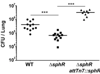
DsphRmutants have a survival defect during mouse lung infection
Oura prioriprediction was that SphR, controlling expression of a strongly induced gene by pulmonary surfactant, would be important for colonization and/or survival in the mammalian lung. To test this hypothesis, we examined bacterial survival 24 hours after infection in the mouse lung. The sphR deletion strain had significantly lower survival than wild type (7.7-fold decrease, Dunnett’s multiple comparisons p,0.001) and the survival defect was complemented by addition of sphR at the Figure 1. Expression ofsphA(PA5325) is induced by sphingosine.(A) Arrangement of thesphAgenomic region inP. aeruginosaPAO1. The genes in green are those discussed further in this study. Data from panels B through F all use a plasmid-bornesphA-lacZreporter (pAL5) to assess regulation with pyruvate (pyr) used as the non-inducing control condition. Fold induction ofsphA-lacZis calculated compared to its pyruvate control. (B)sphAis induced in the presence of pulmonary surfactant (surf), sheep red blood cells (blood), and mouse fibroblasts (fibr). (C) The primarysphA -inducing component of fibroblasts is present in the organic fraction (org), compared to the aqueous fraction (aq) after the mouse fibroblasts (whole) were extracted with chloroform:methanol. (D) The sphingolipids sphingomyelin (sm), ceramide (cer), sphingosine-1-phosphate (S1P), and sphingosine (sph) inducesphA, while likely catabolic products palmitate (pm) and glycine (gly) do not cause induction. Sphingosine causes the highest level of induction. (E) Induction of sphAby sphingosine is dose dependent and occurs at physiologically relevant concentrations of sphingosine. Lines for statistical significance denote groups of samples with the same magnitude of significance, not grouped comparisons. (F) Induction ofsphAby ceramide requires the neutral ceramidase gene,cerN, while sphingosine induction is independent ofcerN. (G) Induction ofsphA
by S1P in a heterologousE. coli sphR-sphA-lacZreporter system (pAL5) requires phosphatase treatment of S1P. Statistical significance determined using one way ANOVA with Dunnett’s post-test for B–E & G with the uninduced condition being the comparator for all other data. In panel F, the wild type andDcerNdata were compared by student t-test within each treatment condition. p-value summaries: n.s. = not significant; * for p,0.05; ** for p,0.01; *** for p,0.001; **** for p,0.0001. All experiments were performed at least three times and data shown is representative of both the scale and statistical significance levels of all experiments.
attTn7site (Fig. 3). In this comparison, wild type andDsphRboth contained the emptyattTn7insertion cassette on the chromosome. The contribution ofsphRto survival in the mouse lung led us to a more in-depth study of SphR and its target genes.
Determination of the SphR regulon
Deletion ofsphRresulted in reducedP. aeruginosasurvival in the mouse lung (Fig. 3), leading us to hypothesize that one or more of the genes in the SphR regulon were likely candidates for this phenotype. To identify SphR-regulated genes in addition tosphA, we conducted microarray transcriptome analyses to compare wild type and thesphRdeletion mutant in the presence and absence of pulmonary surfactant. Using a two-fold change cutoff and a p-value,0.05, there are six genes that differ between wild type and DsphRin the presence of surfactant (Table 1). Transcripts that are induced in wild type but not in thesphRdeletion mutant include sphA, the neutral ceramidase (PA0845), and a three gene operon convergently transcribed towardsphA,PA5328-PA5326. TheargB
gene (PA5323) was induced more strongly in thesphRdeletion than in wild type, which we think is likely due to a cis effect of thesphR (PA5324) deletion, as these genes are convergently transcribed (Fig. 1A). To denote their placement in the SphR regulon, we have renamed the genes in the predicted PA5328-PA5326 operon as sphBCD. The sphB gene encodes a predicted periplasmic
cytochrome and sphC and sphD encode a predicted
flavin-dependent oxidoreductase and a predicted pyridoxalphosphate-containing threonine aldolase-like enzyme, respectively. The predicted functions of SphC and SphD suggest a potential two-step pathway for sphingosine degradation to glycine and a long chain aldehyde by oxidation to an aldol and subsequent cleavage by the aldolase, a prediction we are currently exploring. The neutral ceramidase (PA0845) was previously designated PaCD [37], which does not conform to standard bacterial nomenclature. We propose thatPA0845be renamedcerNfor ceramidase, neutral. The induction ofsphAby surfactant in wild type (17.8-fold) versus the difference ofsphAinduction between wild type andDsphR (5.9-fold) suggested altered regulation ofsphA in the absence of sphR (Table 1). The relative induction of sphA in the sphR mutant compared to wt under pyruvate (non-inducing) conditions supports a de-repression ofsphA transcription in the absence of sphRat baseline. The remaining genes in the operon appear solely regulated by SphR under these conditions, as their induction levels in wild type compared to the difference between wild type and DsphRare not different.
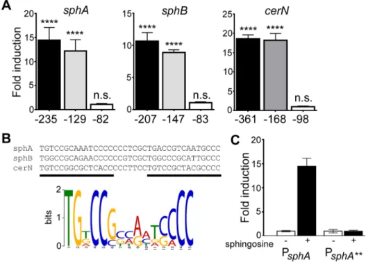
Mapping of thesphA,sphBCD, andcerNpromoters
We used promoter mapping to identify the promoter proximal regions of the sphA, sphBCD, and cerN promoters that were important for sphingosine andsphR-dependent regulation. Using Figure 2. The transcription regulator SphR (PA5324) controls
sphAinduction in response to sphingosine.(A) Sphingosine (sph) inducessphA-lacZ expression (pAL4) compared to the pyruvate (pyr) control in wild-type cells (wild type) but not in sphR mutant cells (DsphR). This regulation is restored by complementation (Comp) ofsphR
at theattTn7locus. (B) In a heterologousE. colisystem, thesphA-lacZ
reporter (pAL4) is not responsive to sphingosine (second bar) unless the
sphRgene is included in the system (pAL5) (fourth bar). Data for these panels were compared by student t-test comparing treatment conditions within each strain. p-value summaries: n.s. = not significant; **** for p,0.0001. All experiments were performed at least three times and data shown is representative of both the scale and statistical significance levels of all experiments.
doi:10.1371/journal.ppat.1003889.g002
Figure 3. Deletion ofsphRreducesP. aeruginosasurvival in the mouse lung. Male C57Bl/6J mice were infected with 26107CFU/
mouse of each strain via oropharyngeal aspiration. Mice were euthanized and lungs harvested 24 hours after infection. Bacterial counts were determined by serial dilution onto Pseudomonas Isolation Agar (PIA). Deletion ofsphR(DsphR) reducedP. aeruginosasurvival 7.7-fold (p,0.001), an effect that was complemented by addition ofsphRat theattTn7site. Wild-type (WT) andDsphRcells carried the emptyattTn7
insertion cassette as described in the methods section. Statistical significance determined using one way ANOVA with Tukey’s post-test comparing all groups to each other. p-value summaries: n.s. = not significant; *** for p,0.001. Data shown is combined from two experiments. The same effect sizes and variance have been seen in at least one additional experiment for each strain, resulting in each strain having been compared to wild type in at least three independent experiments (described further in the Methods section).
lacZ reporter fusions to each upstream region, we identified a portion of each promoter-proximal region required for respon-siveness to sphingosine (Fig. 4A). The regions required for sphingosine responsiveness were aligned using KALIGN [38], which produced an alignment that highlights the general format of an AraC-family binding site (Fig. 4B). The MEME consensus for a single half-site is shown below the alignment (Fig. 4B). Bioinfor-matic search of theP. aeruginosagenome (DNA Motif Search [39]) turned up only one additional predicted binding site (two direct repeats of the consensus (TGNCCSNNRNNSNCC) separated by 6–8 bp) in the genome apart from those present in the three identified promoters. The additional binding site is in the intergenic region betweenPA0428and PA0429, upstream of the PA0428 gene. We did not detect any change in the PA0428 transcript for wild type orDsphRin the presence of surfactant or in either strain in the absence of surfactant. Therefore, based on our microarray data and bioinformatic analysis, we predict thatsphA, sphBCD, and cerN likely comprise the core SphR regulon. The upstream sequences for the SphR regulon members showing the predicted SphR binding sites, promoter elements, and ribosome binding sites are shown in Supplemental Figure S2.
To test both specificity and the importance of conserved consensus sequences we mutated the first two residues in the consensus sequence TG to AA in half-site 1 (sphA**) (Fig. 4B), and tested the ability of the mutant sequence to permit induction of the reporter gene in response to sphingosine. The mutant reporter was unable to support reporter induction in response to sphingosine (Fig. 4C), demonstrating the importance of these conserved binding site residues.
SphR directly binds thesphA,sphBCD, andcerNpromoter proximal regions
We conducted electrophoretic mobility shift assays (EMSAs) with purified MBP-SphR fusion protein to test if SphR directly bound thesphA,sphBCD, and cerNpromoters. The binding of MBP-SphR to the sphApromoter probe was greatly enhanced by the addition of sphingosine to the binding reaction in a concentration-dependent manner, providing evidence that sphingosine was a direct ligand of SphR (Fig. 5A). In the presence of sphingosine, MBP-SphR specifically shifted the sphA, sphBCD, and cerN promoters in a protein concentration-dependent manner and the binding could be competed with unlabeledsphApromoter probe, which gives a sense of the relative affinities for each binding site (Fig. 5B). MBP-SphR did not shift the non-specific plcH probe (Fig. 5B). The plcH probe is a useful negative control and demonstrates the specificity of SphR binding, as it has a known binding site for the AraC-family transcription factor GbdR inP. aeruginosaand its regulation is well described [31,40–43].
To test the predicted SphR binding site, 59-mer oligonucleo-tides containing the proposed SphR binding site from thesphA promoter were annealed and the resultant probe was used in binding reactions. MBP-SphR was able to shift the 59-bp sphA probe (Fig. 5C, left), but only in the presence of sphingosine. Based on the inability of the mutated consensus sequence (sphA**) to support sphingosine-dependent reporter expression (Fig. 4C), we predicted that an oligonucleotide carrying these mutations would also be unable to bind SphR. As shown in the right side of Figure 5C, MBP-SphR was unable to bind this mutated probe. Together with the reporter fusions, these data support both the specificity of SphR binding and the importance of the conserved residues in the consensus.
SphR binds sphingosine
Based on the enhancement of SphR DNA binding in the presence of sphingosine and our genetic evidence, we predicted
that SphR would directly bind sphingosine. We used 3
H-sphingosine to test the ability of SphR to bind H-sphingosine (Fig. 6). The binding assay conditions were similar to those used
for EMSA studies with MBP-SphR in the presence of 3
H-sphingosine. Amylose resin beads were used to pull down the MBP-SphR, and bead-associated sphingosine was assayed by liquid scintillation counting. 3H-sphingosine was substantially
enriched in the fraction containing amylose-bound MBP-SphR, while relatively little remained associated with the amylose beads alone, or beads bound to a non-specific MBP-taggedP. aeruginosa AraC-family transcription factor, CdhR (MBP-CdhR) [44]. These data, in combination with the EMSAs (Fig. 5), demonstrate direct interaction between sphingosine and SphR.
Deletion ofsphAphenocopies the mouse survival phenotype of thesphRdeletion
Because deletion ofsphRled to reduced survival in the mouse lung, we were interested in determining which of the SphR regulon members contributed to survival in the lung. We generated deletions incerN,sphA, andsphCand compared to wild type in our 24 hour lung infection model. Deletion ofsphAled to a significant reduction in bacterial survival in the mouse lung (9-fold decrease, Dunnett’s multiple comparisons p,0.001), while dele-tion ofcerNorsphChad no impact on bacterial survival in vivo (Fig. 7). The sphA mutant phenotype could be complemented by supplying thesphAunder its native promoter control at theattTn7 site (Supplemental Fig. S3). These data suggest an important role forsphAin survival during infection. We did not test deletions of
sphB and sphD in the animal model, given their predicted
coordinate role with sphC in sphingosine metabolism and their Table 1.The SphR (PA5324) regulon inP. aeruginosa.
ORF/Feature Gene name1 Product Function
Fold Change WT Surf over sphR Surf
sphR Pyr over WT pyr
WT Surf over WT Pyr
PA5327 sphC Predicted oxidase 10.6 NC 10.2
PA5325 sphA Predicted porin 5.9 3.9 17.8
PA5328 sphB Predicted cytochrome C (mono-heme) 5.0 NC 5.1
PA0845 cerN Neutral ceramidase 3.1 NC 3.4
PA5326 sphD Predicted threonine aldolase family 2.6 NC 2.9
PA5323 argB Acetylglutamate kinase 22.2 2.1 NC
1gene names, apart from
similar phenotype to ansphCdeletion during in vitro sphingosine killing (Fig. 8 and Figure S4).
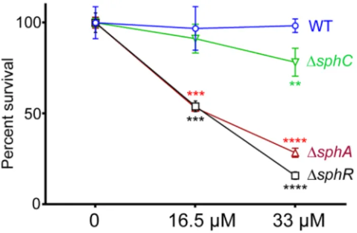
SphR & SphA are important for resistance to sphingosine killing
Sphingosine has previously been shown to have antimicrobial properties and is able to inhibit growth and kill many Gram
positive and Gram negative bacteria [19]. Previous studies suggest thatP. aeruginosais not sensitive to killing by sphingosine [45]. We hypothesized that SphR might play a role in the response ofP. aeruginosato sphingosine and could regulate sphingosine resistance. Using a modified sphingosine killing assay, we show that theDsphR deletion strain is more sensitive to sphingosine compared to wild type (Fig. 8), an effect that could be complemented bysphRon a Figure 4. Determination of the probable SphR binding site from SphR-regulated promoters.(A) Fold-induction of ß-galactosidase activity from each reporter and the truncations compared to a pyruvate non-induced control (not shown). Promoter deletion mapping demonstrated a minimal region required for each SphR-controlled transcript. The negative numbers below each panel refer to the position relative to the translational start of each gene. (B) KALIGN showing the nucleotide alignment of the conserved region from the minimal regulatory regions defined in (A). The black bars denote the two direct repeat half-sites typical of AraC-family transcription factors. Below the alignments is the MEME-generated logo showing the strength of conservation based on the six half-sites from these three promoters. (C) ß-galactosidase assay for wtsphA-lacZand thesphA
-lacZ**promoter mutant (TG at the beginning of the first half-site was changed to AA) to demonstrate the importance of the conserved region for induction. Statistical significance determined using one way ANOVA with Dunnett’s post-test with the uninduced pyruvate condition being the comparator for all other data. p-value summaries: n.s. = not significant; **** for p,0.0001. All experiments were performed at least three times and data shown is representative of both the scale and statistical significance levels of all experiments.
doi:10.1371/journal.ppat.1003889.g004
Figure 5. SphR directly binds to its target regulatory regions and binding is stimulated by sphingosine.The binding of MBP-SphR to target DNA was measured using electrophoretic mobility shift assays (EMSAs). (A) Sphingosine (sph) is required for robust MBP-SphR binding to target DNA and sphingosine stimulates SphR DNA binding in a dose-dependent manner. Based on this data all subsequent EMSAs were conducted with 10mM sphingosine. (B) MBP-SphR (SphR) binds in a dose-dependent manner tosphA,sphB, andcerNupstream regulatory regions, but not to the
sphR-independentplcHregulatory region. The MBP-SphR concentration (mM) is shown below the lanes. Specificity is shown by lack of MBP-SphR binding to theplcHpromoter and by the ability to compete the bulk of the shift with unlabelled competitor DNA (UC) denoted by the+sign under the lanes. (C) MBP-SphR binds a 59-bp oligonucleotide probe containing the predicted SphR binding site upstream ofsphA. The extra shifts seen in panels A & B are absent from the EMSAs with these minimal probes and likely are due to additional interaction sites. Mutation of two conserved residues in half-site#1 (TG to AA) (sphA**) results in substantial reduction in MBP-SphR binding. All experiments were performed at least three times and data shown is representative of all experiments.
plasmid (Supplemental Fig. S4). Most of the sensitivity of the DsphRstrain appears to be due to loss of sphAinduction, as the DsphAstrain is also more sensitive to sphingosine than wild type and is nearly as sensitive asDsphR(Fig. 8). The deletion phenotype of sphA could be complemented by sphA on a plasmid
(Supple-mental Fig. S4). Deletion of sphCand transposon insertions into sphDsurvival on sphingosine, suggesting a minor role for this operon inand sphB also led to small but reproducible decreases in
the response to sphingosine (Fig. 8 and Supplemental Fig. S4). Deletion ofcerN, befitting its known function as an extracellular ceramidase, had no effect on survival in sphingosine (data not shown).
Discussion
The induction of ceramidase activity in response to sphingosine has been demonstrated in a few bacteria [37], but the mechanism of sphingosine detection and conversion into a response had not previously been elucidated. In this study we show that sphingosine is directly detected by the AraC-family transcription factor SphR (PA5324) leading to the induction of sphA, sphBCD, and cerN transcripts. Deletion ofsphRorsphAresulted in survival defects in a mouse model of acute pneumonia, suggesting that the ability to detect and respond to host-derived sphingolipids is important for survival in the lung. Sphingolipids are abundant in mammals, plants, and fungi, constituting a diverse family of molecules that serve as essential structural components of eukaryotic cell membranes and as dynamic signaling molecules that mediate diverse cellular functions [16–19]. In particular, S1P has been implicated as a critical component of mammalian innate and adaptive immune function, particularly in the acute phase response to pathogens [25–28]. Interestingly, orthologs of SphR and some of the SphR-regulon members are present in other opportunistic pathogens including Acinetobacter haemolyticus and Burkholderia pseudomallei, as well as the professional pathogen Mycobacterium tuberculosis.
Sphingolipids play important roles in host-pathogen interac-tions, particularly S1P and ceramide signaling [46–48]. In addition to host modulation of sphingolipid pathways to combat infection, Figure 6. SphR directly binds to sphingosine.The association of
MBP-SphR was determined by measuring binding of 3H-sphingosine and reporting counts per minute (CPM). Sphingosine minimally associates with amylose beads alone (beads) or an un-related AraC-family transcription factor (MBP-CdhR), but approximately 11-fold more
3H-sphingosine binds to MBP-SphR (p
,0.0001). Statistical significance determined using one way ANOVA with Dunnett’s post-test with the beads alone condition being the comparator for all other data. p-value summaries: n.s. = not significant; **** for p,0.0001. This experiment was performed at least three times and data shown is representative of both the scale and statistical significance levels of all experiments. doi:10.1371/journal.ppat.1003889.g006
Figure 7. Deletion of the SphR-regulon member,sphA, reduces P. aeruginosasurvival in the mouse lung.Male C57Bl/6J mice were infected with 26107CFU/mouse of each strain via oropharyngeal
aspiration. Mice were euthanized and lungs harvested 24 hours after infection. Bacterial counts were determined by serial dilution onto PIA. Deletion of sphA (DsphA) reduced P. aeruginosa survival 9-fold (p,0.001). Deletion of the other SphR-regulon members cerN and
sphC (as an sphBCD operon representative) resulted in no survival defect. Statistical analysis determined using one way ANOVA with Dunnett’s post-test with wild type as the comparator. Data shown is combined from two experiments. The same effect sizes and variance have been seen in at least one additional experiment for each strain, resulting in each strain having been compared to wild type in at least three independent experiments (described further in the Methods section).
doi:10.1371/journal.ppat.1003889.g007
Figure 8. Deletion of sphA or sphR renders P. aeruginosa susceptible to killing by sphingosine. Strains with deletions in
sphA(red),sphR(black), andsphC(green) were compared to wild type (blue) using a sphingosine killing assay in neopeptone as described in the Methods section. Cells were exposed to vehicle alone (0) or one of two concentrations of sphingosine for 30 minutes followed by serial dilution and plating. The mean of the vehicle treatment samples for each strain was set as 100% survival for that strain. Statistical significance determined using one way ANOVA with Dunnett’s post-test with wild type at each concentration being the comparator for the mutant strain data at the same concentration. p-value summaries: n.s. = not significant; ** for p,0.01; *** for p,0.001; **** for p,0.0001. This experiment was performed more than three times and data shown is representative of both the scale and statistical significance levels of all experiments.
pathogens can modulate host sphingolipids. M. tuberculosis alters sphingolipid signaling in macrophages by undetermined mecha-nisms [49], and S1P levels in the lungs of patients infected withM. tuberculosis are significantly decreased [50]. Interestingly, M. tuberculosis has an AraC-family transcription factor that is 47% similar along the whole length to SphR (RV1395) that was identified though signature-tagged mutagenesis where theRV1395 transposon mutant strain had an,1.5 log reduced survival in a mouse lung infection model [51]. Similarity between RV1395 and SphR is not restricted to the helix-turn-helix DNA-binding domain, as the two proteins are 44% similar when the DNA-binding domain is removed from the alignment analysis. RV1395 was characterized and found to be an activator of a divergently transcribed cytochrome gene, however the signals that govern RV1395 activation and its direct contribution to virulence have yet to be determined [52]. Based on the similarity of RV1395 to SphR we predict that a sphingolipid, perhaps sphingosine, may be the inducing ligand of RV1395.
The AraC-family transcription regulators are one of the largest groups of regulatory proteins in bacteria, and are often involved in the regulation of catabolism, stress response, and virulence [53]. Many members of the AraC family have been shown to respond to host-derived chemical signals present at the site of infection, but relatively few inducing ligands have been demonstrated to bind directly to their cognate regulator [54]. We found that addition of sphingosine altered the binding of SphR to thesphApromoter in EMSA studies and observed a dose response curve of SphR DNA binding at physiologically relevant concentrations of sphingosine. Bioinformatic analysis suggest similarity of SphR to ToxT (44% similarity and 20% identity), which directly regulates the major virulence factors inVibrio cholerae.ToxT activation is inhibited by unsaturated fatty acids found in bile [55]. Subsequently, the crystal structure of ToxT was solved revealing a bound 16-carbon fatty acid that alters the structure of ToxT to prevent DNA binding in the presence of these bile associated fatty acids [56]. The similar size and hydrophobic nature of the regulatory ligands (palmitate vs. sphingosine) coupled with the sequence similarity allows us to speculate that SphR may bind sphingosine in a manner analogous to ToxT binding of palmitate.
Ito et al. identified a neutral ceramidase encoded by PA0845 (renamedcerNin this study) that was induced in the presence of sphingomyelin, ceramide and sphingosine, however the regulatory mechanism was not reported [37]. The discovery of SphR control of neutral ceramidase allows us to expand a model of bacterial utilization of sphingomyelin by linking it to our previous work on regulation of the phospholipase C/sphingomyelinase PlcH. We previously characterized the AraC-family regulator GbdR that is integral to a positive feedback loop controlling PlcH expression in response to a metabolite of the choline headgroup of sphingomy-elin [31]. Sphingomysphingomy-elin hydrolysis by PlcH yields ceramide [57], which P. aeruginosacan further metabolize through the action of ceramidases [54]. Here we show that CerN is produced as part of an SphR-dependent positive feedback loop in response to the ceramide metabolite sphingosine, in a manner analogous to GbdR control of PlcH. Both of these positive feedback loops link induction of secreted catabolic enzymes not to the availability of the substrate itself, but to metabolic products derived from the substrate. In each case, this ensures that the positive feedback loop will robustly operate only if the substrate is being metabolized at sufficient rates.
Sphingolipids such as sphingosine have long been known to have antimicrobial properties and sphingosine is found in high concentration in the skin where it is thought to be part of the barrier function against microbial infections [58–61]. A variety of
Gram positive and Gram negative bacteria are sensitive to sphingosine, including Staphylococcus aureus and Escherichia coli [62]. The precise bactericidal mechanism of sphingosine remains unknown. However, recent evidence suggests that sphingosine may directly damage bacterial membranes [63].P. aeruginosahas recently been reported to be resistant to the bactericidal effects of sphingosine [45]. While none of the deletion strains generated in this study showed growth defects under normal conditions, we found that both thesphRandsphAdeletion strains were susceptible to the antimicrobial effects of sphingosine compared to wild type in vitro. Strains with deletions insphRandsphAwere also shown to have reduced survival in the mouse lung. We hypothesize that the sensitivity ofsphAandsphRmutants to sphingosine contributes to their observed reduced survival in vivo. It is interesting to note that the double deletionDcerNDsphA strain did not survive better or worse thanDsphA, minimally suggesting that if the defect is due to sphingosine sensitivity, it is not sphingosine derived from P. aeruginosahydrolysis of host-derived ceramide; in other words, they are not causing their own death by sphingosine derived from sphingomyelin and ceramide hydrolysis. Therefore, while the in vitro sphingosine killing correlates well with the in vivo pheno-types, we currently do not know the mechanism governing reduced survival of thesphRandsphAmutants in the lung.
We speculate that SphR responds to sphingosine to induce transcripts encoding proteins that protect P. aeruginosa from the bactericidal effects of sphingosine by induction of membrane stabilizing factors and/or catabolism of sphingosine to non-bactericidal metabolites. Here we show that SphR binds to sphingosine to initiate transcription ofsphA,sphBCDandcerN.sphA encodes a hypothetical protein with some homology to proteins involved in meta-pathway phenol degradation. Protein localization predictions for SphA using the structure similarity-based predic-tion of Phrye2 [64] suggests that SphA is an outer membrane porin. PerhapsP. aeruginosaresponds to sphingosine by providing a porin for sphingosine import and subsequent degradation that could aid in protecting the outer membrane from the damaging effects of free sphingosine. Okino and Ito demonstrated
sphingo-sine utilization by P. aeruginosa by measuring removal of
sphingosine from the culture supernatants and cell fractions [54]. Based on bioinformatic predictions, SphB, SphC and SphD are most likely involved in the metabolism of sphingosine. The sphBgene encodes a predicted cytochrome, whilesphCencodes an FMN-linked oxidoreductase, and sphD encodes a pyridoxalpho-sphate serine-threonine aldolase. The latter two activities could work in concert to oxidize carbon 1, generating an aldol, which SphD could hypothetically act upon, rendering a long chain aldehyde and glycine. Transposon insertion into thesphCcoding sequence (PA5327) resulted in reduced bacterial survival in a chronic rat lung infection model [65], suggesting that while our sphCdeletion strain did not show a phenotype in the acute mouse lung infection (Fig. 7), it nonetheless impacts survival in the mammalian lung.
the choline catabolic pathway were also highly induced in the presence of surfactant (Table S1). In addition to the high proportion of transcripts encoding stress-related proteins (men-tioned in the Results section), there are also a high proportion (,8%) of transcriptional regulators: NalC, BetI, NirG, PsrA, NarL, CgrA, PA3458, and PA4596. It is possible that the effects of induction of these transcription factors is contained in our regulation data, however our transcriptome analyses were a snapshot of transcripts at four hours post-induction and effects from changes in these transcription factors may not have sufficiently accumulated in the transcriptome. Of the reduced transcripts (Table S2), we note that three of the pyrroquinoline quinine biosynthesis genes are down, suggesting a change in requirement for this cofactor between surfactant and pyruvate conditions.
The demonstration of sphingosine detection byP. aeruginosaalso opens up the possibility that this bacterium, and others with similar detection systems, could alter sphingosine and related sphingolipid signals, including S1P in the host. We have not yet examined the contribution of host immune signaling effected by the SphR regulon, but the impact of altering such an important and tightly controlled signaling network by bacterial factors has not been elucidated and may be an important contributing factor to the survival ofP. aeruginosain vivo.
Materials and Methods
Ethics statement
This study was performed in strict accordance with the recommendations in the Guide for the Care and Use of Laboratory Animals of the National Institutes of Health. The protocol for animal infection was approved by the University of Vermont Institutional Animal Care and Use Committee (Permit number A3301-01). All procedures were performed under pentobarbital anesthesia and all efforts were made to minimize animal suffering.
Strains, growth conditions, and chemicals
P. aeruginosaPAO1, isogenic mutant strains, andE. coli(Table 2) were maintained in LB-Lennox (LB) medium. Morpholinepropa-nesulfonic acid (MOPS) medium [66] supplemented with 25 mM sodium pyruvate, 5 mM glucose and 50mg/ml gentamicin (forP. aeruginosa) or MOPS with 10% LB (v/v), 5 mM glucose, and 10mg/ml gentamicin (forE. coli) was used to grow strains prior to
transcriptional induction studies. See LaBauve and Wargo (2012) for further details on P. aeruginosa growth methods [67]. For bactericidal assays, 1% neopeptone was supplemented with varying sphingosine concentrations in ethanol to reach a final concentration of 6.25% (w/v) ethanol in the assay. All lipids were purchased from Avanti Polar Lipids and other chemicals were purchased from Sigma-Aldrich or Fisher.
Mouse lung infection model
We used the oropharyngeal route of mouse lung infection previously described [68,69]. Briefly, P. aeruginosa PAO1 and isogenic strains were streaked onto LB plates from280uC stocks. Colonies from the first plate were restreaked onto a new LB plate after 24 hours and incubated at 37uC for 24 hours. Cells from the second plate were used to start 3 ml cultures in LB that were grown for 16–18 hours at 37uC on a roller drum. From these overnight cultures, cells were collected by centrifugation, washed in Dulbecco’s PBS (DPBS), and resuspended to give ,16107
viableP. aeruginosain 40mL, with actual inoculum determined by
serial dilution and plate counting. Eight to twelve week old male
C57Bl/6J mice (Jackson Labs) were inoculated with 40mL of the
bacterial suspension via oropharyngeal aspiration. Anesthesia, surgery, bronchoalveolar lavage fluid (BALF) collection, organ harvest, and organ homogenization were done as previously described [68,69] at 24 hours post-infection. Viable bacterial counts in organs were determined by serial dilution plating onto Pseudomonas Isolation Agar (PIA) (BD-Difco) followed by incubation at 37uC for 24 hours.
Mouse experiments (Fig. 3 and 7 and Supplemental Fig. S3) show CFU counts from all animals from duplicate experiments with each replicate having 4–6 animals per experimental group. All informative comparisons: mutants versus wild type (both Figures) and mutant versus complementation strain (Fig. 3 and Supplemental Fig. S3) were conducted in at least one additional experiment, included with comparator strains from other studies. Therefore, all informative comparisons were assessed three times. All experiments met the same statistical criteria, i.e. all replicates were consistent with regards to effect size and significance of changes. Inoculation order and harvest order alternated between experiments to eliminate potential issues related to the difference between the duration of inoculation (,20–30 min) and the duration of harvest (,1.5 h). For group comparisons, data (log10
transformed CFU counts) were analyzed by ANOVA followed by Tukey’s (Fig. 3 and Supplemental Fig. S3) or Dunnett’s (Fig. 7) Multiple Comparisons tests. All calculations were done using GraphPad Prism.
RNA extraction and microarray methodology
P. aeruginosaPAO1 wild type andDsphRwere grown overnight in MOPS media supplemented with 20 mM pyruvate and 5 mM glucose. Overnight cultures were collected by centrifugation and resuspended in either MOPS supplemented with 20 mM pyruvate alone or 20 mM pyruvate and a 1:50 dilution of the bovine surfactant preparation Survanta (Abbott) and induced for 4 hours at 37uC. Bacteria were collected by centrifugation, resuspended in MOPS and RNA Protect Bacterial Reagent (Qiagen), and the resultant pellets stored overnight at220uC. RNA was extracted using an RNeasy kit (Qiagen), and eluted samples were treated with DNase I followed by a second round of RNeasy purification including an on-column DNase I treatment. Purified RNA samples were checked for DNA contamination by PCR and RNA integrity scores based on Agilent Bioanalyzer analysis were indicative of little to no DNA contamination.
Construction of deletion strains and complementation
Deletion mutants were generated using the pMQ30 plasmid [70] carrying the flanking regions of each of the four genes,sphR,sphA, sphC, and cerN, using conjugation-mediated deletion as described previously [30,69]. Primers for these constructs are listed in Table S3. Single cross-over mutants were selected on PIA with gentamicin and selection of double crossover deletion mutants were carried out on LB 5% sucrose plates prepared without NaCl. Unmarked deletion mutants were verified using PCR. Complementation was done by integration of the sphRor sphA coding sequence under control of their native promoter at the attTn7 locus using the pUC18-miniTn7T-Gm vector as we described previously [68,69] using the method of Choi and Schweizer [71]. This allowed stable complementation in the absence of antibiotic. For complementation where reporter plasmids were used, the gentamicin resistance cassette was excised by FLP-mediated recombination [71]. All sphR::attTn7 andsphA::attTn7 complementation strains were com-pared with wild type or mutant strains carrying the emptyattTn7 integration region from the pUC18-miniTn7T-Gm vector.
Reporter assay to measuresphAtranscriptional induction
Two reporter constructs were generated in this study using yeast homologous recombination [70] to generate translational fusions to lacZYA. A targetlacZYA-containing vector suitable for yeast cloning (pMW42) was generated by excising thelacZYAregion from pMW5 [31] with HindIII and EcoRI and cloning into the similarly cut pMQ80 backbone [70], which removesegfp-mut3. Either thesphA promoter (pAL4), or the entiresphRgene and the sphApromoter (pAL5) were recombined with pMW42 linearized with KpnI and HindIII. P. aeruginosa strains were electrotransformed with the
reporter constructs and grown overnight in MOPS media supplemented with 20 mM pyruvate, 5 mM glucose, and 50mg/ ml gentamicin prior to induction. Inductions were carried out in MOPS media supplemented with 20 mM pyruvate and the inducing compound and incubated at 37uC for 6 hours. b -galactosidase assays were done as previously described [31,72], using the method of Miller [73]. Studies of heterologous sphA induction inE. coliwere carried by transforming pAL4 and pAL5 intoE. coliNEB5a. ResultingE. colistrains were grown overnight in MOPS media supplemented with 10% LB (v/v), 5 mM glucose and 10mg/ml gentamicin. For induction assays with S1P in E. coli,
2.4mg of S1P or sphingosine were pre-treated with or without 5 U shrimp alkaline phosphatase (SAP) in 100mL of water with 16SAP buffer (USB), and incubated at 37uC for 60 minutes. Induction assays were carried out in MOPS supplemented with 10% LB (v/v), treated inducing compounds, and 10mg/ml gentamicin. AllE. coli strains were induced for 8 hours prior to ß-galactosidase assays.
Promoter mapping ofsphA,sphB, andcerN
Full-length reporter constructs and truncations ofsphA,sphB, and cerNpromoters were cloned into pMW5 [31]. The resultantlacZYA reporter constructs were transformed into wild typeP. aeruginosaand used to identify the region required for response to sphingosine. Inductions were carried out in MOPS media supplemented with 20 mM pyruvate and 150mM sphingosine and incubated at 37uC for 6 hours followed by ß-galactosidase assays.
Cloning, expression and purification of SphR
We constructed a maltose binding protein (MBP) fusion to SphR by using the pMALc2 vector system (NEB). ThesphRgene Table 2.Bacterial strains and plasmids used in this study.
Organism/Plasmid Description/Genotype Name Source
Pseudomonas aeruginosa PAO1 wild type MJ79 [75]
DsphR AL76 This study
DsphA AL82 This study
DsphC AL119 This study
DcerN AL114 This study
Wild type empty::attTn7GmR MJ507 This study
DsphRempty::attTn7GmR MJ508 This study
DsphR sphR::attTn7GmR MJ540 This study
Wild type empty::attTn7GmS AL138 This study
DsphRempty::attTn7GmS AL139 This study
DsphR sphR::attTn7GmS AL140 This study
Escherichia coli NEB5a NEB
S17-1lpir MJ340 [76]
Plasmid sphA-lacZYAreporter pAL4 This study
sphR-sphA-lacZYAreporter pAL5 This study
MBP-SphR expression vector pAL11 This study
Maltose binding protein (MBP) expression vector pMAL-c2 NEB
Integration vector pMQ30 Shanks
P. aeruginosareplicativelacZYAreporter pMW5 Wargo
P. aeruginosa-yeast shuttlelacZYAreporter pMW42 This study
sphR::attTn7integration vector pMW118 This study
Empty::attTn7integration vector pUC18-mini-Tn7T-Gm Schweizer
was amplified from genomic DNA. The PCR product was gel purified and ligated into the pCR Blunt vector (Invitrogen). The insert was excised with KpnI and HindIII, gel purified, and ligated into a similarly digested pMALc2 vector to generate pAL11.E. coli NEB5a(New England Biolabs) carrying the pAL11 plasmid were grown overnight in LB supplemented with 120mg/ml
carbenicil-lin. The overnight culture was transferred to two 500 ml flasks containing 100 ml of LB-carbenicillin and shaken at 220 rpm for 5 hours. Isopropyl-b-D-thiogalactopyranoside (IPTG) was added to a final concentration of 1 mM, and the cells were induced for 3 hours. Cells were collected by centrifugation, lysed in column buffer (20 mM Tris-HCl, pH 7.5, 150 mM NaCl, 1 mM EDTA) supplemented with 3 mg/ml lysozyme and Halt protease inhibitor 16 cocktail (Thermo Scientific). Lysates were clarified by centrifugation, and the soluble fraction was applied to a column containing amylose resin (NEB). The column was washed with ten volumes of column wash buffer (20 mM Tris-HCl, 150 mM NaCl 1 mM EDTA pH 7.4), followed by elution with column wash buffer supplemented with 10 mM maltose. Elution fractions were run on 10% SDS-PAGE gels and visualized by Coomassie staining. Fractions containing the MBP-SphR were pooled and dialyzed against 20 mM Tris-HCl, pH 7.5 at 4uC in a 20,000 kDa cutoff Slide-A-lyzer cassette (Pierce). The full length MBP-SphR fusion protein was used in electrophoretic mobility shift assays, as the MBP tag did not prevent sequence specific DNA binding (Fig. 5) or binding to sphingosine (Fig. 6).
Electrophoretic Mobility Shift Assay (EMSA)
EMSA DNA probes were generated using PCR (Primers in Table S3) and were spot dialyzed against 2.5 mM Tris-HCl, 0.25 mM EDTA, pH 8.0. Labeled probes, generated using a primer with a covalently linked 59biotin tag (IDT), were used at 0.5 fmol/ml, and unlabeled competitor probes were used at a final
concentration of 0.5 pmol/ml. EMSA was carried out using a
Thermo Scientific Thermoshift kit. The final binding buffer was modified to contain 16binding buffer (10 mM Tris-HCl, pH 7.5, 50 mM KCl, 1 mM dithiothreitol), 0.1 mM glycine betaine, and 2mg/ml poly-dI-dC. Various concentrations of sphingosine dis-solved in ethanol were added to reaction tubes and allowed to dry to eliminate ethanol prior to binding reactions. Binding reactions were carried out at 37uC for 15 minutes and electrophoresed on a 5% non-denaturing polyacrylamide gel then transferred to a BioDyne B membrane (Thermo Scientific). Detection was carried out using streptavidin-linked horseradish peroxidase according to the supplied protocol (Thermo Scientific).
Measurements of sphingosine association with SphR
Sphingosine association with SphR was measured by conduct-ing bindconduct-ing reactions usconduct-ing 3H-D-erytho-sphingosine (Perkin-Elmer). Binding reactions were carried out as described for
EMSA except 3H-D-erytho-sphingosine was used at a final
concentration of 50 nM. Samples were incubated with and
without either 10mM MBP-SphR or 10mM MBP-CdhR for
30 minutes then added to amylose resin. The amylose beads were collected by centrifugation and washed 3 times with amylose column wash buffer. After washes, amylose beads were resus-pended in 200ml of amylose wash buffer and transferred to a glass vial containing 10 ml of Biosafe II scintillation cocktail (RPI). Samples were quantified using a Tri-Carb 2910 TR liquid scintillation analyzer (Perkin-Elmer).
Sphingosine killing assay
Killing assays were carried out as previously described [61]. Briefly, overnightP. aeruginosastrains were grown in trypticase soy
broth (TSB) and diluted 1:40. Diluted cultures (100ml) were
added to glass tubes containing 250ml of 1% neopeptone
supplemented with 50ml of the appropriate sphingosine stock in ethanol or ethanol alone as the vehicle control. The cultures were shaken at 170 rpm for one hour. Survival was determined by serial dilution plating on PIA. Colonies were counted after 24 hour incubation and survival calculated by comparison to vehicle only controls.
Supporting Information
Figure S1 Induction of sphA-lacZ in response to induc-ing lipids of varyinduc-ing lengths, structures, and saturation.
Growth, induction, and ß-galactosidase assays conducted as described in the methods section usingP. aeruginosacarrying the pAL5 reporter plasmid. All compounds were used at a final concentration of 150mM with methanol as the vehicle for linoleic acid (final vehicle concentration 0.05%) and ethanol as the vehicle for all other compounds (final vehicle concentration 0.05%).
(TIF)
Figure S2 DNA sequences for the 400-base pairs upstream of thesphA, sphB, andcerN genes. The ATG at the end of each sequence marks the start-codon of the labeled gene. Underlined normal text indicated predicted ribosome binding sites. The teal-blue highlighted bases indicate predicted
210 promoter elements. The potential235 elements were further from consensus in these promoters and are not indicated on this figure. The green highlighted sequences indicate the SphR binding site. For thesphAregion, thesphRstart codon is marked, as is the transcriptional start site (underlined bold italic) as determined by Wurtzel et al. [74]. The other transcriptional start sites were not detected in the Wurtzel study, likely due to the absence of a suitable inducer.
(TIF)
Figure S3 Deletion of sphA reduces P. aeruginosa
survival in the mouse lung. Male C57Bl/6J mice were infected with 26107CFU/mouse of each strain via oropharyngeal aspiration. Mice were euthanized and lungs harvested 24 hours after infection. Bacterial counts were determined by serial dilution onto Pseudomonas Isolation Agar (PIA). Deletion ofsphA(DsphA) reduced P. aeruginosa survival ,7-fold, an effect that was complemented by addition of sphA under control of its native promoter at the attTn7 site. Wild-type (WT) and DsphA cells carried the emptyattTn7insertion cassette (attTn7-E) as described in the methods section. Statistical significance determined using one way ANOVA with Tukey’s post-test comparing all groups to each other. p-value summaries: * for p,0.05, ** for p,0.01. Data shown is combined from two experiments.
(TIF)
three times and data shown is representative of both the scale and statistical significance levels of all experiments.
(TIF)
Table S1 P. aeruginosatranscripts induced more than 3-fold in response to pulmonary surfactant.
(XLSX)
Table S2 P. aeruginosa transcripts reduced more than 3-fold in response to pulmonary surfactant.
(XLSX)
Table S3 Primers used in this study.
(XLSX)
Acknowledgments
We’d like to thank Jenna Bement for technical assistance in the animal handling, infections, and surgeries and Ben Lane for assistance with cloning and reporter assays. Jamie Meadows provided the MBP-CdhR protein and, along with Adam Nock, and Graham Willsey (University of Vermont), provided valuable comments and critically read the manuscript.
Author Contributions
Conceived and designed the experiments: AEL MJW. Performed the experiments: AEL MJW. Analyzed the data: AEL MJW. Contributed reagents/materials/analysis tools: AEL MJW. Wrote the paper: AEL MJW.
References
1. Lieberman D, Lieberman D (2003) Pseudomonal infections in patients with COPD: epidemiology and management. Am J Respir Med 2: 459–468. 2. Chastre J, Fagon JY (2002) Ventilator-associated pneumonia. Am J Respir Crit
Care Med 165: 867–903.
3. Crouch Brewer S, Wunderink RG, Jones CB, Leeper KV, Jr. (1996) Ventilator-associated pneumonia due to Pseudomonas aeruginosa. Chest 109: 1019– 1029.
4. Burns JL, Emerson J, Stapp JR, Yim DL, Krzewinski J, et al. (1998) Microbiology of sputum from patients at cystic fibrosis centers in the United States. Clin Infect Dis 27: 158–163.
5. Briesacher BA, Quittner AL, Fouayzi H, Zhang J, Swensen A (2011) Nationwide trends in the medical care costs of privately insured patients with cystic fibrosis (CF), 2001–2007. Pediatr Pulmonol 46: 770–776.
6. Burns JL, Gibson RL, McNamara S, Yim D, Emerson J, et al. (2001) Longitudinal assessment of Pseudomonas aeruginosa in young children with cystic fibrosis. J Infect Dis 183: 444–452.
7. Rajan S, Saiman L (2002) Pulmonary infections in patients with cystic fibrosis. Semin Respir Infect 17: 47–56.
8. Butorac-Petanjek B, Parnham MJ, Popovic-Grle S (2010) Antibiotic therapy for exacerbations of chronic obstructive pulmonary disease (COPD). J Chemother 22: 291–297.
9. Klockgether J, Munder A, Neugebauer J, Davenport CF, Stanke F, et al. (2010) Genome diversity of Pseudomonas aeruginosa PAO1 laboratory strains. J Bacteriol 192: 1113–1121.
10. Fischbach MA, Walsh CT (2009) Antibiotics for emerging pathogens. Science 325: 1089–1093.
11. Frisk A, Schurr JR, Wang G, Bertucci DC, Marrero L, et al. (2004) Transcriptome analysis ofPseudomonas aeruginosaafter interaction with human airway epithelial cells. Infect Immun 72: 5433–5438.
12. Palmer KL, Mashburn LM, Singh PK, Whiteley M (2005) Cystic fibrosis sputum supports growth and cues key aspects of Pseudomonas aeruginosaphysiology. J Bacteriol 187: 5267–5277.
13. Chroneos ZC, Sever-Chroneos Z, Shepherd VL (2010) Pulmonary surfactant: an immunological perspective. Cell Physiol Biochem 25: 13–26.
14. Glasser JR, Mallampalli RK (2012) Surfactant and its role in the pathobiology of pulmonary infection. Microbes Infect 14: 17–25.
15. Caminiti SP, Young SL (1991) The pulmonary surfactant system. Hosp Pract (Off Ed) 26: 87–90, 94–100.
16. Hannun YA, Obeid LM (2008) Principles of bioactive lipid signalling: lessons from sphingolipids. Nat Rev Mol Cell Biol 9: 139–150.
17. Spiegel S, Milstien S (2011) The outs and the ins of sphingosine-1-phosphate in immunity. Nat Rev Immunol 11: 403–415.
18. Ohanian J, Ohanian V (2001) Sphingolipids in mammalian cell signalling. Cell Mol Life Sci 58: 2053–2068.
19. van Meer G, Voelker DR, Feigenson GW (2008) Membrane lipids: where they are and how they behave. Nat Rev Mol Cell Biol 9: 112–124.
20. Schwab SR, Pereira JP, Matloubian M, Xu Y, Huang Y, et al. (2005) Lymphocyte sequestration through S1P lyase inhibition and disruption of S1P gradients. Science 309: 1735–1739.
21. Vogel P, Donoviel MS, Read R, Hansen GM, Hazlewood J, et al. (2009) Incomplete inhibition of sphingosine 1-phosphate lyase modulates immune system function yet prevents early lethality and non-lymphoid lesions. PLoS One 4: e4112.
22. Walzer T, Chiossone L, Chaix J, Calver A, Carozzo C, et al. (2007) Natural killer cell trafficking in vivo requires a dedicated sphingosine 1-phosphate receptor. Nat Immunol 8: 1337–1344.
23. Wang F, Van Brocklyn JR, Hobson JP, Movafagh S, Zukowska-Grojec Z, et al. (1999) Sphingosine 1-phosphate stimulates cell migration through a G(i)-coupled cell surface receptor. Potential involvement in angiogenesis. J Biol Chem 274: 35343–35350.
24. Postma FR, Jalink K, Hengeveld T, Moolenaar WH (1996) Sphingosine-1-phosphate rapidly induces Rho-dependent neurite retraction: action through a specific cell surface receptor. EMBO J 15: 2388–2392.
25. Roviezzo F, Brancaleone V, De Gruttola L, Vellecco V, Bucci M, et al. (2011) Sphingosine-1-phosphate modulates vascular permeability and cell recruitment in acute inflammation in vivo. J Pharmacol Exp Ther 337: 830–837. 26. Hammad SM (2011) Blood sphingolipids in homeostasis and pathobiology. Adv
Exp Med Biol 721: 57–66.
27. Oskeritzian CA, Price MM, Hait NC, Kapitonov D, Falanga YT, et al. (2010) Essential roles of sphingosine-1-phosphate receptor 2 in human mast cell activation, anaphylaxis, and pulmonary edema. J Exp Med 207: 465–474. 28. Camerer E, Regard JB, Cornelissen I, Srinivasan Y, Duong DN, et al. (2009)
Sphingosine-1-phosphate in the plasma compartment regulates basal and inflammation-induced vascular leak in mice. J Clin Invest 119: 1871– 1879.
29. Jackson AA, Gross MJ, Daniels EF, Hampton TH, Hammond JH, et al. (2013) Anr and Its Activation by PlcH Activity in Pseudomonas aeruginosa Host Colonization and Virulence. J Bacteriol 195: 3093–3104.
30. Wargo MJ, Szwergold BS, Hogan DA (2008) Identification of two gene clusters and a transcriptional regulator required forPseudomonas aeruginosaglycine betaine catabolism. J Bacteriol 190: 2690–2699.
31. Wargo MJ, Ho TC, Gross MJ, Whittaker LA, Hogan DA (2009) GbdR regulates
Pseudomonas aeruginosa plcH and pchP transcription in response to choline catabolites. Infect Immun 77: 1103–1111.
32. Hampel KJ, Labauve AE, Meadows JA, Fitzsimmons LF, Nock AM, et al. (2013) Characterization of the GbdR regulon in Pseudomonas aeruginosa. J Bacteriol 196: 7–15.
33. Nelson GJ (1967) Studies on the lipids of sheep red blood cells. I. Lipid composition in low and high potassium red cells. Lipids 2: 64–71.
34. Lengle E, Geyer RP (1972) Comparison of cellular lipids of serum-free strain L mouse fibroblasts. Biochim Biophys Acta 260: 608–616.
35. Yatomi Y, Igarashi Y, Yang L, Hisano N, Qi R, et al. (1997) Sphingosine 1-phosphate, a bioactive sphingolipid abundantly stored in platelets, is a normal constituent of human plasma and serum. J Biochem 121: 969–973.
36. Law SLS, Squier CA, Wertz PW (1994) Free Sphingosine in Oral Epithelium. Journal of Dental Research 73: 108–108.
37. Okino N, Tani M, Imayama S, Ito M (1998) Purification and characterization of a novel ceramidase from Pseudomonas aeruginosa. J Biol Chem 273: 14368– 14373.
38. Lassmann T, Sonnhammer EL (2005) Kalign–an accurate and fast multiple sequence alignment algorithm. BMC Bioinformatics 6: 298.
39. Winsor GL, Lam DK, Fleming L, Lo R, Whiteside MD, et al. (2011) Pseudomonas Genome Database: improved comparative analysis and popula-tion genomics capability for Pseudomonas genomes. Nucleic Acids Res 39: D596–600.
40. Lucchesi GI, Lisa TA, Domenech CE (1989) Choline and betaine as inducer agents of Pseudomonas aeruginosa phospholipase C activity in high phosphate medium. FEMS Microbiol Lett 48: 335–338.
41. Sage AE, Vasil AI, Vasil ML (1997) Molecular characterization of mutants affected in the osmoprotectant-dependent induction of phospholipase C in
Pseudomonas aeruginosaPAO1. Mol Microbiol 23: 43–56.
42. Sage AE, Vasil ML (1997) Osmoprotectant-dependent expression of plcH, encoding the hemolytic phospholipase C, is subject to novel catabolite repression control inPseudomonas aeruginosaPAO1. J Bacteriol 179: 4874–4881. 43. Shortridge VD, Lazdunski A, Vasil ML (1992) Osmoprotectants and phosphate
regulate expression of phospholipase C inPseudomonas aeruginosa. Mol Microbiol 6: 863–871.
44. Wargo MJ, Hogan DA (2009) Identification of genes required forPseudomonas aeruginosacarnitine catabolism. Microbiology 155: 2411–2419.
45. Fischer CL, Drake D, Dawson DV, Blanchette DR, Brogden KA, et al. (2012) Antibacterial activity of sphingoid bases and fatty acids against gram-positive bacteria and gram-negative bacteria. Antimicrob Agents Chemother 56: 1157– 1161. Epub ahead of print.
47. Park K, Elias PM, Shin KO, Lee YM, Hupe M, et al. (2013) A novel role of a lipid species, sphingosine-1-phosphate, in epithelial innate immunity. Mol Cell Biol 33: 752–762.
48. Garg SK, Volpe E, Palmieri G, Mattei M, Galati D, et al. (2004) Sphingosine 1-phosphate induces antimicrobial activity both in vitro and in vivo. J Infect Dis 189: 2129–2138.
49. Malik ZA, Thompson CR, Hashimi S, Porter B, Iyer SS, et al. (2003) Cutting edge: Mycobacterium tuberculosis blocks Ca2+ signaling and phagosome maturation in human macrophages via specific inhibition of sphingosine kinase. J Immunol 170: 2811–2815.
50. Garg SK, Santucci MB, Panitti M, Pucillo L, Bocchino M, et al. (2006) Does sphingosine 1-phosphate play a protective role in the course of pulmonary tuberculosis? Clin Immunol 121: 260–264.
51. Camacho LR, Ensergueix D, Perez E, Gicquel B, Guilhot C (1999) Identification of a virulence gene cluster of Mycobacterium tuberculosis by signature-tagged transposon mutagenesis. Mol Microbiol 34: 257–267. 52. Recchi C, Sclavi B, Rauzier J, Gicquel B, Reyrat JM (2003) Mycobacterium
tuberculosis Rv1395 is a class III transcriptional regulator of the AraC family involved in cytochrome P450 regulation. J Biol Chem 278: 33763–33773. 53. Gallegos MT, Schleif R, Bairoch A, Hofmann K, Ramos JL (1997) Arac/XylS
family of transcriptional regulators. Microbiol Mol Biol Rev 61: 393–410. 54. Okino N, Ito M (2007) Ceramidase enhances phospholipase C-induced
hemolysis byPseudomonas aeruginosa. J Biol Chem 282: 6021–6030.
55. Chatterjee A, Dutta PK, Chowdhury R (2007) Effect of fatty acids and cholesterol present in bile on expression of virulence factors and motility of Vibrio cholerae. Infect Immun 75: 1946–1953.
56. Lowden MJ, Skorupski K, Pellegrini M, Chiorazzo MG, Taylor RK, et al. (2010) Structure of Vibrio cholerae ToxT reveals a mechanism for fatty acid regulation of virulence genes. Proc Natl Acad Sci U S A 107: 2860–2865. 57. Luberto C, Stonehouse MJ, Collins EA, Marchesini N, El-Bawab S, et al. (2003)
Purification, characterization, and identification of a sphingomyelin synthase fromPseudomonas aeruginosa. PlcH is a multifunctional enzyme. J Biol Chem 278: 32733–32743.
58. Ricketts CR SJ, Topley E, Lilly HA. (1951) Human skin lipids with particular reference to the self-sterilising power of the skin Clinical Science 10: 89–111. 59. Wertz PW, Downing DT (1990) Free sphingosine in human epidermis. J Invest
Dermatol 94: 159–161.
60. Bibel DJ, Aly R, Shah S, Shinefield HR (1993) Sphingosines: antimicrobial barriers of the skin. Acta Derm Venereol 73: 407–411.
61. Bibel DJ, Aly R, Shinefield HR (1992) Antimicrobial activity of sphingosines. J Invest Dermatol 98: 269–273.
62. Fischer CL, Drake DR, Dawson DV, Blanchette DR, Brogden KA, et al. (2012) Antibacterial activity of sphingoid bases and fatty acids against Gram-positive and Gram-negative bacteria. Antimicrob Agents Chemother 56: 1157–1161. 63. Fischer CL, Walters KS, Drake DR, Blanchette DR, Dawson DV, et al. (2013)
Sphingoid Bases Are Taken Up by Escherichia coli and Staphylococcus aureus and Induce Ultrastructural Damage. Skin Pharmacol Physiol 26: 36–44. 64. Kelley LA, Sternberg MJ (2009) Protein structure prediction on the Web: a case
study using the Phyre server. Nat Protoc 4: 363–371.
65. Potvin E, Lehoux DE, Kukavica-Ibrulj I, Richard KL, Sanschagrin F, et al. (2003) In vivo functional genomics of Pseudomonas aeruginosa for high-throughput screening of new virulence factors and antibacterial targets. Environ Microbiol 5: 1294–1308.
66. Neidhardt FC, Bloch PL, Smith DF (1974) Culture medium for enterobacteria. J Bacteriol 119: 736–747.
67. LaBauve AE, Wargo MJ (2012) Growth and laboratory maintenance of Pseudomonas aeruginosa. Curr Protoc Microbiol Chapter 6: Unit 6E 1. 68. Wargo MJ, Gross MJ, Rajamani S, Allard JL, Lundblad LK, et al. (2011)
Hemolytic Phospholipase C Inhibition Protects Lung Function During
Pseudomonas aeruginosaInfection. Am J Respir Crit Care Med 184: 345–354. 69. Wargo MJ (2013) Choline Catabolism to Glycine Betaine Contributes to
Pseudomonas aeruginosa Survival during Murine Lung Infection PLoS One 8: e56850.
70. Shanks RM, Caiazza NC, Hinsa SM, Toutain CM, O’Toole GA (2006)
Saccharomyces cerevisiae-based molecular tool kit for manipulation of genes from gram-negative bacteria. Appl Environ Microbiol 72: 5027–5036.
71. Choi KH, Schweizer HP (2006) mini-Tn7 insertion in bacteria with single attTn7 sites: examplePseudomonas aeruginosa. Nat Protoc 1: 153–161. 72. Fitzsimmons LF, Hampel KJ, Wargo MJ (2012) Cellular choline and glycine
betaine pools impact osmoprotection and phospholipase C production in Pseudomonas aeruginosa. J Bacteriol 194: 4718–4726.
73. Miller JH (1972) Experiments in molecular genetics. Cold Spring, NY: Cold Spring Harbor Laboratory.
74. Wurtzel O, Yoder-Himes DR, Han K, Dandekar AA, Edelheit S, et al. (2012) The single-nucleotide resolution transcriptome of Pseudomonas aeruginosa grown in body temperature. PLoS Pathog 8: e1002945.
75. Jacobs MA, Alwood A, Thaipisuttikul I, Spencer D, Haugen E, et al. (2003) Comprehensive transposon mutant library ofPseudomonas aeruginosa. Proc Natl Acad Sci U S A 100: 14339–14344.