A Combined Enrichment and Aptamer
Pulldown Assay for
Francisella tularensis
Detection in Food and Environmental
Matrices
Elise A. Lamont1., Ping Wang1., Shinichiro Enomoto3
, Klaudyna Borewicz4, Ahmed Abdallah1, Richard E. Isaacson2, Srinand Sreevatsan1,2*
1.Department of Veterinary Population Medicine, University of Minnesota, St. Paul, Minnesota, United States of America,2.Department of Veterinary Biomedical Sciences, University of Minnesota, St. Paul, Minnesota, United States of America,3.Department of Biology, University of Utah, Salt Lake City, Utah, United States of America,4.Molecular Ecology Group, Wageningen University, Dreijenplen 10, 6703HB, Wageningen, Netherlands
*sreev001@umn.edu
.These authors contributed equally to this work.
Abstract
Francisella tularensis, a Gram-negative bacterium and causative agent of tularemia, is categorized as a Class A select agent by the Centers for Disease Control and Prevention due to its ease of dissemination and ability to cause disease. Oropharyngeal and gastrointestinal tularemia may occur due to ingestion of contaminated food and water. Despite the concern to public health, little research is focused onF. tularensis detection in food and environmental matrices. Current diagnostics rely on host responses and amplification of F. tularensisgenetic elements via Polymerase Chain Reaction; however, both tools are limited by development of an antibody response and limit of detection, respectively. During our investigation to develop an improved culture medium to aidF. tularensis
diagnostics, we found enhancedF. tularensisgrowth using the spent culture filtrate. Addition of the spent culture filtrate allowed for increased detection ofF. tularensis
in mixed cultures of food and environmental matrices. Ultraperformance liquid chromatography (UPLC)/MS analysis identified several unique chemicals within the spent culture supernatant of which carnosine had a matchingm/zratio. Addition of 0.625 mg/mL of carnosine to conventional F. tularensismedium increased the growth ofF. tularensisat low inoculums. In order to further enrichF. tularensiscells, we developed a DNA aptamer cocktail to physically separate F. tularensisfrom other bacteria present in food and environmental matrices. The combined enrichment steps resulted in a detection range of 1–106CFU/mL (starting OPEN ACCESS
Citation:Lamont EA, Wang P, Enomoto S, Borewicz K, Abdallah A, et al. (2014) A Combined Enrichment and Aptamer Pulldown Assay for
Francisella tularensisDetection in Food and Environmental Matrices. PLoS ONE 9(12): e114622. doi:10.1371/journal.pone.0114622
Editor:Katerina Kourentzi, University of Houston, United States of America
Received:July 29, 2014
Accepted:November 11, 2014
Published:December 23, 2014
Copyright:ß2014 Lamont et al. This is an
open-access article distributed under the terms of the
Creative Commons Attribution License, which permits unrestricted use, distribution, and repro-duction in any medium, provided the original author and source are credited.
Data Availability:The authors confirm that all data underlying the findings are fully available without restriction. All relevant data are within the paper and its Supporting Information files.
Funding:This study was funded by the Department of Homeland Security’s National Center for Food Protection and Defense (#3002-11364-00022511) awarded to SS. The funders had no role in study design, data collection and analysis, decision to publish, or preparation of the manuscript.
inoculums) in both soil and lettuce backgrounds. We propose that the two-step enrichment process may be utilized for easy field diagnostics and subtyping of suspectedF. tularensiscontamination as well as a tool to aid in basic research ofF. tularensis ecology.
Introduction
Increased global processing and distribution of food has raised awareness of food safety in regards to accidental or purposeful introduction of a biological
contaminates into the food network [1,2].Francisella tularensis subsp. tularensis, a Gram-negative bacterium responsible for tularemia in a wide range of hosts, is categorized as a class A agent by the Centers for Disease Control and Prevention (CDC) due to its high infectivity, ease of dissemination and ability to cause disease [3,4]. Current models of F. tularensisinfectivity and dissemination concern aerosolization leading to pneumonic tularemia; however, tularemia may exist as oropharyngeal and gastrointestinal clinical forms due to oral exposure and/or ingestion of contaminated food or water [4–7]. Clinical presentation of oropharyngeal and gastrointestinal tularemia may include lesions in the
oropharynx, draining lymph nodes, and gastrointestinal tract [5,8]. Progression from oropharyngeal to pneumonic tularemia (aspiration) may occur due to bacteremic spread into the lungs [9,10].
Traditional diagnostic tools forF. tularensis have been developed for patient samples and consequently rely on host responses, including serum antibodies [11– 15]. Serodiagnostics forF. tularensisrequire antibody levels that are achieved after 10 or more days of disease and would provide minimal information about the source of infection and how to best manage a potential outbreak [5]. Availability ofFrancisella tularensisgenomes and comparative analyses against other members of theFrancisellagenus have allowed researchers to use specificF. tularensisgenes in diagnostic formats such as Polymerase Chain Reaction (PCR) and real-time PCR [16–22]. It is important to note that the gold-standard to validate F. tularensisdetection using serology and various PCR platforms remains cultivation of the organism, which requires growth on cysteine or thioglycolate enriched medium and incubation times of 2–4 days at 37
˚
C [5,23]. Studies utilizing these tools have been widely applied to F. tularensisdetection in patients and animal carcasses; however, few techniques have been reported for identification of F. tularensisin food and environmental matrices [24–27]. Inasmuch as the potential for biocontamination with F. tularensis and the presence of resident microbes, which may outcompete F. tularensisgrowth and act as PCR inhibitors, there remains a critical need for improved cultivation and unambiguous detection ofF. tularensis in food and environmental matrices.process first utilizes logarithmic-phase F. tularensisspent culture filtrate to supplement standard culture medium to enhance F. tularensis growth in the presence of resident bacteria from food and environmental matrices. Next, F. tularensis is further concentrated by physical separation from resident bacteria using a DNA aptamer cocktail capture assay.
Initial characterization of unique chemical entities found within the spent culture filtrate was carried out using UPLC/MS analysis with automated and manual database searches. Manual database searches identified carnosine as a chemical target that had similar m/zand retention time characteristics to those found by UPLC/MS. Addition of 0.625 mg/mL to conventional growth medium resulted in increased growth of F. tularensis.We propose that the application of the two-step enrichment process may be utilized in field diagnostics and
molecular subtyping as well as extended to basic research in understanding the ecology of tularemia.
Results
F. tularensis
spent medium increases the growth and detection of
F. tularensis
in mixed cultures of bacteria from food matrices
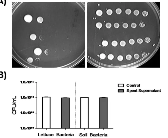
Intracellular pathogens frequently grow slower, have a long lag time, or fail to grow in vitro compared to replication within the host cell [28–31]. This poses a significant challenge for disease and bioweapon diagnostics, which rely on specific detection ranges of pathogen cells, toxin and/or other protein concentrations. Detectable levels from low starting inoculums of F. tularensis are difficult to achieve in vitro. Various studies have shown that the spent culture medium of pathogenic bacteria, such as Mycobacterium tuberculosis, stimulated enhanced growth of dormant and small inoculum cultures [32–34]. More importantly, studies conducted by Halmann and colleagues have described the presence of a growth-initiation substance (GIS) in the culture filtrate using small inocula of virulent strains of F. tularensis [35,36]. Therefore, we tested if the spent culture filtrate ofF. tularensisvaccine strain would stimulate and enhance the growth of a small number of F. tularensiscell. Overnight cultures of F. tularensiswere serially diluted and spotted on standard culture medium (TSA and 0.1% L-cysteine) supplemented with and without spent culture filtrate (Fig. 1A).F. tularensis
cultured on spent culture filtrate resulted in robust growth at all dilutions (1021– 1026) in contrast to standard culture medium, which only supported the growth of 1021–1022 dilutions (Fig. 1A).
The main challenge in diagnostics and pre-analytical processing ofF. tularensis
in food and environmental matrices is the presence of other bacteria, which may interfere withF. tularensisgrowth [7]. Furthermore, the possibility remained that
soil on spent filtrate supplemented medium and standard medium alone (Fig. 1B). We observed no difference in CFU/mL between standard medium supplemented with or without spent filtrate (Fig. 1B). However, the possibility remains that the spent culture filtrate may inhibit the growth of certain bacterial species, while other bacterial communities may continue to exist and fill the vacant niche. In fact, we did observe changes to colony phenotype when the spent culture filtrate was added to lettuce homogenates (data not shown). Next, we investigated our ability to detect F. tularensis by real-time PCR. Specifically, does the addition of spent culture filtrate in relation to the limit of detection and does the inclusion of soil and lettuce bacteria interfere with (Fig. 2)? F. tularensis
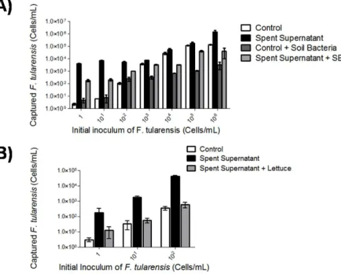
cultured with spent filtrate showed a 2.5–3.5 fold increase in cells per mL at a starting inoculum of 1 bacterium per mL compared to standard medium alone (Fig. 2). Addition of soil bacteria to F. tularensis cultured with spent filtrate reduced the overnight growth of F. tularensisan average of 1.5 fold; however, a 2 Fig. 1.F. tularensisspent medium enhances growth and is specific forF. tularensis. A)F. tularensiswas cultured for 24 h and diluted on TSA containing 0.1% L-cysteine (left) and TSA containing 0.1% L-cysteine and 10% spent medium (right). A ten-fold dilution series ofF. tularensiswas created (1021–1026, left to right).B)Lettuce bacteria were obtained from stomacher processing of 10 g of lettuce in 50 mL of TSB containing 0.1% L-cysteine. Soil
bacteria were obtained from culturing of 1 g of soil, hay and dust in TSB containing 0.1% L-cysteine overnight at 37˚C with shaking at 120 rpm. Bacteria from lettuce and soil sources were diluted to 1026and plated on TSA supplemented with 0.1% L-cysteine (control) and control agar supplemented with 10%F.
tularensisspent filtrate. Samples were incubated overnight at 37˚C and the CFU/mL was calculated. Legend: Control5white bar, Spent filtrate5medium grey bar.
fold increase was observed when compared against F. tularensis mixed with soil bacteria in standard medium (Fig. 2A). Given that the spent culture filtrate resulted in enhanced detection at low inoculums (1–102cells/mL), we focused on detection of F. tularensis at 1–102 cells/mL spiked in lettuce bacteria (Fig. 2B). Supplementation of TSB and 0.1% L-cysteine with spent culture filtrate resulted in increased growth of F. tularensis mixed with lettuce bacteria at all inoculums (1–102cells/mL) in comparison to standard medium (Fig. 2B). It is interesting to note that unlike soil bacteria, inclusion of lettuce bacteria inhibitedF. tularensisin control samples (Fig. 2B). Together these data confirm the findings presented by Halmann et al. and suggest that the increased growth of F. tularensis due to the addition of spent culture filtrate is unhindered by the presence of other bacteria [35].
Fig. 2. Spent filtrate supplementation results in increased detection ofF. tularensisin mixed cultures. A)F. tularensiswas spiked in a ten-fold serial dilution in 109CFU/mL of soil bacteria or inoculated as a pure
culture and incubated in control broth (TSB with 0.1% L-cysteine) or spent filtrate (composition: TSB and 0.1% L-cysteine and 10% spent culture filtrate) overnight at 37˚C with 120 rpm shaking speed.B)F. tularensis(1– 102cells/mL) was spiked in 109CFU/mL of lettuce bacteria. Supplementation of TSB and 0.1% L-cysteine with spent culture filtrate resulted inF. tularensisdetection at all inoculums (1–102cells/mL). Control medium combined with lettuce showed Cp values greater than 34 and were considered negative forF. tularensis. All samples were conducted in triplicate with technical triplicates. Legend: Control5white bar, Spent
filtrate5black bar, Control and soil bacteria5dark grey bar, and Spent filtrate and soil/lettuce bacteria5light grey bar.
DNA aptamer cocktail captures
F. tularensis
in mixed cultures of
bacteria
In order to further lower the range of detection, we generated DNA aptamers against F. tularensis, which may be applied to diagnostics in food matrices to achieve physical separation of F. tularensisfrom background bacteria. Aptamers, short RNA and single stranded DNA sequences, have high affinities to their selected receptors, show limited cross-reactivity to homologous targets, and may serve as pre-analytical tools and biosensors for food surveillance [37–41]. DNA aptamers againstF. tularensiswere enriched through 11 iterations of SELEX and 4 iterations of counter-SELEX. Confirmation ofF. tularensisbinding to the selected aptamer pool (from SELEX round 8) was determined by Southwestern blot analysis. The selected aptamer pool showed binding to 103cells ofF. tularensis as opposed to the unselected aptamer library which could not detect the presence of
F. tularensis at this concentration (data not shown). Sequencing of 188 transformants containing aptamer inserts revealed 10 redundant DNA motif sequence groups (S1 Fig.). A single aptamer (those with the highest number of repeat clones) were selected from each motif group (Table 1) for further testing against F. tularensis.
Individual aptamers were tested forF. tularensis binding usingfopA real-time PCR analysis. Cp values did not significantly differ from one another; therefore, the 10 aptamers were combined into a cocktail to increase binding efficiency (data
not shown). The aptamer cocktail bound to M-280 Dynabeads was separately
incubated with F. tularensis control and spiked lettuce or soil solutions. Using pure cultures of F. tularensis as well as mixed cultures (both soil and lettuce solutions), the range ofF. tularensiscell capture was 102–106cells/mL (Fig. 3A and B). Furthermore, F. tularensis spiked with lettuce bacteria was fully recovered at 102–106 cells/mL (Fig. 3B) while bead controls did not capture F. tularensisin pure or mixed cultures (Fig. 3). However, aptamers failed to captureF. tularensis
at low inoculums (1–10 cells/mL) (Fig. 3). In summary, we have developed a DNA aptamer cocktail to enrich for F. tularensis from resident bacteria in food and environmental matrices at 102–106 cells/mL.
Two-step enrichment process provides optimal recovery of
F.
tularensis
in mixed cultures
spent culture filtrate followed by physical separation and capture by the DNA aptamer cocktail can be utilized alongside current diagnostics as a pre-analytical processing tool.
Table 1.Repeat DNA sequences and number of clones from sequenced aptamer poola.
DNA Aptamer Sequence (59---39) No. of Repeats
FT17 CGCGTCAGAGGTGTGTCGGGGCTGTGTAGATCTACATGGG 17
FT12 CAGTCGCTTTCCGTTCTCCGGCAGGTTCATTGTGGTTTCG 12
FT11 CATATCAGGTCGTCACCGTAACAGAGCTCTCGCAATCACG 11
FT9 CAAAGGCAGCAGTAGCATGGCGATTTACATCAATTATTGG 9
FT8 CAATATCAGAAGTAGCGCGAAGGACGACATGTCAGGAAGG 8
FT7 CACACATCCTCGCAGCCTCGTACCTGATTCCAGTCTATTG 7
FT6 CCAGGAAGGCGAGAGCCGAGAAGCGATCCTTGGGTATAGG 6
FT5 CGGCATCCGTTCACGTACCTGTCCTAGTTATCACCGTTTG 5
FT5.1 CAAGCAGAGTTCCGAGACACAGTACCACACGCATATCCGG 5
FT4 CACACAGAGACGGTGAAGGCGCCGACCAGTTCCTAAAGAG 4
aSequence aptamer pool belonged to the 11thround of SELEX.
doi:10.1371/journal.pone.0114622.t001
Fig. 3. DNA aptamers againstF. tularensisspecifically captureF. tularensisin mixed bacteria samples. A biotinylated aptamer cocktail composed of 10 aptamers (0.4 pmol each) was bound to M-280 streptavidin Dynabeads.F. tularensis(1–106cells/mL) was separately mixed withA)109CFU/mL of soil bacteria orB)
lettuce bacteria and aptamer cocktail beads or control beads (beads without aptamers) and incubated for 1 h at room temperature. Samples were denatured and the supernatant was subjected to real-time PCR analysis. Bead controls showed Cp values greater than 34 and were considered negative forF. tularensis. Legend: Aptamers5black bar, Aptamers and Soil Bacteria5dark grey bar.
Carnosine yields enhanced growth for
F. tularensis
growth
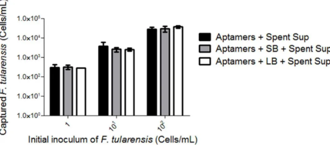
Although the two-step enrichment process is an easy method that does not require specialized equipment forF. tularensisdetection, it is not advisable to collect liter amounts of F. tularensisspent culture filtrate for multiple sample testing. Therefore, we sought to identify the GIS component within theF. tularensisspent supernatant to enable chemical synthesis and subsequent supplementation to control medium. We hypothesized that the GIS accumulates overtime; therefore, we selected spent culture filtrates at 0.5, 2, 8 and 24 h for UPLC/MS analyses. The analytical workflow to identify chemical targets specific growth enhancement entailed untargeted UPLC/MS profiling of spent culture filtrates; PCA analyses, chemometrics OPLS-DA; evaluation of extracted ion chromatograms (XICs) for each GIS specific ion; database searches to provide chemical identities of GIS features; mass accuracy confirmation and evaluation of fragmentation patterns for each target of MS simultaneously acquired high-collision energy mass spectra. PCA showed that the greatest separation between time points for both positive and negative ionization was achieved when comparing 0.5 and 2 h separately to 8 and 24 h post incubation (S2 Fig.). Therefore, the following comparisons were evaluated by OPLS-DA: 0.5 h v 8 or 24 h and 2 h v 8 or 24 h. OPLS-DA data were visualized by S-plots and those features unique to early (0.5 and 2 h) or late (8 or 24 h) were selected for further investigation. S-plot examples for positive and negative ionization and target selection for 2 v. 24 h are shown inS3 Fig. Similar S-plots were generated for 0.5 v 24 h and early time points v. 8 h (data not shown). Chemical entities with high confidence (high correlation) were defined as being unique to either early (21.0) or late (1.0) time points and selected in lower Fig. 4. Combined enrichment with spent culture supernatant and DNA aptamer capture leads to complete isolation ofF. tularensis.F. tularensis(1–102cells/mL) mixed samples in soil and/or lettuce
homogenates were enriched for by overnight incubation with 10% spent culture supernatant andF. tularensis
cells were subsequently captured by the DNA aptamer pool. Samples were denatured and the supernatant was subjected to real-time PCR analysis. Aptamers combined with either soil or lettuce bacteria alone (controls) showed Cp values greater than 34 and were considered negative forF. tularensis. Legend: Aptamers and Spent Sup (Spent supernatant)5black bar, Aptamers and SB and Spent Sup5grey bar, and Aptamers and LB and Spent Sup5white bar.
left and upper right quadrants, respectively (S3 Fig.; selection represented by fuchsia boxes). Trend plots for each selected feature were created to determine abundance at comparison time points (S4A Fig.) with manual evaluation of XICs (S4B Fig.). Manual validation of tentative chemical entities (mined using Yeast, ChEBI, ChemSpider, KEGG, and LIPID MAPS databases) using UPLC/MS raw data and chemical standards failed. However, manual database searches (limited to parent ions and mass fractionation patterns of m/zof 397 and 297) as well as extensive literature based searches uncovered two potential pathways for
enhanced growth 1) an autoinducer (such as N-acetyl-homoserine lactones (AHLs) or 2) carnosine, a chemical previously reported to cause enhanced growth and biofilm formation inE. coli[42]. Addition of commercially available AHLs to control broth did not yield enhanced growth of overnight cultures ofF. tularensis
(data not shown). Carnosine supplementation at concentrations of 0.625–2.5 mg/ mL to control broth resulted in increased growth of F. tularensisalbeit to a lesser extent (10 fold) than the spent culture filtrate (Fig. 5). Higher concentrations of carnosine, particularly at 10 mg/mL, had a bacteriocidal effect on F. tularensis
(Fig. 5).
Discussion
Reliable and time-sensitive detection of a biological contaminant within the food network is essential to advise management and treatment procedures during a potential outbreak. The gold standard for microbe detection is successful
cultivation of the agent in vitro; however, this remains a challenge for intracellular pathogens as many are either slow-growers or dormant in vitro. F. tularensis, a possible aerosol and biocontaminate in food, requires incubation for 2–4 days at 37
˚
C when supplemented with cysteine, thioglycolate and/or blood. Furthermore, culture of F. tularensisin biological samples is also difficult to achieve in broth culture, unless a high starting inoculum is used.During the course of our investigation to create an improved cultivation medium for F. tularensis, we rediscovered initial experiments and subsequent observations reported by Halmann et al. [35,36]. Halmann et al. report the presence of a Growth Initiating Substance (GIS) from low inocula ofF. tularensis
(strains Schu, Schu-M16, Vavenly, 425-F4G, 301I0, and LVS) that enhanced the growth of dormant F. tularensiscells when supplemented in traditional culture medium [36]. Similar to the Halmann studies, we have also shown that
supplementation of standard medium with 10% spent culture filtrate results inF.
tularensis enhanced growth in pure and mixed (lettuce and soil homogenates)
cultures (Fig. 1Aand Fig. 2). This data suggests that the majority of F. tularensis
cells are composed of dormant cells capable of resuscitation with the addition of spent culture filtrate. Through a series of Sephadex gel filtration and ion-exchange chromatography, Halmann et al. characterized the GIS as having a low-molecular weight and heat stable at neutral pH but destroyable if heated in an acidic solution [36]. Furthermore, GIS was shown to form metal complexes (iron and copper complexes) and have enhanced activity when supplemented with ornithine [36]. Halmann and colleagues conjectured that ornithine may be a precursor to the GIS and that the GIS may be a sideramine or related to sideramine production. However, our analysis of the spent culture filtrate using UPLC/MS analysis and subsequent extensive database searches did not find any chemical entities related to sideramine function. UPLC/MS trend plots uncovered unique chemical signatures that had m/z ratios of 297 and 397; however, database searches were unable to provide a definitive identification. Manual database and literature searches related to bacterial growth and chemical entities with a m/zof 297–397 revealed AHLs or carnosine as suitable candidates. While available AHLs did not result in enhanced F. tularensis growth, carnosine (0.625 mg–2.5 mg/mL) promoted a 10-fold increase inF. tularensiscells/mL (Fig. 5). However, increased growth by carnosine did not fully recapitulate growth seen by the spent culture Fig. 5. Carnosine increasesF. tularensisgrowth.F. tularensis(102cells/mL) were incubated overnight at 37
˚C with agitation in control broth alone or supplemented with 10% spent culture filtrate or various concentrations of carnosine (0.625 mg/mL–10 mg/mL). DNA was extracted from all samples and analyzed byfopAreal-time PCR. All samples were conducted in triplicate with technical triplicates. Legend: Control5white bar, Spent filtrate5dark grey bar, [carnosine510 mg/mL (light grey bar), 5.0 mg/mL (medium grey bar), 2.5 mg/mL (checkered pattern bar), 1.25 mg/mL (diagonal pattern bar) and 0.625 mg/ mL (diamond pattern bar)].
filtrate (Fig. 5). It is possible that other chemicals found within the spent filtrate act in concert with carnosine to increase F. tularensis growth or that the GIS component is a chemical related to carnosine and that additional side-chain components are responsible for the 10-fold difference. Higher concentrations of carnosine, specifically at 10 mg/mL, had a bacteriocidal effect (Fig. 5). This may be due to its ability to bind zinc and ferrous ions, which high carnosine
concentrations may lead to iron toxicity or depletion of iron from the media [43]. Therefore, it is likely that F. tularensis may have a ‘‘Goldilocks’’ mechanism to regulate an appropriate amount of carnosine. These data suggest that carnosine functions both as a growth promoter and bactericide and that careful regulation by the bacterium must be conducted to ensure persistence. Further studies will need to revisit chemical entities identified by UPLC/MS to determine the exact GIS compound found in the spent supernatant andF. tularensisgene regulation in response to carnosine.
spent culture filtrate and real-time PCR had an increased detection range for F. tularensis (Fig. 4).
The combination of spent culture filtrate or carnosine and DNA aptamer capture as a two-part enrichment method forF. tularensis may be amendable for field diagnostics. For example, this method may be used in tandem with a portable real-time PCR device for rapid detection of F. tularensis. Furthermore, this method is advantageous to concentrateF. tularensiscells for subtyping, which may impact decision making in a potential outbreak. Benefits of such a diagnostic tool may be further extended in bench research investigating the ecology of F. tularensisby screening of animal carcass samples collected from multiple sites and varying years. Current investigations in our laboratory seek to determine if aptamer captured F. tularensis can be re-cultured. Pure cultures ofF. tularensis
isolates from the environment would allow investigators to pursue downstream studies that go beyond diagnostics.
Materials and Methods
Bacterial Culture
F. tularensis subsp. holarctica Live Vaccine Strain (LVS) NR-14 (Biodefense and Emerging Infections Research Resources Repositories, NAID, NIH; herein referred to as F. tularensis) was maintained in tryptic soy agar (TSA) supplemented with 0.1% L-cysteine at 37
˚
C. F. tularensiswas subcultured in tryptic soy broth (TSB) supplemented with 0.1% L-cysteine (Sigma-Aldrich, St. Louis, MO) at 37˚
C with 120 rpm shaking. Environmental bacteria (herein referred to as soil bacteria) were obtained from one gram (g) of soil, hay and dust and cultured in TSB and diluted to 1021 in TSB containing 0.1% MgSO4. 7H2O to represent low and highnutrition conditions at room temperature and 37
˚
C under constant shaking at 120 rpm. Lettuce bacteria were cultured from bagged lettuce purchased from a local grocery store. Approximately 10 g of lettuce were mixed with 50 mL of TSB supplemented with 0.1% L-cysteine inside a sterile Stomacher 400 classic bag (Seward Laboratory Systems Inc., Port Saint Lucie, FL) and homogenized for 2 min at high speed in a Stomacher. Homogenate was subcultured at 1021in TSB supplemented with 0.1% L-cysteine at 37˚
C with 120 rpm shaking.F. tularensis
Growth Enrichment
F. tularensis was grown as described overnight at 37
˚
C with shaking at 120 rpm. Upon completion of incubation, F. tularensis was pelleted at 70006gfor 10 minand the supernatant was filtered through a 0.22 mM syringe-driven filter (EMD
Millipore, Billerica, MA) to remove any residual bacteria. An overnight culture of
were incubated overnight at 37
˚
C and differences in growth were recorded the following day. The number ofF. tularensis cells per mL were determined byfopAreal-time PCR analysis described below.
Selection of DNA aptamers against
F. tularensis
using Systemic
Evolution of Ligands by Exponential Enrichment (SELEX)
Aptamer candidates against F. tularensiswere selected using a SELEX protocol. Briefly, a modified single-stranded aptamer library (WAP40m) consisting of a 40-mer randomized regions flanked by constant-pri40-mer binding regions (Integrated DNA Technologies, Inc., Coralville, IA) underwent four rounds of counter-SELEX iterations. The aptamer library was incubated for 30 min with 108CFU/mL of soil bacteria (previously cultured overnight at both 25
˚
C or 37˚
C) in TSB containing 0.1% L-cysteine at room temperature with gentle shaking (Labquake tube shaker) [45,49]. Aptamers bound to soil bacteria were discarded and the filtrate was collected, which was used for positive rounds of SELEX against F. tularensis. Positive SELEX iterations involved exposure of the remaining aptamer library to 108cells/mL ofF. tularensisat room temperature with gentle shaking for 30 min. Unbound or weak-binding aptamers were removed from further rounds of SELEX by washing cells 6 times in 1X PBS containing 0.025% Tween-80 and twice with nuclease-free water. Aptamers bound to F. tularensiscells were resuspended in 100 mL of nuclease-free water and subjected to Polymerase Chain Reaction (PCR)after every round of SELEX to amplify aptamer candidates. PCR was performed using a biotin-labeled on the primer’s anti-sense strand (WP20R1; 5 pmol), an unlabeled primer (WP20F1; 5 pmol), 2X Hotstar taq polymerase (Qiagen, Valencia, CA) and the following program: 95
˚
C for 15 min, followed by 15 cycles of 95˚
C for 30 s, 63˚
C for 30 s, and 72˚
C for 7 min (Table 1). In order to enrich for aptamer sense strands needed for further rounds of SELEX, amplicons were applied as templates at 1:10 and 1:25 dilutions for asymmetric-touchdown PCR using 30 pmol of WP20F1 and 1.2 pmol of biotin labeled WP20R1. Theasymmetric-touchdown program was 95
˚
C for 15 min, followed by 9 cycles of 95˚
C for 15 s, 72˚
C for 15 s (gradually decreasing by 1˚
C each cycle), and 72˚
C for 15 s, followed by 11 cycles of 95˚
C for 15 s, 63˚
C for 15 s, and 72˚
C for 15 s, and a final extension at 72˚
C for 3 min. PCR amplicons were pooled and anti-sense strands were removed using a snap cool method with 50 mL of Dynabeads M-280streptavidin coated beads (Invitrogen, Carlsbad, CA). The amplicon sense strand was applied to the remaining rounds of SELEX. A total of 11 SELEX iterations were performed.
Cloning, sequencing and characterization of DNA aptamers
against
F. tularensis
Four hundredmL of PCR amplicons from the 11thround of SELEX were purified
Carlsbad, CA) and later transformed into TOP10 chemically competent E. coli
cells per manufacturer’s instructions. Transformed TOP10 E. colicells (100 mL)
were plated on Luria-Bertani agar containing ampicillin (50 mg/mL) and X-gal.
Successful transformants were selected based on blue/white screening for
disruption of lacZ. Colony PCR was performed on selected transformants using M13 primers and the following PCR conditions: 95
˚
C for 15 min and 30 cycles of 95˚
C for 30 s, 56˚
C for 30 s, 72˚
C for 1 min, and final extension for 72˚
C for 5 min (Table 2). One hundred and eighty-eight transformants with the correct amplicon size were sequenced and analyzed for redundancy using Sequencher (GeneCodes, Ann Arbor, MI) and CLUSTAL W in MEGA 4.0 (http://www. megasoftware.net/mega4/mega.html) (Table 2and S1 Fig.). Redundant aptamers were further characterized using the m-fold program (http://mfold.rna.albany. edu/?q5mfold/DNA-Folding-Form) available from the RNA Institute.Spike and recovery of
F. tularensis
An overnight broth culture ofF. tularensiswas washed three times in PBS, serially diluted ten-fold (1–106cells/mL) in TSB supplemented with 0.1% L-cysteine and spiked in 109CFU/mL of soil or lettuce bacteria (homogenized solutions) or medium alone. Dilutions in medium alone and those mixed with soil or lettuce bacteria were supplemented with 10% filtered spent medium. Control samples did not include supplementation with spent filtrate. All samples were incubated overnight at 37
˚
C with shaking at 120 rpm. After incubation, samples were pelleted at 8,000 rpm and filtrates were discarded. Genomic DNA was extracted from samples using the DNeasy blood and tissue kit (Qiagen, Valencia, CA) per manufacturer’s instructions. DNA was treated with 4.0 mL of RNase A (Qiagen,Valencia, CA) for 2 min at room temperature. Genomic DNA was suspended in 50 mL of nuclease-free water. All treatments were conducted in triplicate. All
experiments were conducted in triplicate. Genomic DNA was stored at 220
˚
C until real-time PCR analysis.Spike and aptamer cocktail recovery of
F. tularensis
An overnight broth culture of F. tularensiswas prepared and spiked in soil or lettuce bacteria (homogenized solutions) or medium alone as described above. Approximately 0.4 pmol of each selected biotin tagged aptamer (Table 1) was mixed together as a cocktail and bound to Dynabeads M-280 streptavidin coated beads (Invitrogen, Carlsbad, CA) using a modified protocol [38]. Briefly, 25mL of
M-280 streptavidin beads were loaded into individual wells in a 96 well PCR plate, washed 3 times in PBS and incubated with the biotin labeled aptamer cocktail overnight at 37
˚
C with shaking at 200 rpm. After incubation, aptamers bound to M-280 streptavidin beads were washed thrice in 200 mL of PBS and blocked for2 h at room temperature with shaking at 200 rpm using 100 mL of PBS containing
of nuclease-free water. One hundred mL of F. tularensis control and spiked
samples (soil or lettuce bacteria) were mixed separately in individual wells containing aptamer M-280 beads and incubated for 1 h at room temperature with 200 rpm shaking. Aptamer M-280 beads were washed 5 times in 200 mL of PBS
containing 0.05% Tween-80 and thrice in 200 mL of nuclease-free water and
resuspended in 100 mL of nuclease-free water. In order to release the genomic
DNA from captured bacterial cells, they were heated to 95
˚
C for 10 min. All treatments were conducted in triplicate and each experiment was performed thrice.Two-step enrichment of
F. tularensis
in Mixed Bacterial Cultures
An overnight culture of F. tularensiswas washed thrice in PBS and diluted to 1– 102 cells/mL in TSB supplemented with 0.1% L-cysteine. F. tularensis dilutions were either mixed in medium with or without 10% spent culture filtrate alone or with 109CFU/mL of lettuce or soil bacteria. All samples were incubated overnight at 37
˚
C with shaking at 120 rpm and later mixed separately with aptamer M-280 streptavidin beads. All treatments were conducted in triplicate and eachexperiment was performed thrice.
Real-time PCR Analysis of
F. tularensis
in Pure and Mixed
Bacterial Cultures
Real-time PCR of thefopAgene was performed usingF. tularensisspiked with soil or lettuce bacteria and control samples using a Lightcycler 480 Probes Master Mix and Universal ProbeLibrary Probe #10 (Roche, Indianapolis, IN) per manufac-turer’s recommendations (Table 2). All samples were analyzed on the Roche Lightcycler 480II and corresponding software (Roche, Indianapolis, IN). The following real-time PCR conditions were applied: 95
˚
C for 5 min and 45 cycles of 95˚
C for 10 s, 50˚
C for 30 s and 72˚
C for 1 s. Primers were designed using the Universal ProbeLibrary Assay Design Center (https://www.roche-applied-science. com/). The number of cells/mL was calculated for each sample based on a F. tularensiscells/mL standard curve. The standard curve was generated by a ten-fold serial dilution ofF. tularensisgenomic DNA with a known cells/mL (10–107cells/ mL). Cross-point (Cp) values were calculated using the second derivative Table 2.Primers used in this study.Primer Sequence (59---39)
WP20F1 AGTGCAAGCAGTATTCGGTC
WP20R1 TAAAGCTGATGCGTGATGCC
M13F GTTTTCCCAGTCACGAC
M13R CAGGAAACAGCTATGAC
fopA_F GTTCAAGGTGCTTGGATG
fopA_R TGCAACAGCGCTAAGAGTTTT
maximum method available in the 480 II lightcycler software (Roche,
Indianapolis, IN) for each dilution in the standard curve and for all test samples [50]. Cp values from the standard curved ranged 13–34 cycles. Sample Cp values outside of the standard curve range were designated as negative for F. tularensis. TmCalling analysis was used to verify negative samples [50]. In the case of a
negative sample, TmCalling results showed two-shouldered or no melting peaks.
The former indicated the production of primer dimers. All samples were conducted in triplicate.
Supporting Information
S1 Fig. DNA Aptamer Sequence Motifs. The DNA aptamer pool was sequenced
after the 11thround of SELEX and sequences were aligned using CLUSTAL W in MEGA 4.0. Ten repeated motifs were identified and 1 DNA aptamer was selected from each motif group for further characterization.
doi:10.1371/journal.pone.0114622.s001 (PDF)
S2 Fig. Principal Component Analyses (PCA) of F. tularensis spent culture
filtrate. MarkerLynx software was used to generate PCA plots of UPLC/MS data.
PCA plots include positive (right) and (negative) ionization at 0.5 h (blue circles) v 24 h (seafoam green) and 2 h (blue circles) v 24 h.
doi:10.1371/journal.pone.0114622.s002 (TIFF)
S3 Fig. OPLS-DA Generated S-Plots. Chemometric S plot resulting from
OPLS-DA of UPLC/MS analysis ofF. tularensis spent culture filtrates at 2 h versus 24 h with positive and negative ionization. Similar S-plots were generated for 0.5 h versus 24 h. The y-axis represents correlation (confidence; time point specific), while the x-axis represents the coefficient (specificity). The selection boxes represent features that are highly specific to each time point. Selection boxes are noted in fuchsia.
doi:10.1371/journal.pone.0114622.s003 (TIFF)
S4 Fig. UPLC/MS Characterization. A) A chemometric trend plot depicting the
relative abundance of the chemical entity found at 0.5 h and 24 h. B) Extracted ion chromatogram (XIC) for m/z397.2205.
doi:10.1371/journal.pone.0114622.s004 (TIFF)
Acknowledgments
We thank Dr. Joesph J. Dalluge (University of Minnesota) for help in UPLC/MS sample preparation and data analysis. We thank the Biomedical Genomics Center at the University of Minnesota for sequencing services.
Author Contributions
Contributed to the writing of the manuscript: SS EAL PW SE KB AA REI. Voted on the manuscript: EAL SS.
References
1. Hartnett E, Paoli GM, Schaffner DW(2009) Modeling the public health system response to a terrorist event in the food supply. Risk Anal 29: 1506–1520.
2. Hennessy DA(2008) Economic aspects of agricultural and food biosecurity. Biosecur Bioterror 6: 66– 77.
3. Day JB, Nguyen H, Sharma SK, Al-Khaldi SF, Hao YY(2009) Effect of dehydrated storage on the survival of Francisella tularensis in infant formula. Food Microbiol 26: 932–935.
4. Willke A, Meric M, Grunow R, Sayan M, Finke EJ, et al. (2009) An outbreak of oropharyngeal tularaemia linked to natural spring water. J Med Microbiol 58: 112–116.
5. Dennis DT, Inglesby TV, Henderson DA, Bartlett JG, Ascher MS, et al. (2001) Tularemia as a biological weapon: medical and public health management. JAMA 285: 2763–2773.
6. Egan JR, Hall IM, Leach S(2011) Modeling inhalational tularemia: deliberate release and public health response. Biosecur Bioterror 9: 331–343.
7. Oyston PC, Sjostedt A, Titball RW (2004) Tularaemia: bioterrorism defence renews interest in Francisella tularensis. Nat Rev Microbiol 2: 967–978.
8. Foley JE, Nieto NC(2010) Tularemia. Vet Microbiol 140: 332–338.
9. Gill V, Cunha BA(1997) Tularemia pneumonia. Semin Respir Infect 12: 61–67.
10. Ojeda SS, Wang ZJ, Mares CA, Chang TA, Li Q, et al.(2008) Rapid dissemination of Francisella tularensis and the effect of route of infection. BMC Microbiol 8: 215.
11. Bevanger L, Maeland JA, Naess AI(1988) Agglutinins and antibodies to Francisella tularensis outer membrane antigens in the early diagnosis of disease during an outbreak of tularemia. J Clin Microbiol 26: 433–437.
12. Sharma N, Hotta A, Yamamoto Y, Fujita O, Uda A, et al.(2013) Detection of Francisella tularensis-specific antibodies in patients with tularemia by a novel competitive enzyme-linked immunosorbent assay. Clin Vaccine Immunol 20: 9–16.
13. Massey ED, Mangiafico JA (1974) Microagglutination test for detecting and measuring serum agglutinins of Francisella tularensis. Appl Microbiol 27: 25–27.
14. Bevanger L, Maeland JA, Kvan AI(1994) Comparative analysis of antibodies to Francisella tularensis antigens during the acute phase of tularemia and eight years later. Clin Diagn Lab Immunol 1: 238–240.
15. Martin-Serradilla JI, Sanchez Navarro JJ, Arias Giralda M, San Jose Alonso J(2004) [Evolution of serological characteristics in 26 patients with tularemia three years after the outbreak]. Rev Clin Esp 204: 351–354.
16. Antwerpen MH, Schacht E, Kaysser P, Splettstoesser WD(2013) Complete Genome Sequence of a Francisella tularensis subsp. holarctica Strain from Germany Causing Lethal Infection in Common Marmosets. Genome Announc 1.
17. Chaudhuri RR, Ren CP, Desmond L, Vincent GA, Silman NJ, et al. (2007) Genome sequencing shows that European isolates of Francisella tularensis subspecies tularensis are almost identical to US laboratory strain Schu S4. PLoS One 2: e352.
18. Broekhuijsen M, Larsson P, Johansson A, Bystrom M, Eriksson U, et al.(2003) Genome-wide DNA microarray analysis of Francisella tularensis strains demonstrates extensive genetic conservation within the species but identifies regions that are unique to the highly virulent F. tularensis subsp. tularensis. J Clin Microbiol 41: 2924–2931.
20. Sridhar S, Sharma A, Kongshaug H, Nilsen F, Jonassen I(2012) Whole genome sequencing of the fish pathogen Francisella noatunensis subsp. orientalis Toba04 gives novel insights into Francisella evolution and pathogenecity. BMC Genomics 13: 598.
21. Fulop M, Leslie D, Titball R(1996) A rapid, highly sensitive method for the detection of Francisella tularensis in clinical samples using the polymerase chain reaction. Am J Trop Med Hyg 54: 364–366.
22. Versage JL, Severin DD, Chu MC, Petersen JM(2003) Development of a multitarget real-time TaqMan PCR assay for enhanced detection of Francisella tularensis in complex specimens. J Clin Microbiol 41: 5492–5499.
23. Ellis J, Oyston PC, Green M, Titball RW(2002) Tularemia. Clin Microbiol Rev 15: 631–646.
24. Day JB, Whiting RC(2009) Development of a macrophage cell culture method to isolate and enrich Francisella tularensis from food matrices for subsequent detection by real-time PCR. J Food Prot 72: 1156–1164.
25. Whitehouse CA, Kesterson KE, Duncan DD, Eshoo MW, Wolcott M (2012) Identification and characterization of Francisella species from natural warm springs in Utah, USA. Lett Appl Microbiol 54: 313–324.
26. Humrighouse BW, Adcock NJ, Rice EW(2011) Use of acid treatment and a selective medium to enhance the recovery of Francisella tularensis from water. Appl Environ Microbiol 77: 6729–6732.
27. Petersen JM, Carlson J, Yockey B, Pillai S, Kuske C, et al.(2009) Direct isolation of Francisella spp. from environmental samples. Lett Appl Microbiol 48: 663–667.
28. Storrs EE, Walsh GP, Burchfield HP, Binford CH (1974) Leprosy in the armadillo: new model for biomedical research. Science 183: 851–852.
29. Merriott J, Shoemaker A, Downs CM(1961) Growth of Pasteurella tularensis in cultured cells. J Infect Dis 108: 136–150.
30. Abd H, Johansson T, Golovliov I, Sandstrom G, Forsman M(2003) Survival and growth of Francisella tularensis in Acanthamoeba castellanii. Appl Environ Microbiol 69: 600–606.
31. Sansonetti PJ, Ryter A, Clerc P, Maurelli AT, Mounier J(1986) Multiplication of Shigella flexneri within HeLa cells: lysis of the phagocytic vacuole and plasmid-mediated contact hemolysis. Infect Immun 51: 461–469.
32. Sun Z, Zhang Y (1999) Spent culture supernatant of Mycobacterium tuberculosis H37Ra improves viability of aged cultures of this strain and allows small inocula to initiate growth. J Bacteriol 181: 7626– 7628.
33. Shleeva MO, Bagramyan K, Telkov MV, Mukamolova GV, Young M, et al.(2002) Formation and resuscitation of ‘‘non-culturable’’ cells of Rhodococcus rhodochrous and Mycobacterium tuberculosis in prolonged stationary phase. Microbiology 148: 1581–1591.
34. Mukamolova GV, Kormer SS, Kell DB, Kaprelyants AS(1999) Stimulation of the multiplication of Micrococcus luteus by an autocrine growth factor. Arch Microbiol 172: 9–14.
35. Halmann M, Benedict M, Mager J(1967) Nutritional requirements of Pasteurella tularensis for growth from small inocula. Journal of General Microbiology 49: 451–460.
36. Halmann M, Mager J(1967) An endogenously produced substance essential for growth initiation of Pasteurella tularensis. Journal of General Microbiology 49: 461–468.
37. Joshi R, Janagama H, Dwivedi HP, Senthil Kumar TM, Jaykus LA, et al. (2009) Selection, characterization, and application of DNA aptamers for the capture and detection of Salmonella enterica serovars. Mol Cell Probes 23: 20–28.
38. Lamont EA, He L, Warriner K, Labuza TP, Sreevatsan S(2011) A single DNA aptamer functions as a biosensor for ricin. Analyst 136: 3884–3895.
39. Ellington AD, Szostak JW(1990) In vitro selection of RNA molecules that bind specific ligands. Nature 346: 818–822.
40. Gold L (1995) The SELEX process: a surprising source of therapeutic and diagnostic compounds. Harvey Lect 91: 47–57.
42. Brombacher E, Dorel C, Zehnder AJ, Landini P (2003) The curli biosynthesis regulator CsgD co-ordinates the expression of both positive and negative determinants for biofilm formation in Escherichia coli. Microbiology 149: 2847–2857.
43. Guiotto A, Calderan A, Ruzza P, Borin G (2005) Carnosine and carnosine-related antioxidants: a review. Current medicinal chemistry 12: 2293–2315.
44. Tian Y, Adya N, Wagner S, Giam CZ, Green MR, et al.(1995) Dissecting protein:protein interactions between transcription factors with an RNA aptamer. RNA 1: 317–326.
45. Wongphatcharachai M, Wang P, Enomoto S, Webby RJ, Gramer MR, et al.(2013) Neutralizing DNA aptamers against swine influenza H3N2 viruses. J Clin Microbiol 51: 46–54.
46. Zelada-Guillen GA, Riu J, Duzgun A, Rius FX(2009) Immediate detection of living bacteria at ultralow concentrations using a carbon nanotube based potentiometric aptasensor. Angew Chem Int Ed Engl 48: 7334–7337.
47. Duan N, Ding X, Wu S, Xia Y, Ma X, et al.(2013) In vitro selection of a DNA aptamer targeted against Shigella dysenteriae. J Microbiol Methods.
48. Vivekananda J, Kiel JL(2006) Anti-Francisella tularensis DNA aptamers detect tularemia antigen from different subspecies by Aptamer-Linked Immobilized Sorbent Assay. Lab Invest 86: 610–618.
49. Wang P, Hatcher KL, Bartz JC, Chen SG, Skinner P, et al.(2011) Selection and characterization of DNA aptamers against PrP(Sc). Exp Biol Med (Maywood) 236: 466–476.


