Cytogenetic analysis of the effects of 2.5 and 10.5 GHz microwaves on
human lymphocytes
André B.S. Figueiredo
1, Rex N. Alves
1and Adriana T. Ramalho
2 1Instituto Militar de Engenharia, Rio de Janeiro, RJ, Brazil.
2Instituto de Radioproteção e Dosimetria, CNEN, Rio de Janeiro, RJ, Brazil.
Abstract
The biological effects of microwaves on living organisms remain highly controversial. Although some reports have suggested that microwaves may be directly or indirectly genotoxic, a direct action is unlikely because the low energy of microwave photons makes them unable to cause single-strand breaks in DNA. In this work, we examined the possible clastogenic properties of microwaves (2.5 and 10.5 GHz) on blood lymphocytesin vitro by monitoring the frequency of chromosomal aberrations. We also investigated whether blood cells showed increased radiosensitivity or radioresistance when pretreated with the microwaves and then irradiated with gamma radiation. There was no significant difference in the frequency of chromosomal aberrations between cells which had or had not been treated with microwaves. Control cells had a mean frequency of 0.013 aberrations per cell compared to 0.010 and 0.011 aberrations per cell in the microwave-exposed samples. Nor was there any alteration in the radiosensitivity of cells pretreated with microwaves. Gamma irradiated cells showed a mean frequency of 0.279 aberrations per cell compared to 0.343 and 0.310 aberrations per cell in samples pretreated with microwaves. However, cell mortality increased markedly after exposure to microwaves. The results suggest that microwaves do not interact directly or indirectly with chromosomes, although they may target other cell structures, such as cell membranes.
Key words:chromosomal aberrations, human lymphocytes, microwaves.
Received: August 15, 2003; Accepted: November 14, 2003.
Introduction
In recent decades, there have been considerable ad-vances in the development of sources of non-ionizing radi-ation, such as microwaves. The widespread use of such energy sources and the increase in the number of devices emitting microwaves and radiofrequencies (RF), including mobile phones, has become a matter of concern for regula-tory authorities and non-regularegula-tory bodies (IEGMP, 2000).
Structural chromosomal aberrations may involve the chromosomes or chromatids. Most chemical mutagens and non-ionizing mutagenic radiations are unable to cause dou-ble-strand breaks in DNA and act mainly in the S phase of the cell cycle. Such agents are only indirectly clastogenic and produce mainly chromatid-type aberrations (OECD, 1997). In contrast, chromosome-type aberrations are in-duced directly by agents such as ionizing radiation that can produce double strand breaks in DNA (IAEA, 2001).
The biological effects of microwaves on living organ-isms are highly controversial (Maeset al.,1993). A direct
genotoxic action is unlikely because of the low energy of microwave photons which are unable to cause strand breaks in DNA. However, despite this general conviction that microwaves are not sufficiently energetic to be able to directly damage DNA, there is considerable evidence indi-cating that microwaves can be directly and indirectly clastogenic, with a significant increase in chromosome damage (Sagripanti and Swicord, 1986; Garaj-Vrhovacet al., 1991, 1992; Maes et al. 1993; Haidler et al., 1994; Sarkaret al.,1994; Lai and Singh 1995, 1996; Timchenko and Ianchevskaia, 1995; Balode, 1996; Verschaeveet al., 1994; Vijayalaxmiet al.,1997; Phillipset al.,1998; Ticeet al.,1999). In addition, cell phone radiation can alter proto-oncogene activity (Ivaschuket al.,1997; Goswamiet al., 1999). However, a similar number of studies have failed to detect obvious clastogenic effects following microwave ir-radiation of isolated animal cellsin vitro(Alamet al.,1978; Lloydet al.,1984, 1986; Wolffet al.,1985; Meltzet al., 1987, 1989, 1990; Kerbacheret al.,1990; Maeset al.,1997, 2001). Thus, there is still no conclusive answer as to whether exposure to microwaves is clastogenic, i.e., whether they can direct or indirectly increase the frequency of chromosomal aberrations.
Send correspondence to Adriana T. Ramalho. Instituto de Radio-proteção e Dosimetria (IRD), Av. Salvador Allende, Caixa Postal 37750, Barra da Tijuca, 22780-160 Rio de Janeiro, RJ, Brazil. E-mail: [email protected].
A further question is whether microwaves can act as epigenetic factors to influence the genotoxicity of other en-vironmental “pollutants” (Maes et al., 2001). Cancer is generally considered to be initiated by alterations in DNA. However, some non-genotoxic chemicals and processes (known as epigenetic carcinogens) are unable to damage DNA and are usually not clastogenicin vitro, but can en-hance the progress of cells towards malignancy in vivo. Several studies have suggested that radiofrequency radia-tion (RF) has an epigenetic effectin vivo, and can enhance the genotoxic effects of ionizing radiation or cancer-inducing substances, or potentiate other epigenetic factors (ICNIRP, 1998).
The aim of this study was to investigate the clastogenic effects of 2.5 and 10.5 GHz microwave fields, alone and in combination with ionizing radiation, on pe-ripheral blood lymphocytes. The combination of micro-waves with ionizing radiation (“synergy” test) was designed to screen for joint effects of those two types of ra-diation. For this, we assessed whether blood samples pretreated with microwaves would be more sensitive (or re-sistant) to damage by gamma radiation. In all cases, the chromosomal damage was assessed using conventional cytogenetic techniques. The chromosome aberration test is often used to identify physical or chemical agents that cause structural chromosomal aberrations in cultured mam-malian cells (OECD, 1997).
Materials and Methods
The microwave sources used were: (a) a 2450 MHz microwave thermal oven (model MARS 5, CEM Corpora-tion) with a power output of up to 1200 W and controls for regulating power and temperature by ventilation, and (b) a 10.5 GHz, 15 mW, linearly polarized, non-thermal micro-wave source (model WA-9314B, PASCO). The gamma ra-diation source was cobalt-60 (0.034 Gy.min-1), with the absorbed dose being 1.5 Gy (4.5 mJ) per blood aliquot.
Initially, whole blood samples were exposed to the microwave sources for varying periods of time in order to determine the best exposure time for each source. Long pe-riods of exposure resulted in a high cell mortality, seen as a high rate of cell lysis and a low number of metaphases after culturing. Based on these preliminary experiments, expo-sure times of 40 s at 3 W for the 2.5 GHz oven, and 5 min for the 10.5 GHz device were used. To prevent overheating in the 2.5 GHz oven, the temperature of the blood samples was kept below 36 °C, (starting temperature was 28 °C and reached 33 °C after 40 s of exposure). In the case of the 10.5 GHz device, initial tests showed that there was no in-crease in the temperature of water samples, even after hours of exposure to this device.
The energies transmitted to each blood sample were calculated to be 75,310 and 230 mJ for the 2.5 and 10.5 GHz sources, respectively. When expressed as the specific energy absorption rate (SAR), these energies
cor-responded to 626.67 W.kg-1and 0.25 W.kg-1, respectively.
The SAR expresses the energy absorbed and is a function of the power absorbed in the sample (in Watts) per kg of sam-ple mass.
A 10 mL blood sample was collected into a heparinized vacutainer and immediately divided into six blood aliquots. An equal volume of culture medium (1.5 mL) without phytohemaglutinin (PHA) was added to each blood aliquot before the treatment (irradiation with microwaves and/or gamma radiation). One 3 mL aliquot served as the untreated control, another served as the 1.5 Gy gamma-irradiated control, and the remaining aliquots were treated with microwaves, with or without subsequent 1.5 Gy gamma irradiation. All aliquots were held at 37 °C during the irradiations and incubations. A 2 h interval was allowed between the treatment with microwaves and expo-sure to gamma radiation. All control samples were handled in the same way as the exposed ones, but without exposure to microwaves or radiation.
Lymphocytes from all blood samples were cultured under identical conditions using standard methods (IAEA, 2001), with modifications. Briefly, 10 mL of Ham’s F-10 medium (Cultilab, Campinas, SP, Brazil) supplemented with 25% fetal calf serum (Cultilab) and 0.5 mL of phytohemagglutinin M (Gibco - Invitrogen Corporation, Carlsbad, CA, USA) was used. The cells were incubated for 48 h and 0.04 mg of colchicine (Sigma Chemical Co., St. Louis, MO, USA) was added 3 h before harvesting. After treatment with hypotonic saline solution (0.075 M KCl) for 15 min, the lymphocytes were fixed in methanol:acetic acid (3:1, v/v) and transferred to clean microscope slides fol-lowed by staining with 3% Giemsa.
The chromosome aberration test was done using blood samples from four healthy volunteers, both sexes, different ages and not under the use of medications (age and sex in parentheses): donor 1 (44 y, F), donor 2 (28 y, M), donor 3 (23 y, F) and donor 4 (36 y, F). After exposure of the blood aliquots to the different treatments, phytohemagglutinin-stimulated (48 h) lymphocyte cultures were started to obtain chromosomal preparations. The sam-ples were scored blind, except during the initial experi-ments to estimate the appropriate exposure times, during which the viability of the cultures was evaluated.
In all of the experiments, the maximum possible num-ber of cells per sample was scored using a Nikon Labophot light microscope. Based on the guidelines for thein vitro mammalian chromosome aberration test issued by the OECD (1997), at least 200 well-spread metaphases per sample were scored for structural chromosome- and chromatid-type aberrations. The frequency of polyploid cells was also examined since an increase in polyploidy may indicate that a chemical or physical agent has the po-tential to induce numerical aberrations.
The results were expressed as the aberration yield (Y
distribution. The distribution of aberrations among the scored cells was tested for conformity to the Poisson distri-bution. Values ofUhigher than 1.96 indicated that the dis-tribution was overdispersed (IAEA, 2001). All statistical comparisons were done using the Kruskal-WallisH-test, within 95% confidence limits.
Results
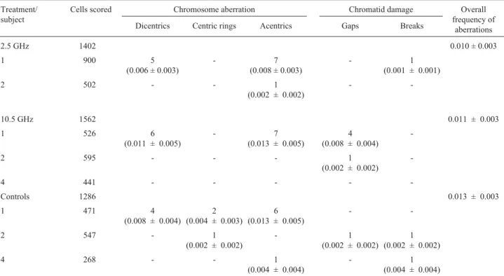
Table 1 shows the chromosome- and chromatid-type aberrations seen among lymphocytes from blood samples exposed to the microwave fields. There was no significant difference between control cells and those exposed to mi-crowave fields. Control cells had a mean frequency of 0.013 aberrations per cell compared to 0.010 and 0.011 aberrations per cell in the microwave-exposed samples. Statistical comparison of these results using the Kruskal-Wallis H-test revealed no significant differences within 95% confidence limits. The distribution of the aberrations among cells is shown in Table 2. Subject 1 had a somewhat higher than normal and overdispersed frequency of aberra-tions, probably because of previous partial-body irradia-tion. This donor was therefore not used in subsequent experiments (Table 3).
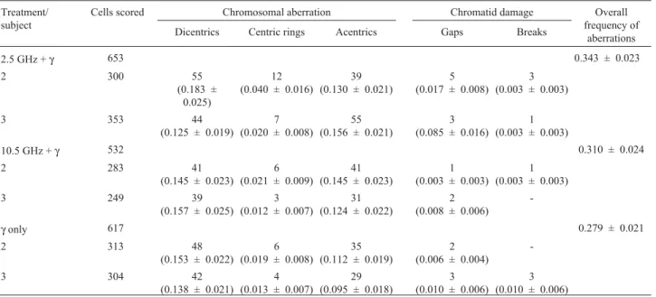
Table 3 shows the results of the “synergy” test. Cells were exposed to microwaves and subsequently to 1.5 Gy of
60Co gamma radiation. There was no significant difference
between microwave-treated or non-treated cells. Gamma irradiated cells showed a mean frequency of 0.279 aberra-tions per cell compared to 0.343 and 0.310 aberraaberra-tions per cell in samples pretreated with microwaves. Statistical
comparison of these results using the Kruskal-WallisH-test revealed no significant differences within 95% confidence limits. The distribution of the aberrations among the cells scored is shown in Table 4. Acentric fragments tended to be slightly overdispersed, as normal (IAEA, 2001).
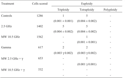
Table 5 summarizes the results of the numerical aber-rations observed according to the different treatments. Again, there were no significant differences among the var-ious groups, according to the Kruskal-WallisH-test, within 95% confidence limits.
Discussion
Following exposure to microwaves from both sources, there was a high rate of cell mortality that in-creased with the amount of energy transferred to the cells. This mortality was reflected in the high degree of cell lysis and the low number of metaphases after culturing. For blood samples treated in the 2450 MHz oven, the cell lysis was attributed to thermal effects (“cooking”). This phe-nomenon was also observed by Lloydet al.(1984) in simi-lar experiments. To prevent hyperthemia in the present experiments, the temperature of the blood samples was kept below 36 °C (the starting temperature was 28 °C and reached 33 °C after 40 s of exposure). However, in the case of the 10.5 GHz device, no thermal effects were observed since there was no increase in the temperature of the water samples, even after hours of exposure to this device. Thus, the high level of cell lysis and mortality seen following ex-posure to the 10.5 GHz device was attributable to other non-thermal processes.
Table 1- Number and frequencies of structural chromosomal aberrations in human lymphocytes exposed to microwave fields.
Treatment/ subject
Cells scored Chromosome aberration Chromatid damage Overall
frequency of aberrations
Dicentrics Centric rings Acentrics Gaps Breaks
2.5 GHz 1402 0.010 ± 0.003
1 900 5
(0.006 ± 0.003)
- 7
(0.008 ± 0.003)
- 1
(0.001 ± 0.001)
2 502 - - 1
(0.002 ± 0.002)
-
-10.5 GHz 1562 0.011 ± 0.003
1 526 6
(0.011 ± 0.005)
- 7
(0.013 ± 0.005)
4 (0.008 ± 0.004)
-2 595 - - - 1
(0.002 ± 0.002)
-4 441 - - - -
-Controls 1286 0.013 ± 0.003
1 471 4
(0.008 ± 0.004) 2 (0.004 ± 0.003)
6 (0.013 ± 0.005)
-
-2 547 - 1
(0.002 ± 0.002)
- 1
(0.002 ± 0.002) 1 (0.002 ± 0.002)
4 268 - - 1
(0.004 ± 0.004)
- 1
Table 2- Distribution of the chromosome-type structural aberrations among the scored cells indicated in Table 1 and according to the treatment given.
Treatment/ subject
Aberrations Number of cells with X
chromosomal aberrations
Uvalue
0 1 2 3
2.5 GHz 1
Dicentrics 896 3 1 - 9.39a
Centric rings 900 - - -
-Acentrics 893 7 - - -0.15
2.5 GHz 2
Dicentrics 502 - - -
-Centric rings 502 - - -
-Acentrics 501 1 - -
-10.5 GHz 1
Dicentrics 520 6 - - -0.17
Centric rings 526 - - -
-Acentrics 521 3 2 - 9.82a
10.5 GHz 2
Dicentrics 595 - -
-Centric rings 595 - - -
-Acentrics 595 - - -
-10.5 GHz 4
Dicentrics 441 - - -
-Centric rings 441 - - -
-Acentrics 441 - - -
-Control 1
Dicentrics 468 2 1 - 8.76a
Centric rings 469 2 - - -0.05
Acentrics 467 2 2 - 11.04a
Control 2
Dicentrics 547 - - -
-Centric rings 546 1 - -
-Acentrics 547 - - -
-Control 4
Dicentrics 268 - - -
-Centric rings 268 - - -
-Acentrics 267 1 - -
-aOverdispersed.
Table 3- Number and frequencies of structural chromosomal aberrations in human lymphocytes exposed to microwave fields and subsequently to 1.5 Gy of gamma radiation.
Treatment/ subject
Cells scored Chromosomal aberration Chromatid damage Overall
frequency of aberrations
Dicentrics Centric rings Acentrics Gaps Breaks
2.5 GHz +γ 653 0.343 ± 0.023
2 300 55
(0.183 ± 0.025)
12 (0.040 ± 0.016)
39 (0.130 ± 0.021)
5 (0.017 ± 0.008)
3 (0.003 ± 0.003)
3 353 44
(0.125 ± 0.019) 7 (0.020 ± 0.008)
55 (0.156 ± 0.021)
3 (0.085 ± 0.016)
1 (0.003 ± 0.003)
10.5 GHz +γ 532 0.310 ± 0.024
2 283 41
(0.145 ± 0.023) 6 (0.021 ± 0.009)
41 (0.145 ± 0.023)
1 (0.003 ± 0.003)
1 (0.003 ± 0.003)
3 249 39
(0.157 ± 0.025) 3 (0.012 ± 0.007)
31 (0.124 ± 0.022)
2 (0.008 ± 0.006)
-γonly 617 0.279 ± 0.021
2 313 48
(0.153 ± 0.022) 6 (0.019 ± 0.008)
35 (0.112 ± 0.019)
2 (0.006 ± 0.004)
-3 304 42
(0.138 ± 0.021) 4 (0.013 ± 0.007)
29 (0.095 ± 0.018)
3 (0.010 ± 0.006)
3 (0.010 ± 0.006)
Table 4- Distribution of the chromosome-type structural aberrations among the scored cells in Table 3.
Treatment/ subject
Aberrations Number of cells with X
chromosomal aberrations
Uvalue
0 1 2 3 4
2.5 GHz +γ
2
Dicentrics 250 45 5 - - 0.02
Centric rings 288 12 - - - -0.47
Acentrics 267 29 3 - 1 4.16a
2.5 GHz +γ
3
Dicentrics 313 36 4 - - 0.81
Centric rings 346 7 - - - -0.24
Acentrics 305 41 7 - - 1.36
10.5 GHz +γ
2
Dicentrics 243 39 1 - - -1.12
Centric rings 277 6 - - - -0.23
Acentrics 247 32 3 1 - 1.83
10.5 GHz +γ
3
Dicentrics 212 35 2 - - -0.57
Centric rings 246 3 - - - -0.11
Acentrics 233 12 3 1 - 5.63a
γonly
2
Dicentrics 267 44 2 - - -0.85
Centric rings 307 6 - - - -0.22
Acentrics 283 26 3 1 - 2.98a
γonly
3
Dicentrics 264 38 2 - - -0.50
Centric rings 300 4 - - - -0.14
Acentrics 277 25 2 - - 0.58
There were no significant differences in the frequen-cies of chromosomal aberrations between micro-wave-treated or untreated samples, despite the fact that samples treated with microwaves received huge amounts of transferred energy that were 50-17,000 times greater than the energy transferred by 1.5 Gy of ionizing radiation. The intracellular targets for ionizing radiation are the chromo-somes in the nucleus (IAEA, 2001). The results shown here suggest that microwaves do not interact directly or indi-rectly with chromosomes, although they may target other cell structures, such as cell membranes. This would explain the high degree of lysis seen in the microwave experiments. Although direct genetic effects from microwave ex-posure were not expected to occur, indirect effects of mi-crowaves would be more likely, because of the influence of eletromagnetic fields on the free radical system (Maeset al.,1997), but this was not seen in the present chromosome aberration test. These findings agree with other reports showing that the microwave irradiation of human lympho-cytesin vitrohas no direct or indirect clastogenic effects (Lloydet al.,1984; Maeset al.,1997). In addition to this lack of a direct or indirect effect of microwaves on chromo-somes, pretreating cells with microwaves also failed to af-fect their sensitivity to ionizing radiation.
A long-standing dogma in radiation science has been that energy from radiation must be deposited in the nucleus to elicit its biological effects. In recent years, a number of epigenetic effects have been described that challenge this dogma. Epigenetic factors, although not themselves genotoxic, act synergistically to enhance the carcinogenic effects of other agents. Several studies (Szmigielskiet al., 1982; Scarfiet al., 1996; Maeset al., 1997; Pakhomovaet al., 1997) have suggested that microwaves can have an epigenetic effectin vivo, and that they can exacerbate the genotoxicity of ionizing radiation or cancer-inducing
sub-stances, or potentiate other epigenetic factors (IEGMP, 2000). However, the evidence for an epigenetic effect of microwaves is equivocal since some studies have failed to reproduce the positive results reported by others (Ciaravino et al., 1987, 1991; Meltz et al., 1989, 1990; Cainet al., 1997).
In conclusion, the results described here do not sup-port the hypothesis that microwaves enhance the direct ef-fect of gamma radiation or cause cells to respond differently to ionizing radiationin vitro. It is possible that some of the epigenetic responses to microwaves in vivo could be the result of thermal effects (IEGMP, 2000), as concluded by Pakhomova et al. (1997), who found that high frequency microwaves (61 GHz) enhanced DNA re-combination, but not mutagenesis, in yeast cells exposed to ultraviolet radiation. Our findings indicate that further in-vestigations are needed to examine the influence of micro-wavesin vivoandin vitro.
Acknowledgements
The authors thank colleagues at the IME and IRD/CNEN for their help and suggestions, and also Dr. Da-vid Lloyd (NRPB, UK) for useful comments.
References
Alam MT, Barthakur N, Lambert NG and Kasatiya SS (1978) Cy-tological effects of microwave radiation in Chinese hamster cellsin vitro. Can J Genet Cytol 20:23-28.
Balode Z (1996) Assessment of radio-frequency electromagnetic radiation by the micronucleus test in bovine peripheral erythrocytes. Sci Total Environ 180:81-86.
Cain CD,Thomas DL and Adey WR (1997) Focus formation of C3H/10T1/2cells and exposure to a 836.55 MHz modulated
radiofrequency field. Bioelectromagnetics 18:237-243.
Table 5- Number and frequencies of chromosomal aberrations in human lymphocytes according to the different treatments. Samples from the different subjects were pooled for each treatment.
Treatment Cells scored Euploidy
Triploidy Tetraploidy Polyploidy
Controls 1286 1 5
-(0.001 ± 0.001) (0.004 ± 0.002)
-2.5 GHz 1402 5 5
-(0.004 ± 0.002) (0.004 ± 0.002)
-MW 10.5 GHz 1562 - 1
-- (0.001 ± 0.001)
-Gamma 617 2 2
-(0.003γ0.002) (0.003γ0.002)
-MW 2.5 GHz +γ 653 - 1
-- (0.001γ0.001)
-MW 10.5 GHz +γ 532 - -
-Ciaravino V, Meltz ML and Erwin DN (1987) Effects of radiofre-quency radiation and simultaneous exposure with mytomicin C on the frequency of sister chromatid ex-changes in Chinese hamster ovary cells. Environ Mutagen 9:393-398.
Ciaravino V, Meltz ML and Erwin DN (1991) Absence of a syner-gistic effect between moderate-power-radiofrequency electromagnetic radiation and adriamycin on cell-cycle pro-gression and sister-chromatid exchange. Bioelectromagnetics 12:289-294.
Garaj-Vrhovac V and Fucic A (1993) The rate of elimination of chromosomal aberrations after accidental exposure to mi-crowave radiation. Bioelectrochem Bioenerg 30:319-325. Garaj-Vrhovac V, Fucic A and Horvat D (1992) The correlation
between the frequency of micronuclei and specific aberra-tions in human lymphocytes exposed to microwave radia-tionin vitro. Mutat Res 281:181-186.
Garaj-Vrhovac V, Horvat D and Koren Z (1991) The relationship between colony-forming ability, chromosome aberrations and incidence of micronuclei in V79 Chinese hamster cells exposed to microwave radiation. Mutat Res 263:143-149. Goswami PC, Albee LD, Parsian AJ, Baty JD, Moros EG, Pickard
WF, Roti Roti JL and Hunt CR (1999) Proto-oncogene mRNA levels and activities of multiple transcription factors in C3H 10T 1/2 murine embryonic fibroblasts exposed to 835.62 and 847.74 MHz cellular telephone communication frequency radiation. Radiat Res 151:300-309.
Haidler T, Knasmueller S, Kundi M and Haidler M (1994) Clastogenic effects of radiofrequency radiations on chromo-somes of Tradescantia. Mutat Res 324:65-71.
IAEA (2001) International Atomic Energy Agency. Cytogenetic Analysis for Radiation Dose Assessment: A Manual. Tech-nical Report Series 405, Vienna, 127 pp.
ICNIRP (1998) International Committee on Non Ionising Radia-tion ProtecRadia-tion. Guidelines for limiting exposure to time-varying electric, magnetic, and electromagnetic fields (up to 300 GHz). Health Phys 74:494-522.
IEGMP (2000) Independent Expert Group on Mobile Phones. Mobile Phones and Health. NRPB publication, Chilton, UK, 159 pp.
Ivaschuk OI, Jones RA, Ishida-Jones T, Haggren Q, Adey WR and Phillips JL (1997) Exposure of nerve growth factor-treated PC12 rat pheochromocytoma cells to a modulated radiofrequency field at 836.55 MHz: Effects on c-jun and c-fos expression. Bioelectromagnetics 18:223-229. Kerbacher JJ, Meltz ML and Erwin DN (1990) Influence of
radio-frequency radiation on chromosome aberrations in CHO cells and its interaction with DNA-damaging agents. Radiat Res 123:311-317.
Lai H and Singh NP (1995) Acute low-intensity microwave expo-sure increases DNA single-strand breaks in rat brain cells. Bioelectromagnetics 16:207-210.
Lai H and Singh NP (1996) Single- and double-strand DNA breaks in rat brain cells after acute exposure to radiofrequen-cy electromagnetic radiation. Int J Radiat Biol 69:513-521. Lloyd DC, Saunders RD, Finnon P and Kowalczuk CI (1984) No
clastogenic effect fromin vitromicrowave irradiation of G0
human lymphocytes. Int J Radiat Biol 46:135-141. Lloyd DC, Saunders RD, Moquet JE and Kowalczuk CI (1986)
Absence of chromosomal damage in human lymphocytes
exposed to microwave radiation with hyperthermia. Bioelectromagnetics 7:235-240.
Maes A, Collier M, van Gorp U, Vandoninck S and Verschaeve L (1997) Cytogenetic effects of 935.2-MHz (GSM) micro-waves alone and in combination with mitomycin C. Mutat Res 393:151-156.
Maes A, Collier M and Verschaeve L (2001) Cytogenetic effects of 900 MHz (GSM) microwaves on human lymphocytes. Bioelectromagnetics 22:91-96.
Maes A, Verschaeve L, Arroyo A, de Wagner C and Vercruyssen L (1993)In vitrocytogenetic effects of 2450 MHz waves on
human peripheral blood lymphocytes. Bioelectromagnetics 14:495-501.
Meltz ML, Eagan P and Erwin DN (1989) Absence of mutagenic interaction between microwaves and mitomycin C in mam-malian cells. Environ Mol Mutagen 13:294-299.
Meltz ML, Eagan P and Erwin DN (1990) Proflavin and micro-wave radiation: Absence of a mutagenic interaction. Bioelectromagnetics 11:149-154.
Meltz ML, Walker KA and Erwin DN (1987) Radiofrequency (microwave) radiation exposure of mammalian cells during UV-induced DNA repair synthesis. Radiat Res 110:255-260.
OECD (1997) Organisation for Economic Cooperation and De-velopment. OECD Guidelines for the Testing of Chemicals, Guideline 473:In vitroMammalian Chromosome
Aberra-tion Test, Paris, 10 pp.
Pakhomova ON, Pakhomov AG and Akyel Y (1997) Effect of millimeter waves on UV-induced recombination and muta-genesis in yeast. Bioelectrochem Bioenerg 43:227. Phillips JL, Ivaschuk O, Ishida-Jones T, Jones RA,
Campbell-Beachler M and Haggnen W (1998) DNA damage in molt-4 T-lymphoblastoid cells exposed to cellular telephone radio-frequency fieldsin vitro. Bioelectrochem Bioenerg
45:103-110.
Sagripanti J and Swicord ML (1986) DNA structural changes caused by microwave radiation. Int J of Radiat Biol 50:47-50.
Sarkar S, Ali S and Behari J (1994) Effect of low power micro-wave on the mouse genome: A direct DNA analysis. Mutat Res 320:141-147.
Scarfi MR, Lioi MB, dÁmbrosio G, Massa R, Zeni O, De pietro R and De Berardino D (1996) Genotoxic effects of mytomicin-C and microwave radiation on bovine lympho-cytes. Electro-Magnetobiology 15:99-104.
Szmigielski S, Szudzinski A, Pietraszek A, Bielec M andWrembel JK (1982) Accelerated development of spontaneous and benzopyrene-induced skin cancer in mice exposed to 2450-MHz microwave-radiation. Bioelectromagnetics 3:179-185.
Tice R, Hook ± and McRee DI (1999) Chromosome aberrations from exposure to cell phone radiation. Microwave News 7-8:7.
Timchenko OI and Ianchevskaia NV (1995) The cytogenetic ac-tion of electromagnetic fields in the short-wave range. Psychopharmacol Ser 7-8:37-39.
Field: Instrumentation and Measurements in Bioelectromagnetics Research. D. Simunic (ed), Informa-tion Ventures Inc., Plzen, Chech Republic, pp 74-83.
Vijayalaxmi BZ, Frei MR, Dusch SJ, Guel V, Meltz ML and Jauchem JR (1997) Frequency of micronuclei in the periph-eral blood and bone marrow of cancer-prone mice
chroni-cally exposed to 2450 MHz radiofrequency radiation. Radiat Res 147:495-500.
Wolff S, James TL, Young GB, Margulis AR, Bodycote J and Afzal V (1985). Magnetic resonance imaging: Absence ofin vitrocytogenetic damage. Radiology 155:163-169.