The Effect of NF-
κ
B Signalling Pathway on
Expression and Regulation of Nacrein in Pearl
Oyster,
Pinctada fucata
Juan Sun1, Guangrui Xu1, Zeshi Wang1, Qing Li1, Yu Cui1, Liping Xie1,2*, Rongqing Zhang1,2*
1Institute of Marine Biotechnology, School of Life Science, Tsinghua University, Beijing, China,2Protein Science Laboratory of the Ministry of Education, Tsinghua University, Beijing, China
*[email protected](LX);[email protected](RZ)
Abstract
Nacrein is the first identified and widely investigated molluscan matrix protein and is consid-ered to play an important role in the shell formation of the pearl oyster,Pinctada fucata. Here, we investigate the effect of the NF-κB signalling pathway onNacreingene expression inP.fucatato elucidate the mechanisms involved in shell formation. Inhibition of NF-κB signalling decreasedNacreinpromoter-dependent luciferase activity. However, co-trans-fection of theNacreinpromoter vector with Pf-IKK or Pf-Rel expression plasmids could enhance luciferase activity, thus proving NF-κB signalling could regulate the transcriptional activity of theNacreinpromoter. Gene silencing by RNA interference and subsequent observation of the inner surface of the nacreous layer of oyster shells by SEM, showed that suppression of the genePf-Rellead to a partial inhibition of Nacrein expression, not only at the mRNA level but also at the protein level. The inner surface of the shells became abnor-mal. Electrophoretic mobility shift assays (EMSAs) revealed that Pf-Rel could directly bind to the relative sites of theNacreinpromoter. These results confirm that an important compo-nent of the NF-κB signalling pathway, Pf-Rel, can directly bind theNacreinpromoter inP.
fucata, and regulate its transcription and shell formation.
Introduction
Pearl is an example of biomineralization product that has a complicated nacre layer structure. Although accounting for less than 5% of the nacre, matrix proteins control the size and shape of calcium carbonate crystals in pearl and shell formation [1].
By subtle interactions with mineral ion precursors of calcification, such as calcium, bicar-bonate and other elements, organic matrix proteins secreted from the mantle are critical for the development of shells in molluscs [2]. These proteins not only participate in the construction of the organic nacre framework but also control the nucleation and growth of aragonitic crys-tals [3], determining the polymorph specificity of calcium carbonate in nacreous layers. Nacrein was the first matrix protein purified from the nacreous layer within the shell of the OPEN ACCESS
Citation:Sun J, Xu G, Wang Z, Li Q, Cui Y, Xie L, et al. (2015) The Effect of NF-κB Signalling Pathway on Expression and Regulation of Nacrein in Pearl Oyster,Pinctada fucata. PLoS ONE 10(7): e0131711. doi:10.1371/journal.pone.0131711
Editor:Yiguo Hong, CAS, CHINA
Received:March 12, 2015
Accepted:June 4, 2015
Published:July 9, 2015
Copyright:© 2015 Sun et al. This is an open access article distributed under the terms of theCreative Commons Attribution License, which permits unrestricted use, distribution, and reproduction in any medium, provided the original author and source are credited.
Data Availability Statement:All relevant data are within the paper and its Supporting Information files.
Funding:This work was supported by National Basic Research Program of China Grant 2010CB126405, National High Technology Research and Development Program of China Grant
pearl oyster. Nacrein was considered to play an important role in the biomineralization process of the shell ofPinctada fucataowing to its unique composition, including a domain with homology to carbonic anhydrase and an acidic Gly-Xaa-Asn (Xaa = Asp, Asn, or Glu) calcium binding domain [4]. Nacrein acts as a negative regulator in calcification by inhibiting the pre-cipitation of CaCO3in vitro[5] and was involved in ACC and nacreous layer formation in the early formation of pearls [6].
The nuclear factor-κB (NF-κB) signalling pathway, consisting of the core IKK complex, inhibitor IκB protein and transcription factor NF-κB. NF-κB transcription factor was found both in vertebrates [7] and invertebrates [8]. It is known as a classic, evolutionarily conserved, mediator of immune responses in vertebrates [9]. IKK is activated through the effects of many extracellular stimuli and catalyses the phosphorylation, ubiquitination and degradation of IκB proteins, resulting in translocation of the released Rel/NF-κB dimer to the nucleus. After enter-ing the nucleus, the NF-κB/Rel transcription factor binds to specific DNA sequences to regu-late gene transcription [10,11].
Since initial discovery as a B-cell-specific transcription factor [12], Previous researches have shown that in mammals, the NF-κB family of transcription factors regulates the expression of a wide array of genes involved in various physiological processes [13–16]. The NF-κB signalling pathway was found not only regulates genes involved in the inflammatory and immune responses, but also plays an important role in bone homeostasis, osteoclast dif-ferentiation and vertebrate bone formation [17–20]. Therefore, the NF-κB signalling path-way was thought to be the bridge linking the immune response and bone formation in mammals.
Genes from the pearl oysterP.fucata, with significant sequence homology to important components of the NF-κB signalling pathway were cloned and namedPf-IKK,Pf-Reland
poIκB[21–23]. Using software[24], we predicted two putative binding sites of NF-κB in theNacreinpromoter, suggesting that NF-κB signalling may be involved in regulating
Nacreingene transcription. Thus, elucidating any potential regulation ofNacreingene expression by NF-κB signalling could facilitate a better understanding of pearl formation and any interaction that may exist between the immune system and biomineralization in molluscs.
In this study, we investigate the effect of NF-κB signalling onNacreingene expression inP.
fucata, to further understand the mechanism involved in shell and pearl formation. The trans-fection ofPf-IKKorPf-Relcould increaseNacreinpromoter-dependent luciferase activity, while the NF-κB inhibitor pyrrolidine dithiocarbamate (PDTC) could decrease luciferase activ-ity. Additionally, RNA interference knockdown ofPf-ReldecreasedNacreinmRNA levels. Additionally, scanning electron microscopy (SEM) analysis showed a large number of scattered crystal particles appeared on the surface of the nacreous layer after knockdown ofPf-Rel, fol-lowed by the formation of irregular multi-layer stacking. Electrophoretic mobility shift assays (EMSAs) showed that Pf-Rel could bind directly to theNacreinpromoter. These results are consistent with NF-κB regulation ofNacreinexpression and the shell biomineralization pro-cesses inP.fucata.
Materials and Methods
Ethics Statement
This study was approved by the Animal Ethics Committee of Tsinghua University, China.
Molecular cloning of the
Nacrein
promoter from oyster
We obtained live adultP.fucatafrom the Guofa Pearl Farm in Beihai, Guangxi Province, China. The mantle was separated from the oyster with a sterile knife and then ground into very fine powder in liquid nitrogen. Genomic DNA was extracted with the Tissue/Cell genome iso-lation kit (Tiangen, China) according to the manufacturer’s instructions. A pair of degenerate oligonucleotide primers Nacrein-F and Nacrein-R (Table 1) was synthesized. Using the mantle genome DNA as a template, PCR cycles were carried out in the following steps: denaturation at 95°C for 5 min, followed by 35 cycles of 95°C for 0.5 min, 63°C for 1 min, and 72°C for 4 min. A final extension step was conducted at 72°C for 10 min. The PCR product with the expected size of 1381 bp was confirmed by sequence analysis.
Transient transfection and Luciferase Reporter Assay
Construction of vector. We inserted the 1.3 kb PCR-amplified fragment into the multiple cloning site of the pGL3-Basic vector (Promega) to construct theNacreinpromoter-luciferase reporter, designated pGL3-Nacrein. We used this as a reporter for investigating the activity of
Nacreinpromoter fromP.fucata. Both pcDNA4.0A/Pf-IKKand pcDNA4.0A/Pf-Releukaryotic expression plasmids were constructed following the previously described method for the con-struction of pcDNA4.0A/Pf-IKKplasmid. [21].
Transient transfection. 24 hours prior to transfection, Hela cells were seeded into 60 mm plates (2–6×10⁴cells/plate). Cells were transfected with different doses of the reporter lucifer-ase plasmid (pGL3-Nacrein), or transfected with the same amount of reporter luciferase plas-mid and various amounts of the pcDNA4.0A/Pf-IKKor pcDNA4.0A/Pf-Relexpression plasmid, with renilla used as an internal reference. The total amount of transfected plasmid was kept constant with the empty expression vector, pGL3-Basic. Transient transfection was carried out using Lipofectamine 2000 Transfection Reagent (Invitrogen) according to the man-ufacturer’s instructions.
Luciferase Reporter Assay. We measured all luciferase activity with a TD-20/20 Lumin-ometer (Promega) according to the manufacturer’s instructions. Reactions were completed in triplicate and statistically significant differences identified by One-way analysis of variance (ANOVA).
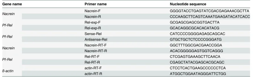
Table 1. Nucleotide sequences of primers used in this study.
Gene name Primer name Nucleotide sequence
Nacrein Nacrein-F GGGGTACCTGAGTATCGACGAGAAACGCTTA
Nacrein-R CCCAAGCTTCAGTCAAATGAAGATACATCACC
Pf-Rel Rel-exp-F GCGAGCGAGCGGTGACTTA
Rel-exp-R GCACAGGCGCACACATACG
Pf-Rel Sense-Rel CATCCCCGGGGAGAGCAGCAC
Antisense-Rel GTGCTGCTCTCCCCGGGATG
Nacrein Nacrein-RT-F GGCTTTGGCGACGAACCGGA
Nacrein-RT-R ACACGGGGGAGTGGTCAGGG
Pf-Rel Rel-RT-F CTCGAGTGAAAGCTTCAACA
Rel-RT-R CGAGCTATACGAGCACGCAGC
ß-actin actin-RT-F CTCCTCACTGAAGCCCCCCTCA
actin-RT-R ATGGCTGGAATAGGGATTCTGG
PDTC inhibition experiments. 24 hours after transfection with the reporter luciferase plasmid (pGL3-Nacrein), Hela cells were treated with 0–200μM of PDTC (Sigma-Aldrich) for 24 h before the detection of luciferase activity.
RNA interference (RNAi)
We used an NCBI blast search to compare the mRNA sequences ofPf-Relfor homology with other species.Pf-Relhas higher similarity with molluscs than with other unrelated species. A 100 bp sequence in theRelhomologous region was highly conservative across all species.Pf-Rel
silencing probes were designed by GENEIOUS software (Biomatters) as Sense-Rel and Anti-sense-Rel (Table 1).
Probes were synthesized by Invitrogen, and were diluted in RNase free dH2O.P.fucatawith a shell length of 5–6 cm were used in all experiments. 15 ng of dsRNA was injected into the adductor muscle of each oyster by syringe and needle while control samples were injected with 150 mM NaCl. Five individuals were used for each treatment.
Total RNA extraction and Real-time quantitative PCR
Total RNA was extracted from the oyster mantle 3 or 6 days after injection. Extracted RNA was quantified by absorbance at 260 nm. Quantitative real-time PCR analysis was carried out using 2μg of total RNA and a Quant Reverse Transcriptase Kit (Tiangen), as per the manufac-turer’s instructions.
Real-time PCR analysis was carried out using an Mx3000P RT-PCR System (Stratagene). Target genes were normalized toß-actinmRNA expression levels. The nucleotide sequence of each primer used for real-time PCR is shown inTable 1. PCR amplification was carried out as 95°C for 10 s, followed by 35 cycles of 95°C for 5 s and 60°C for 20 s in duplicate. SYBR Green Real-time PCR Master Mix Kit (TaKaRa) was used for the detection.
All of the real time PCR reactions were repeated in triplicate. The gene expression levels were calculated using the 2–ΔΔCtmethod [25] and normalized relative toß-actinmRNA at the same time point. The data from the experiments were analysed by ANOVA in Origin 7.0 (Ori-ginLab Corporation).
Scanning electron microscopic observations
Shells taken from RNAi experiment samples were soaked in 5% NaOH for 8 h to remove organic compounds. Samples were then washed in distilled water several times and air-dried. The nacreous layers were sputter-coated with gold and observed under a QUANTA 200 scan-ning electron microscope (FEI).
EMSA
Expression and Purification ofPf-Rel. Pf-RelcDNA was amplified with a pair of specific primers: Rel-exp-F and Rel-exp-R (Table 1). The PCR products, incorporating BamHI and XhoI restriction sites, were purified, digested and inserted into the prokaryotic expression vec-tor pET28b (Novagen). The recombinant plasmids were confirmed by sequencing.
manufacturer’s instructions. The titer was determined using a standard enzyme-linked immu-nosorbent assay.
Nuclear protein extraction. Nuclear protein was extracted from the gills ofP.fucata oys-ters using a total nuclear protein extraction kit (Xinghan) according to the manufacturer’s instructions. Quantification of nuclear protein was carried out using the bicinchoninic acid method [26].
EMSA. EMSAs were carried out using DIG Gel Shift Kit, 2nd Generation (Roche), accord-ing to the manufacturer’s instructions. Briefly, the probe containing the consensus Pf-Rel bind-ing site (underlined letters) KBWTL-5: GATCACTGCAACAGGGTCTGGTTGGAGAT CCCCTCCCTTCTTGTAAGA was synthesized and labelled with DIG-11-ddUTP. 1 mg of nuclear extract was incubated with the DNA probe at 30°C for 30 min and separated on a 5% native polyacrylamide gel. After electrophoresis, the DNA-protein complexes were blotted onto a nylon+membrane by electro-blotting. The signals were visualized by chemiluminescent detection on X-ray film.
Results
Nacrein
promoter is transcriptionally active
We confirmed transcription of the constructedNacreinpromoter plasmid through luciferase activity. Different amounts of pGL3-Nacreinluciferase plasmids were transfected into Hela cells and the activity of theNacreinpromoter was measured (Fig 1). Luciferase activity increased as the dose of pGL3-Nacreinluciferase vector increased, confirming that the con-structed pGL3-Nacreinpromoter plasmid is transcriptionally active and can be used for further transfection experiments.
Fig 1.Nacreinpromoter is transcriptionally active.Different amounts of pGL3-Nacreinplasmid (0, 0.4μg, 0.8μg, 1.6μg) was transfected into Hela cells and promoter-dependent Luciferase activity was measured. We used Renilla as an internal reference. Empty pGL-3 Basic plasmid was used to keep the total amount of plasmid the same in each group. Each reaction was completed in triplicate. Luciferase activity increased as the dose of pGL3-Nacreinluciferase vector increased, confirming that the constructed pGL3-Nacrein promoter plasmid is transcriptionally active. Significant differences were identified by One-way ANOVA. The symbol“*”indicates a significant reduction (P<0.05), compared to control, which was transfected with pGL-3 Basic alone.
Pf-IKK and Pf-Rel increased the
Nacrein
promoter activity
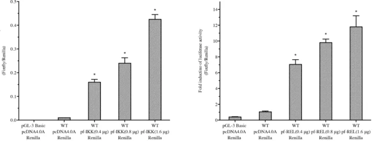
Both IKK and Rel are important components in the NF-κB pathway, and their homologues, Pf-IKK and Pf-Rel fromP.fucata, have been cloned previously [21,22]. To investigate whether the NF-κB pathway regulatesNacreingene transcription, pGL3-Nacreinpromoter reporter plasmid was transfect into Hela cells together with pcDNA4.0A/Pf-IKKor pcDNA4.0A/Pf-Rel
plasmids, and Luciferase activity was measured. Increasing concentrations of both thePf-IKK
andPf-Relplasmids increased the activity of theNacreinpromoter significantly (Fig 2). This dose dependent increase in luciferase activity shows that the NF-κB signalling is capable of reg-ulating the transcription activity of theNacreinpromoter.
The NF-
κ
B inhibitor PDTC could inhibit the
Nacrein
promoter activity
PDTC, a metal chelator and antioxidant, can specifically inhibit the activation of NF-κB by suppressing the release of the inhibitory subunit IκB from the latent cytoplasmic form of
NF-κB [27]. In order to study the effect of NF-κB signalling on activation of theNacreinpromoter, PDTC was added into cultured Hela cells that were co-transfected with the Nacrein-Luciferase reporter plasmid pGL3-Nacreinand the plasmid pcDNA4.0A/Pf-IKK. The pcDNA4.0A/ Pf-IKKplasmid was used to enhance the basic activity of the NF-κB promoter. Interestingly, increasing amounts of PDTC lead to a decrease in luciferase activity (Fig 3). This result sug-gested that the NF-κB signalling pathway was involved in regulatingNacreingene expression.
Pf-Rel
knockdown decreased the
Nacrein
gene expression level
The luciferase reporter assay showed that key components in the NF-κB signalling pathway could affect the activity of theNacreinpromoter. In order to further clarify if and how NF-κB signalling regulates theNacreingene transcription, we performed a knockdown ofRelgene by RNAi.Pf-Reltargeted double strand RNA (dsRNA) was injected into the adductor muscle ofP.
fucata. 3 and 6 days after injection, the expression levels ofPf-RelandNacreinmRNA in the
Fig 2. Pf-IKK and Pf-Rel increased theNacreinpromoter activity.Increasing doses (0, 0.4μg, 0.8μg and 1.6μg) of pcDNA4.0A/Pf-IKK(A) or pcDNA4.0A/Pf-Rel(B) were co-transfected with pGL3-Nacreinpromoter Luciferase plasmids into Hela cells. 0.5 ng of Renilla was used as an internal reference in each group. Empty pcDNA4.0A plasmid was used to keep the total amount of plasmid the same between treatments. Control cells were transfected with pGL3 Basic alone. The luciferase activity, which reports theNacreinpromoter activity, increased significantly as the does of (left)Pf-IKKor Pf-Rel(right) plasmids increased. Significant difference was identified by One-way ANOVA. The symbol“*”indicates a significant reduction (P<0.05), compared to control.
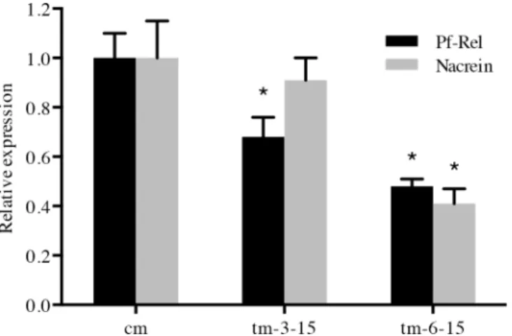
oyster mantle were measured by real-time PCR. The expression levels ofNacreinslightly decreased after dsRNA injection (Fig 4), compared to the control group (treated with NaCl solution). 6 days after injection, the expression level of bothPf-RelandNacreinwere sup-pressed by nearly 50%. These results demonstrate that silencing of thePf-Relgene is capable of decreasingNacreinmRNA transcription levels.
Fig 3. PDTC inhibits theNacreinpromoter activity.Equal amounts of pGL3-NacreinLuciferase and pcDNA4.0A/Pf-IKKplasmids were co-transfected into Hela cells that were 24 h previously treated with increasing concentrations of PDTC (0, 25μM, 50μM, 100μM and 200μM). 0.5 ng of Renilla was used as an internal reference. Each reaction was repeated in triplicate. Increasing amounts of PDTC lead to a decrease in luciferase activity, suggesting a decrease inNacreinpromoter activity. Significant differences were identified by One-way ANOVA. The symbol“*”indicates a significant reduction (P<0.05), compared to the control.
doi:10.1371/journal.pone.0131711.g003
Fig 4.Pf-Relknockdown decreased theNacreingene expression level.The expression levels of Nacrein(grey columns) andPf-RelmRNA (black columns) in oyster mantle were measured by Real-time PCR, 3 or 6 days after injection ofPf-ReldsRNA. Five oysters (n = 5) were used in each experiment. Reactions were completed in triplicate. cm: NaCl control solution. BothNacreinandPf-RelmRNA expression level in controls are attributed a relative value of 1.0. tm-3-15 and tm-6-15: samples injected with15 ngPif-Rel dsRNA 3 or 6 days respectively. The expression levels ofNacreinslightly decreased after dsRNA injection, compared to the control group. 6 days after injection, the expression level of bothPf-RelandNacreinwere suppressed by nearly 50%. Significant difference was identified by One-way ANOVA. The
symbol“*”indicates a significant reduction (P<0.05), compared to control oysters.
Pf-Rel
knockdown disturbed the shell biomineralization
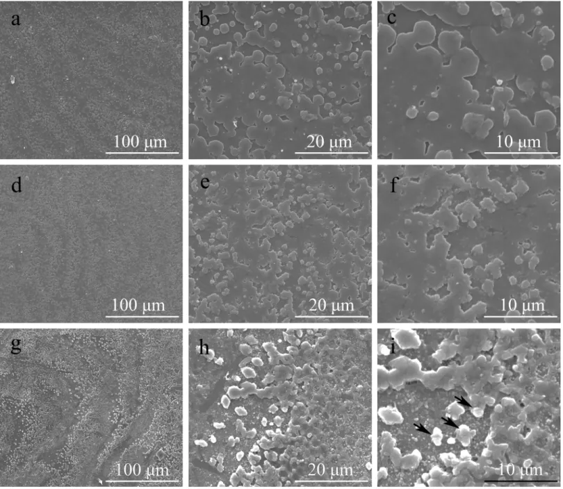
As Nacrein plays an important role in pearl biomineralization, a decrease in Nacrein expres-sion could possibly cause changes in the crystal morphology of oyster shells. Therefore, we observed the surface structure of the nacreous layer in each dsRNA injection group using SEM. Compared to NaCl injected controls, oysters with decreasedNacreintranscription had an obvi-ous change on the surface of nacreobvi-ous shell, which was shown 3 and 6 days after injection with 15μg ofPf-ReldsRNA (Fig 5). After 3 days injection (Fig 5D, 5E and 5F), crystal particles became more intensive then the controls (Fig 5A, 5B and 5C). Their distribution were scattered (Fig 5D and 5E). And the edges of these particles also become irregular (Fig 5F). After 6 days injection, the situation got more serious (Fig 5G, 5H and 5I). A large number of scattered crys-tal particles appeared on the surface of the nacreous shell, followed by the formation of irregu-lar, multi-layer stacking (Fig 5I), leading to the complete interruption of the normal layered structure. These results are similar to the SEM pictures ofP.fucatanacre shells in which Nacrein was directly inhibited by application of Nacrein monoclonal antibodies [28,29].
Pf-Rel could bind to the promoter of
Nacrein
Through sequence analysis, we identified two possible NF-κB binding sites in theNacreingene promoter (seeS1 Fig). As a putative Rel/NF-κB homolog, Pf-Rel may bind these possible
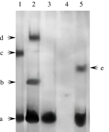
NF-κB binding sites. We performed a series of EMSAs to determine whether Pf-Rel is involved in the nuclear translocation of NF-κB. Nuclear proteins extracted from the gills ofP.fucatawere incubated with DIG-labelledNacreinpromoter probes and Pf-Rel antibodies. The samples were separated on a non-denaturing PAGE gel and bands were visualized by chemiluminescent detection on X-ray film. As shown inFig 6, besides Band a (free DNA), no band was detected in Lane 3 and Lane 4. Compared to Lane 2, there is a super shift in Lane 1 after Pf-Rel antibody was added, suggesting that Pf-Rel present in the total nuclear protein extracts is capable of binding to theNacreinpromoter probes.
Discussion
Nacrein is an important matrix protein capable of regulating the formation of oyster shells [4]. In this study we describe how the oysterP.fucataregulates this important matrix protein both
in vitroandin vivo, and describe the morphological effect of decreased Nacrein expression on oyster shell structure.
Xionget al. found that Pf-IKK activated the expression of NF-κB-controlled reporter genes and induced NF-κB translocation [21]. Sequence analysis of Pf-Rel shows that it shares high similarity with other Rel/NF-κB family proteins, especially within conserved domains [22]. A conserved degradation motif and six ankyrin repeats were identified in the poIκB, which shares significant homology with other IκB proteins [23]. Here, we have shown that the NF-κB signal-ling pathway exists and functions to regulateNacreintranscription inP.fucata.
Anin silicoanalysis of theNacreinpromoter sequence identified two possible NF-κB bind-ing sites between nucleotides 429–438 and 882–891 (seeS1 Fig). We were able to confirm that the NF-κB signalling pathway could regulate the activity of theNacreinpromoter inP.fucata
by co-transfection PGL3-Nacrein and pcDNA4.0A/Pf-IKKor pcDNA4.0A/Pf-Rel. Expression of both of these proteins promotedNacreintranscription, while the NF-κB inhibitor PDTC inhibitedNacreintranscription.
Significantly, SEM images of the inner nacreous layer structure of shells after dsRNAPf-Rel
knockdown showed similar morphological changes to the nacreous layer when Nacrein was inhibited directly by its antibody [28]. EMSAs were able to demonstrate direct binding between Pf-Rel and theNacreinpromoter. It therefore appears likely that Pf-Rel regulatesNacrein tran-scription by binding to its promoter.
Fig 5.Pf-Relknockdown disturbed the shell biomineralization (SEM).The oysters used were treated for Pf-Rel RNAi by being injected with either NaCl solution or 15 ng Pf-Rel dsRNA. a, b, c: control group with NaCl solution. d, e, f: 3 days after injection with 15 ng dsRNA. g, h, i: 6 days after injection with 15 ng dsRNA. b, e, h: magnified images of a, d, g. c, f, i: magnified images of b, e, h. The black arrows in I indicate the examples of the irregular, multi-layer stacking particles. After 3 days injection (d, e, f), crystal particles became more intensive then the controls. The distribution and the edges of these particles become irregular. After 6 days injection, a large number of scattered crystal particles appeared on the surface of the nacreous shell, followed by the formation of irregular, multi-layer stacking, leading to the complete interruption of the normal layered structure. Scal bars: a, d, g: 100μm; b, e, h: 20μm; c, f, i: 10μm.
The NF-κB signalling pathway has been extensively studied in mammals. Here, we focused on the effect of one component of NF-κB signalling in regulatingNacreintranscription in pearl oysters. In the future, it will be interesting to investigate if an interaction exists between the different NF-κB signalling components Pf-IKK, poIκB, and Pf-Rel. Because of a lack of mollusc cell lines, transfection experiments have to be performed on mammalian cells instead. It would be interesting to confirm these results experimentally within primary cell cultures of oyster tissue.
Conclusion
In conclusion, we have shown how the important NF-κB signalling pathway protein Pf-Rel regulates the transcription ofNacrein. We described novel NF-κB signalling inP.fucata, which has the ability to regulate the pearl biomineralization process.
Supporting Information
S1 Fig. Schematic representation of the two NF-κB binding sites in the Nacrein promoter.
The black boxes indicates the two NF-κB binding sites in the Nacrein promoter (GeneBank Number AB274024). The binding sites were predicted by the software TF SEARCH[30]. The
Fig 6. Pf-Rel could bind to the promoter ofNacrein(EMSA).Nuclear proteins extracted from the gills ofP. fucatawere incubated with oligonucleotide probes labelled with DIG-ddNTP to perform EMSAs. Lane 1: labelled DNA probes, nuclear protein and antibody of Pf-Rel. Lane 2: labelled DNA probes and nuclear protein. Lane 3: labelled DNA probes. Lane 4: unlabelled DNA probes and nuclear protein. Lane 5: positive control. Band a: free DNA. Band b: DNA and Pf-Rel. Band c: DNA, Pf-Rel and antibody of Pf-Rel. Band d: DNA, Pf-Rel and other interactive nuclear protein. Band e: positive control band. Compared to Lane 2, there is a super shift after Pf-Rel antibody was added in Lane 1, suggesting that Pf-Rel present in the total nuclear protein extracts is capable of binding to theNacreinpromoter probes.
scores were both 86.8. (TIF)
Acknowledgments
This work was supported by National Basic Research Program of China Grant 2010CB126405, National High Technology Research and Development Program of China Grant
2010AA09Z405, National Natural Science Foundation of China Grants 31172382 and
U0831001 (Joint Fund with Guangdong), and Independent Research Projects of Tsinghua Uni-versity Grant 20111080964. Grant 2011ZX08011-006 is a Major Project of Ministry of
Agriculture.
Author Contributions
Conceived and designed the experiments: JS ZW LX RZ. Performed the experiments: JS GX YC QL ZW. Analyzed the data: JS QL GX. Contributed reagents/materials/analysis tools: ZW GX. Wrote the paper: JS YC LX RZ.
References
1. Lowenstam H, Weiner S (1989) On biomineralization. USA: Oxford University Press.
2. Marie B, Marin F, Marie A, Bédouet L, Dubost L, Alcaraz G, et al. (2009) Evolution of Nacre: Biochemis-try and Proteomics of the Shell Organic Matrix of the Cephalopod Nautilus macromphalus. ChemBio-Chem 10: 1495–1506. doi:10.1002/cbic.200900009PMID:19472248
3. Hou W, Feng Q (2006) Morphologies and Growth Model of Biomimetic Fabricated Calcite Crystals Using Amino Acids and Insoluble Matrix Membranes ofMytilus edulis. Crystal growth & design 6: 1086–1090.
4. Miyamoto H, Miyashita T, Okushima M, Nakano S, Morita T, Matsushiro A (1996) A carbonic anhydrase from the nacreous layer in oyster pearls. Proceedings of the National Academy of Sciences 93: 9657–
9660.
5. Miyamoto H, Miyoshi F, Kohno J (2005) The carbonic anhydrase domain protein nacrein is expressed in the epithelial cells of the mantle and acts as a negative regulator in calcification in the mollusc Pinc-tada fucata. Zoological science 22: 311–315. PMID:15795493
6. Liu X, Li J, Xiang L, Sun J, Zheng G, Zhang G, et al. (2012) The role of matrix proteins in the control of nacreous layer deposition during pearl formation. Proceedings of the Royal Society B, Biological Sci-ence 279: 1000–1007. doi:10.1098/rspb.2011.1661PMID:21900328
7. Baldwin AS (1996) The NF-kappa B and I kappa B proteins: new discoveries and insights. Annu Rev Immunol 14: 649–681. PMID:8717528
8. Hetru C, Hoffmann JA (2009) NF-kappaB in the immune response of Drosophila. Cold Spring Harbor perspectives in biology 1.
9. Ghosh S, May MJ, Kopp EB (1998) NF-κB and Rel Proteins: Evolutionarily Conserved Mediators of Immune Responses. Annual Review of Immunology 16: 225–260. PMID:9597130
10. Whiteside ST, Israël A (1997) IκB proteins: structure, function and regulation. Seminars in cancer biol-ogy 8: 75–82. PMID:9299585
11. Hayden MS, Ghosh S (2008) Shared principles in NF-κB signaling. Cell 132: 344–362. doi:10.1016/j. cell.2008.01.020PMID:18267068
12. Sen R, Baltimore D (1986) Multiple Nuclear Factors Interact with the Immunoglobulin Enhancer Sequences. Cell 46: 705–716. PMID:3091258
13. Salles A, Romano A, Freudenthal R Synaptic NF-kappa B pathway in neuronal plasticity and memory. Journal of Physiology-Paris.
14. DiDonato JA, Mercurio F, Karin M (2012) NF-κB and the link between inflammation and cancer. Immu-nological Reviews 246: 379–400. doi:10.1111/j.1600-065X.2012.01099.xPMID:22435567
16. Batra S, Balamayooran G, Sahoo M (2011) Nuclear Factor-κB: a Key Regulator in Health and Disease of Lungs. Archivum Immunologiae et Therapiae Experimentalis 59: 335–351. doi: 10.1007/s00005-011-0136-zPMID:21786215
17. Yamaguchi A, Katagiri T, Ikeda T, Wozney JM, Rosen V, Wang EA, et al. (1991) Recombinant human bone morphogenetic protein-2 stimulates osteoblastic maturation and inhibits myogenic differentiation in vitro. The Journal of cell biology 113: 681–687. PMID:1849907
18. Nakashima T, Penninger JM (2003) RANKL and RANK as novel therapeutic targets for arthritis. Cur-rent opinion in rheumatology 15: 280–287. PMID:12707582
19. Boyce B, Xing L, Franzoso G, Siebenlist U (1999) Required and nonessential functions of nuclear fac-tor-kappa B in bone cells. Bone 25: 137–139. PMID:10423039
20. Kim K, Kim JH, Youn BU, Jin HM, Kim N (2010) Pim-1 Regulates RANKL-Induced Osteoclastogenesis via NF-κB Activation and NFATc1 Induction. The Journal of Immunology 185: 7460–7466. doi:10. 4049/jimmunol.1000885PMID:21068407
21. Xiong X, Feng Q, Chen L, Xie L, Zhang R (2008) Cloning and characterization of an IKK homologue from pearl oyster,Pinctada fucata. Developmental & Comparative Immunology 32: 15–25.
22. Wu X, Xiong X, Xie L, Zhang R (2007) Pf-Rel, a Rel/nuclear factor-kappaB homolog identified from the pearl oyster,Pinctada fucata. Acta Biochim Biophys Sin 39: 533–539. PMID:17622473
23. Zhang D, Jiang S, Qiu L, Su T, Wu K, Li Y, et al. (2009) Molecular characterization and expression anal-ysis of the IκB gene from pearl oysterPinctada fucata. Fish & shellfish immunology 26: 84–90.
24. Heinemeyer T, Wingender E, Reuter I, Hermjakob H, Kel AE, Kel OV, et al. (1998) Databases on Tran-scriptional Regulation: TRANSFAC, TRRD, and COMPEL. Nucleic acids research 26: 362–367. PMID:9399875
25. Livak KJ, Schmittgen TD (2001) Analysis of Relative Gene Expression Data Using Real-Time Quantita-tive PCR and the 2−ΔΔCT Method. methods 25: 402–408. PMID:11846609
26. Osnes T, Sandstad O, Skar V, Osnes M, Kierulf P (1993) Total protein in common duct bile measured by acetonitrile precipitation and a micro bicinchoninic acid (BCA) method. Scand J Clin Lab Invest 53: 757–763. PMID:8272764
27. Schreck R, Meier B, Männel DN, Dröge W, Baeuerle PA (1992) Dithiocarbamates as potent inhibitors of nuclear factor kappa B activation in intact cells. The Journal of experimental medicine 175: 1181–
1194. PMID:1314883
28. Gong N, Shangguan J, Liu X, Yan Z, Ma Z, Xie L, et al. (2008) Immunolocalization of matrix proteins in nacre lamellae and theirin vivoeffects on aragonitic tablet growth. Journal of structural biology 164: 33–40. doi:10.1016/j.jsb.2008.05.009PMID:18620869
29. Fang D, Xu G, Hu Y, Pan C, Xie L, Zhang R (2011) Identification of genes directly involved in shell for-mation and their functions in pearl oyster,Pinctada fucata. PLoS One 6: e21860. doi:10.1371/journal. pone.0021860PMID:21747964