Potential Cross-Talk between Alternative and
Classical NF-κB Pathways in Prostate Cancer
Tissues as Measured by a Multi-Staining
Immunofluorescence Co-Localization Assay
Ingrid Labouba1☯, Cécile Le Page1☯, Laudine Communal1, Torbjoern Kristessen2,
Xiaotian You1, Benjamin Péant1, Véronique Barrès1, Philippe O. Gannon1,
Anne-Marie Mes-Masson1,3, Fred Saad1,4 *
1Institut du cancer de Montréal / Centre de recherche du Centre hospitalier de l’Université de Montréal (CRCHUM), 900 rue St-Denis, Montreal, Canada,2Visiopharm, Argen Allé 3, Hoersholm, Denmark,
3Department of Medicine, Université de Montréal, Montreal, Canada,4Division of Urology, CHUM and Department of Surgery, Université de Montréal, Montreal, Canada
☯These authors contributed equally to this work.
Abstract
Background
While the classical NF-κB/p65 pathway is known to be involved in prostate cancer progres-sion and is associated with poor patient outcome, the role of the NF-κB /RelB alternative protein is not well defined. Here we analyzed the activation of both NF-κB pathways in pros-tate cancer tissues and correlate this activation with clinical features of the disease.
Methods
A multiple immunofluorescence technique was employed to concomitantly and quantita-tively visualize the nuclear localization of p65 and RelB in 200 paraffin embedded samples. Epithelia were defined using appropriate fluorochrome markers and the resulting immuno-fluorescent signals were quantified with an automated scoring system.
Results
The nuclear frequency of p65 was found to be significantly increased in tumor tissues as compared with normal adjacent tissue, whereas the frequency for RelB was decreased (p< 0.001, Wilcoxon test). As previously reported, p65 nuclear frequency was associated with a risk of biochemical recurrence. Although, RelB nuclear frequency alone did not predict recurrence, the presence of activated RelB reduced the risk of recurrence associated with the activation of p65.
OPEN ACCESS
Citation:Labouba I, Le Page C, Communal L, Kristessen T, You X, Péant B, et al. (2015) Potential Cross-Talk between Alternative and Classical NF-κB Pathways in Prostate Cancer Tissues as Measured by a Multi-Staining Immunofluorescence Co-Localization Assay. PLoS ONE 10(7): e0131024. doi:10.1371/journal.pone.0131024
Editor:Natasha Kyprianou, University of Kentucky College of Medicine, UNITED STATES
Received:February 9, 2015
Accepted:May 26, 2015
Published:July 17, 2015
Copyright:© 2015 Labouba et al. This is an open access article distributed under the terms of the
Creative Commons Attribution License, which permits unrestricted use, distribution, and reproduction in any medium, provided the original author and source are credited.
Data Availability Statement:All relevant data are within the paper and its supporting files.
Conclusion
For the first time p65/RelB co-distribution was assessed in prostate cancer tissues and sug-gested a negative crosstalk between the two NF-κB pathways in prostate cancer
progression.
Introduction
Prostate cancer is the most frequently diagnosed cancer in North American men and is the sec-ond leading cause of cancer-related death in males after lung and colon cancers [1]. Typically, prostate cancer remains localized within the prostate gland. However, it may also grows aggres-sively beyond the prostate capsule and lead to metastasis formation [2]. While some prostate cancer patients only require active monitoring of the disease, others need more radical treat-ments [3,4]. At present, the majority of patients with localized prostate cancer undergo sur-gery, radiotherapy or androgen deprivation therapy as first line treatment [5]. However, despite the high success rates obtained with these treatments, 25% of patients develop bio-chemical recurrence (BCR) after hormone-therapy treatment and progress to a more aggres-sive disease [6,7]. Thus, a major challenge in prostate cancer is the proper discrimination of patients with a latent or slow progressing disease from those with a more aggressive form of cancer. Such discrimination will help tailor treatment appropriately for individual patients. To better stratify prostate cancer patients prognosis, the identification of specific molecular bio-markers able to predict outcome would represent an important advancement over existing clinical tools. In this context, our group has been the first to address the prognostic potential of the p65 subunit of Nuclear Factor (NF)-κB in prostate cancer [8] and highlight a potential role of Nuclear Factor (NF)-κB in prostate cancer progression [9,10].
The NF-κB family includes five transcription factor proteins characterized by their Rel-homology domain. There are subdivided into two main groups: Class I (NFκB1 and NFκB2) and class II (RelA/p65, RelB, and c-Rel). The NFκB1 and NF-κB2 are translated in the p100 and p105 proteins that are processed in their respective p50 and p52 subunits [11]. Two major pathways control NF-κB signaling: the classical pathway, in which the heterodimer p65/p50 is the main active dimer, and the alternative pathway where the RelB/p52 is the active dimer. Under normal conditions, NF-κB is retained inactive in the cytosol by the IκB or p100 pro-teins. During the activation, in the classical pathway (IKKβ-dependent), the IKK complex phosphorylates IκB, inducing its ubiquitination and degradation by the proteasome. This leads to the nuclear translocation of the p65/p50 or p50/c-rel dimers. In the alternative pathway (IKKα-dependent), the IKK complex regulates the processing of the p100 precursor, as opposed to IκB [12–15], which generates p52 and the nuclear translocation of the p52/RelB dimer.
Numerous molecular and biological functions, such as inflammation [16,17], angiogenesis [18], survival, migration and invasion [19] have been shown to be associated with the activity of the classical NF-κB nuclear factors in cancer progression (reviewed in [20]). While signifi-cant work has focused on the study of the classical NF-κB pathway, recently, attention has been directed towards the alternative NF-κB pathway, and particularly in regards to its influ-ence on inflammation. The biological functions of NF-κB also involve crosstalk between the classical and alternative pathways at different levels, from upstream signaling to nuclear inter-actions via the formation of diverse NF-κB dimers. Depending on the context and stimuli, the two pathways can cooperate, or interfere negatively, in the regulation of gene expression.
publish, or preparation of the manuscript. The specific role of this author is articulated in the‘author contributions’section.
Furthermore, the biological functions directed by the crosstalk of both pathways remain poorly understood and has never been investigated in prostate cancer cells.
In prostate cancer, several studies have reported that both RelB and p65 may be putative prognostic biomarkers associated with disease progression. We, and others, have previously observed by immunohistochemistry (IHC) in prostate cancer tissue, that elevated amounts of nuclear p65 were associated with more aggressive disease [8,21]. Subsequently, it was shown that the nuclear localization of p65 is highly predictive of lymph node invasion [9] and was associated with biochemical recurrence (BCR), either alone or in combination with activated ErbB/Akt signaling [21–25]. More recently, we observed an increased presence of nuclear RelB in prostate cancer tissues compared to non-neoplastic tissues, thereby suggesting that the alter-native NF-κB pathway is also activated during the course of disease progression [10]. In addi-tion, it was shown in anin vitromodel that the inhibition of RelB expression increases the proliferation of 22Rv1 castrate-resistant prostate cancer cells and stimulates cellular autophagy [26]. Other studies found that RelB increases radiosensitivity of the castrate-resistant cells [27–
30] seemingly by up-regulating IL-8 [31]. In line with these first observations, it was also shown that RelB is overexpressed in prostate glands in response to androgen deprivation [32] and radiation treatment [33], and also induces expression of ofβ-galactosideα 2,3-sialyltrans-ferase, a gene involved in the gangliosides expression and cancer progression [34]. More recently, a negative crosstalk was shown between RelB and AR, and association with survival and metastatic cancer [35].
In order to estimate the role of RelB and the crosstalk between the classical and the alterna-tive NF-κB pathways, we implemented a multi-staining fluorescence technique so as to quanti-tatively analyze the simultaneous presence of nuclear p65 and RelB in the same prostate cancer tissue cores. The quantitation of the fluorescent signal was supported by an automated system. To evaluate the biological role of both NF-κB pathway activities, we correlated the nuclear localization of p65 or RelB with clinical parameters and risk of biochemical recurrence to better undrstand the potential role of the NF-κB pathway crosstalk in prostate cancer progression.
Materials and Methods
Cohort of patients
Gleason score<7; 18 cases with seminal vesicles involvement against 170 without; 26% with
extraprostatic extension, 32% with positive surgical margin; 3% with lymph node invasion; 142 with pathological stage 2 and 47 with pathological stage 3; the cancer specific rate is 3% and the biochemical recurrence is 28%.
Tissue microarrays
From FFPE human tissue samples, tumor areas were selected based on reviews of Hematoxy-lin/Eosin (H&E)-stained slides by a pathologist. FFPE tumor blocks were then biopsied using a 0.6 mm diameter tissue arrayer needle and the resultant cores were arrayed onto a grid in a recipient paraffin block. Each TMA set contained, one core of a tumor sample and one core of a normal adjacent prostatic glandular tissue for a total of 100 patients. These TMAs were built in duplicate to analysis two independent cores of each tissue type per cases. TMA samples were sectioned, stained by both H&E-and immunohistochemistry (IHC) for High Molecular weight Cytokeratin (HMCK), and then subsequently underwent an independent pathology review to confirm and annotate the histology of each core.
Immunohistochemistry (IHC)
IHC was performed using a Benchmark XT automated stainer (Ventana Medical System Inc. VMSI), Tucson, USA). Antigen retrieval was carried out with the Ventana Cell Conditioning 1 reagent (VMSI #950–124). The primary antibodies used were: anti-p65 (sc-8008), anti-RelB (sc-226), anti-cytokeratin (CK) 18 (sc-6259) and anti-PSA (sc-7638), all obtained from Santa Cruz Biotechnology Inc. (Santa Cruz, CA) and anti-CK19 (ms-198-P0) from Thermo Scientific Lab Vision (Ottawa, ON, Canada). The specificity of the above antibodies against p65 and RelB was previously verified by western-blot [26,36]. Antibodies were diluted from 1:25 to 1:1000 in an antibody diluent buffer (VMSI #ADB250), except for anti-PSA that was diluted in phos-phate buffered saline (PBS). Diluted antibodies were manually dispensed on the slides and incubated at 37°C for 60 min. Reactions were carried out using the UltraView DAB detection kit (VMSI #760–500) and slides were then counterstained with hematoxylin and bluing reagent (VMSI #760–2021 and #760–2037). All sections were scanned with a 20x 0.75NA objective with a resolution of 0.3225 mm, using the VS-110 slide scanner (Olympus, Richmond Hill, ON, Canada) and analyzed offside by side with HW cytokeratin staining to account for benign glands.
Immunofluorescence (IF)
We produced an epithelial mask using several specific epithelial antigens to distinguish stromal and epithelial components within tissue. To ensure coverage of all prostate cancer cells, even in their most undifferentiated state, we used a cocktail of CK18, CK19 and PSA that were all labeled to emit in the orange channel. Slides were also stained with DAPI (blue) to identify nuclei. Secondary antibodies against p65 and RelB were labeled to emit in the red and green channels, respectively. This allowed us to distinguish specific staining of p65 and RelB in the nucleus and cytoplasm of epithelial cells.
Technologies Inc.) for RelB, were both diluted 1:250 in 1X PBS. After two successive washes with 1X PBS, TMA slides were blocked for 60 min with Mouse-On-Mouse blocking reagent (1 drop in 250μL PBS, MKB-2213, Vector Laboratories, CA, USA) and then incubated for 90 min at RT with anti-PSA (1:100 in PBS). The TMA slides were washed twice and incubated for 45 min at RT with a secondary fluorescent anti-goat Cy3 (#705-165-003, Jackson ImmunoRe-search Laboratories Inc., PA, USA) diluted at 1:250 in 1X PBS. Slides were then blocked once again with Mouse-On-Mouse blocking reagent overnight at 4°C. Subsequently, they were incu-bated for 90 min at RT with a mix of anti-CK18 and anti-CK19 (both at 1:100 in 1X PBS) and then 45 min at RT with secondary fluorescent anti-mouse Alexa Fluor 546 (A546 antibody) (#A10036, Life Technologies Inc.). Finally, slides were incubated for 15 min at RT with a 0.1% (w/v) solution of Sudan Black in 70% ethanol to quench the tissue auto-fluorescence. The slides were mounted with ProLong Gold Antifade Mountant with DAPI (P-36931, Life Technologies Inc.) for nuclei staining. Between each step, TMA slides were washed twice with 1X PBS. Slides were stored at 4°C and scanned the following day. A negative control TMA slide was also done in parallel and incubated with 1X PBS in place of all primary antibodies.
Staining quantification
The staining and scoring were performed blinded to the study end-point. The tissue sections were scanned with an Olympus microscope and VS110 slide scanner linked to an OlyVia image viewer software (xvViewer.exe). Fluorescent staining was then quantified with the Visio-morphDP software (Visiopharm, Denmark) allowing for an automated image analysis. The staining via CK18/CK19/PSA markers was used to limit the analysis to epithelial areas as the region of interest #1 (ROI #1), while DAPI served to assess the fluorescence only in nuclei (ROI #2) (S1 Fig). Cores with defined ROI #1 and #2 were manually reviewed to ensure proper selection of epithelial cells and to remove surrounding tissue with necrosis or inflammatory zones from the ROI. Continuous values of fluorescence intensity for both ROI #1 and #2 were then collected by the VisiomorphDP software and transferred to Excel to define epithelial and nuclear NF-κB scores. The positive and negative signals were based on designated threshold values (mean intensities + standard deviation) and used to calculate the frequency of positive nuclei per core. The average of tumor cores from the same patient was used for analysis. The intra class correlation (ICC) between the duplicate cores was 0.68 and 0.70 (p<0.001,
Spear-man) for nuclear p65 and nuclear RelB respectively.
Statistical analyses
applying the multivariate model (n = number of events, k = number of variables). The covari-ables were not time dependent. Additional clinicopathological varicovari-ables included pre-operatory PSA, Gleason score, surgical margin status, extra-prostatic extension and seminal vesicle involvement.
Ethics Statement
An Ethical approval for the study was obtained from the appropriate review board (Comité d’éthique de la recherche du CHUM). A written informed consent was obtained from all participants.
Results
Immunofluorescent multi-staining analysis of p65 and RelB
The aim of the study is to analyze p65 and RelB in normal adjacent or malignant epithelial prostate cancer tissues on the same TMA slide using a quantitative and semi-automated pro-cess. A quantitative analysis provides continuous data, allowing for a wider range of detection and more accurate determination of low and high intensity staining. In order to facilitate this process and achieve maximal specificity, we chose to implement a multi-immunofluorescent staining technique (IF).
First, we selected epithelial areas to be analyzed. Because of its known constitutive expres-sion in prostatic epithelial cells, cytokeratin 18 (CK18) was chosen to identify epithelial cells in our specimens [37]. However, we observed that tissue samples stained by IHC produced vari-able CK18 expression levels and that staining was weaker in poorly differentiated tumors (data not shown), thus reducing the accuracy of tumor cell identification in advanced prostate cancer tissues. To overcome this, we selected CK19 and PSA as epithelial antigens, since they are known to be highly expressed in prostatic epithelial cells [37,38]. Visual verification of the simultaneous staining of CK18, CK19 and PSA with orange fluorescent dyes (A546 for CK18 and CK19, and Cy3 for PSA) confirmed full epithelial coverage by this tri-antigen staining cocktail, providing the sensitivity and specificity required to identify prostatic epithelial cells regardless of the level of tissue differentiation (S1 Fig).
The orange epithelial mask was used with the DAPI nuclear staining to restrict quantifica-tion of p65 and RelB to epithelial nuclei. The subunits p65 and RelB were stained with Cy5 (red) and A488 (green) dyes, respectively (Fig1). After this quadruple IF staining, we per-formed a quantitative analysis of p65 and RelB from scanned images using the immunofluores-cence image analysis program VisiomorphDP. A threshold value of fluoresimmunofluores-cence intensity was defined to discriminate positive and negative fluorescent signals, which was used to determine the specific frequency (%) of p65- or RelB- positive nuclei in each tissue core (including double positive nuclei) (Fig 1).
Comparison of p65 expression using immunohistochemistry and
quantitative automated immunofluorescence analysis
To evaluate the performance of this IF technique, implemented for quantification of nuclear markers in prostate cancer tissue, we compared quantification results from the automated anal-ysis with results obtained using traditional IHC staining. In both cases, the frequency of p65 stained nuclei was evaluated. We obtained a significant correlation between both datasets (r = 0.410,p<0.001 Spearman), which is in the same range as the correlation between
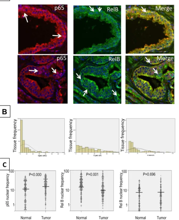
dupli-cate cores within the given technique. The traditional IHC method, based on visual scoring, Fig 1. Nuclear co-localization of p65 and RelB by a quadruple multiple staining approach. A.Epithelial staining with CK18, CK19 and PSA defines the epithelial mask using orange fluorochromes (A546, Cy3).B.Identification of epithelial area (presented in grey for optimal contrast in subsequent analyses) as ROI #1 (region of interest #1). Identification of nuclei (grey surrounded by red tracing) as ROI#2 from pre-defined ROI#1. P65 and RelB fluorescence were subsequently evaluated separately in ROI#1 and #2.C.RelB staining with green fluorescent dye (A488) and p65 staining with red fluorescent dye (Cy5). Steps 1, 2 and 3 correspond to the fluorescence analysis process followed using Visiomorph DP software.
was unable to distinguish between low (or no) staining, thereby leading to an overestimation of negative p65 expression cores (Fig 2B). Using the Kaplan-Meier estimation curves, we also Fig 2. Example of immunofluorescence of p65 and RelB. A.Magnification (40X) highlighting glandular structures in prostate cancer tissues. Merge: superimposed images of RelB (A488) in green, p65 (Cy5) in red and nuclei (DAPI) in blue, in normal adjacent (top) or cancer tissues (bottom). Arrows show stained nuclei.B. Comparative quantification from IHC (left) and IF (right) staining of p65. Graphs show the frequency distribution of nuclear p65 as evaluated visually (IHC) or automatically (IF).C. Frequency of nuclear p65 (left), RelB (middle) and double stained nuclei (right) in normal adjacent and tumor tissues. The comparison between adjacent normal and tumor cores was conducted using a Wilcoxon‘s test.
compared the survival association between nuclear p65 frequency and risk of BCR, as previ-ously analyzed in numerous other prostate cancer cohorts [21,22,25,39]. Nuclear p65 was found to be similarly and significantly associated with risk of BCR in both the traditional IHC and software-analyzed IF samples (Log rank = 4.38,p= 0.036 and Log rank = 4.83,p= 0.0028, respectively). Altogether, these results validate the use of automated analysis by IF for the eval-uation of NF-κB subunit localization and quantification in prostate cancer tissues.
Analysis of nuclear distribution of p65 and RelB in prostate cancer
tissues
While p65 nuclear localization was increased in prostate tumors compared to normal adjacent tissues (Fig 2C,p<0.000, Wilcoxon test), RelB exhibited a higher expression in normal
adja-cent tissue vs. tumor tissue (p = 0.001, Wilcoxon test,Fig 2C). The percentage of double stained p65/RelB nuclei did not show any significant variation between normal and tumor tissues
(Fig 2C).
In tumor cores, the activation of the classical pathway, as seen by nuclear p65, was signifi-cantly correlated with the activation of the alternative pathway, as represented by nuclear RelB localization (r = 0.522, p<0.001 Spearman). To estimate the amount of interaction between
the two NF-κB pathways, we compared the nuclear frequency of each subunit in the presence or absence of the other subunit. When nuclear RelB was expressed, there was a concomitant increase in p65 nuclear localization frequency compared to cores without nuclear RelB (27% and 19%, respectively). Similarly, the presence of nuclear p65 was associated with an increase in nuclear localization of RelB (18% and 10%, respectively).
Correlation of NF-
κ
B variables with clinico-pathological parameters
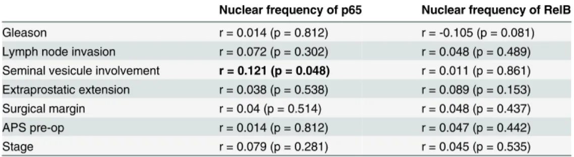
Next we evaluated the correlation between nuclear p65 (representing activation of the classical pathway) or RelB (representing activation of the alternative pathway) and clinicopathological parameters. Although nuclear expression of these two proteins was not significantly correlated with most prostate cancer aggressiveness parameters, there was a significant correlation between nuclear p65 and seminal vesicle involvement (r = 0.121, p = 0.048, Spearman,Table 1).Furthermore, there was a trend between nuclear RelB and patient Gleason score (r =
-0.105, p = 0.081, Spearman,Table 1).
Table 1. Spearman correlation test between NF-κB and clinic-pathological parameters.
Nuclear frequency of p65 Nuclear frequency of RelB
Gleason r = 0.014 (p = 0.812) r = -0.105 (p = 0.081)
Lymph node invasion r = 0.072 (p = 0.302) r = 0.048 (p = 0.489) Seminal vesicule involvement r = 0.121 (p = 0.048) r = 0.011 (p = 0.861) Extraprostatic extension r = 0.038 (p = 0.538) r = 0.089 (p = 0.153) Surgical margin r = 0.04 (p = 0.514) r = 0.048 (p = 0.437)
APS pre-op r = 0.014 (p = 0.812) r = 0.047 (p = 0.442)
Stage r = 0.079 (p = 0.281) r = 0.045 (p = 0.535)
Significant Spearman correlations are indicated in bold. Spearman correlations were considered significant atp<0.05.
Analysis of the co-expression of p65 and RelB
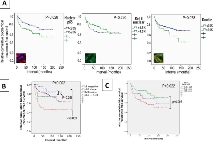
To assess the role of each NF-κB pathway in prostate cancer progression and the potential crosstalk between both pathways, we used biochemical recurrence (BCR) as a functional indicator of these activities. The nuclear frequency of p65 was associated with overall BCR (p= 0.028, Log rank = 4.843,Fig 3A). A Cox regression analyses confirmed the association between p65 and BCR in univariate (HR = 1.93,p= 0.017) and multivariate models
(HR = 3.15,p= 0.003), including clinicopathological parameters such as Gleason score, extra-prostatic extension, lymph node invasion, seminal vesicle involvement, and surgical margins
(Table 2). In contrast to p65, Kaplan-Meier estimation and univariate Cox regression analyses
showed that the RelB status alone was not associated with BCR (p= 0.301,Fig 3BandTable 2). Surprisingly, the frequency of p65/RelB double positive nuclei was not significantly predictive of BCR but a trend towards a worse prognosis was observed (p= 0.078,Fig 3C).
The activity and interaction between the p65 and RelB pathways can vary widely within a single tissue which can contains a mixed pattern of expression with cells showing only p65 or
Fig 3. Association between nuclear p65 and/or RelB and biochemical recurrence in prostate cancerpatients.Kaplan–Meier biochemical recurrence-free survival curvesA.High (>20%) and low (<20%) frequency of nuclear p65 in the epithelia of cancer tissues.B.High (>5%) and low (<5%) frequency of nuclear RelB in the epithelia of cancer tissues. Significance (p) is indicated by log rank.C.Double nuclear epithelial staining of p65 and RelB.D.Epithelial
and nuclear staining of p65 and RelB. TheκB negative variable represents patients without positive tissue cores for p65 and RelB. The variable p65 alone or RelB alone represents patients positive for only nuclear p65 or nuclear RelB. The variable p65 + RelB are patients with presence of both nuclear subunits in the same core.E.Analysis of the epithelial proportion of nuclear p65 to RelB in tissue cores within both p65 and RelB staining. The ratio p65/RelB represents the frequency of p65 to the frequency of RelB. The variable p65>RelB represents a ratio of at least 2 fold more p65 than RelB; p65-RelB represents a ratio between 0.50 and 2 fold, and RelB>p65 represents a ratio of at least 2 fold more RelB than p65.
only RelB while others being double positive. To determine the individual roles of p65 and RelB, we compared patient tissues with activation of a single pathway, represented by nuclear frequency of either p65 or RelB, and patients with no NF-κB activity (Fig 3D). Patients with only p65 activity showed a significantly shorter time of recurrence as compared to patients with no NF-κB activity (138 months and 98 months respectively, Log rank = 8.8,p= 0.002,) or compared to patients with RelB only (p= 0.021, Log Rank = 5.4,Fig 3D). This confirms that the classical NF-κB pathway strongly promotes the progression of prostate cancer. Conversely, in our patient cohort, RelB activity alone was not found to affect recurrence when compared to patients without NF-κB activation(p= 0.741,Fig 3D). Interestingly, the nuclear presence of both p65 and RelB in the same core was not significantly associated with BCR (p= 0.252), sug-gesting that RelB may counteract the biological effect of p65.
To define the potential of the role of RelB on p65 activity, we investigated the impact of each pathway on the other in varying proportions. The ratio of nuclear p65 and RelB was used to segregate patient tissues with double staining of p65 and RelB (Fig 3E). Three categories were made: patients with a higher proportion of nuclear p65 than RelB (greater than 2 fold), patients with a higher proportion of nuclear RelB than p65, and patients with relatively equal numbers of p65- and RelB-positive nuclei (up to 2 fold difference). As expected, a high level of nuclear p65 was associated with a significant risk of BCR (Log rank = 6.8,p= 0.026Fig 3E) compared to cases similar level of p65 and RelB. However, when nuclear RelB was also present, the p65-dependent increase in risk was abolished (p= 0.344), supporting the hypothesis that RelB interferes with the cancer-related biological activity of p65.
Discussion
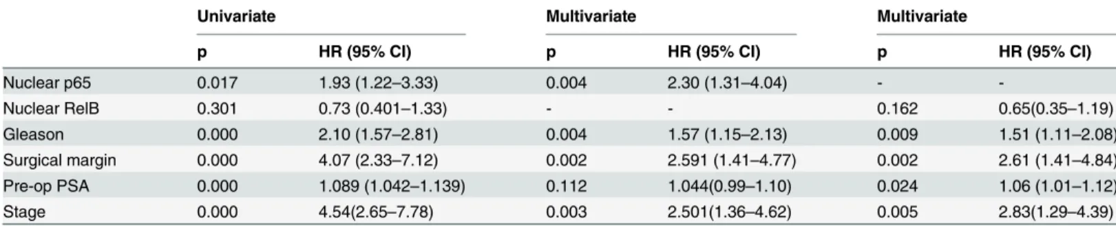
One of the multiple advantages of a semi-automated approach to antigen scoring is the ability to generate of robust and continuous data allowing for a more precise quantification of bio-marker expression as compared to the traditional categorical IHC scoring. Comparison of IHC and IF data showed superior sensitivity and accuracy for the latter in the quantification of low frequency cores. In addition, we used the Kaplan-Meier estimator to compare the efficiency of the two methods in the evaluation of risk association. While IF staining and image analysis was a slightly better discriminator of recurrence risk between the two groups compared to IHC, the difference in accuracy was minimal (Log rank = 4.38,p= 0.036 and Log rank = 4.83, p= 0.0028, respectively,S2 Fig) and did not overwhelmingly favor the use of IF for this pur-pose. In addition we found that IF approach is time consuming and expensive compare to the standard IHC approach. It should be noted that an evaluation of the automated approach for Table 2. Univariate and multivariate Cox regression analysis.
Univariate Multivariate Multivariate
p HR (95% CI) p HR (95% CI) p HR (95% CI)
Nuclear p65 0.017 1.93 (1.22–3.33) 0.004 2.30 (1.31–4.04) -
-Nuclear RelB 0.301 0.73 (0.401–1.33) - - 0.162 0.65(0.35–1.19)
Gleason 0.000 2.10 (1.57–2.81) 0.004 1.57 (1.15–2.13) 0.009 1.51 (1.11–2.08)
Surgical margin 0.000 4.07 (2.33–7.12) 0.002 2.591 (1.41–4.77) 0.002 2.61 (1.41–4.84)
Pre-op PSA 0.000 1.089 (1.042–1.139) 0.112 1.044(0.99–1.10) 0.024 1.06 (1.01–1.12)
Stage 0.000 4.54(2.65–7.78) 0.003 2.501(1.36–4.62) 0.005 2.83(1.29–4.39)
Cox regression models: significance (p<0.05) and Hazard ratio (HR) are indicated. CI: Confidence interval. PSA: Prostate Specific Antigen. NF-κB variables (p65 and RelB): status in cancer tissues. Significance is considered at p<0.5.
sensitivity, efficiency and reproducibility and experimental variability (including intra- and inter-assay assay) within whole tissue sections would also have to be performed before any con-sideration is given to applications in clinical settings. However, the consistency of the compari-son, lends confidence to an automated analysis approach to evaluate the role of NF-κB in prostate cancer progression.
An advantage of the IF technique is the ability to stain multiple proteins on a single tissue slide. Coupled with an automated detection system, IF multi-staining allows for the accurate detection and distinction of several fluorochromes that would otherwise not be discernable by the human eye. However it allows using continuous quantification of biomarker which give a better representation of their biological expression. Here, we were able to detect the co-localiza-tion of p65 and RelB specifically in epithelial cell nuclei by combining Cy5 dye and Alexa Fluor 488. Since prostate cancer tissue is heterogeneous and contains differing amounts of stromal tissue, we combined the detection of NF-κB subunits with antibodies against structural mark-ers of epithelium coupled to Cy3 and Alexa Fluor 546 dyes in order to restrict quantification to the epithelial compartment. In our IF analysis we could not account for the presence of benign glands in tissue cores due to technical limitations based on the number of simoutaneous fluoro-chromes we could use. This is the main limitation to evaluation of NF-κB subunits as potential clinical prognostic biomarkers in prostate tissues in this work.
While immunofluorescence is now well developed and is often used in translational research, particularly for FISH assays, there are still some drawbacks to consider before apply-ing any immunofluorescence data in the clinical settapply-ing. First, auto-fluorescence is a major problem for background staining which is difficult to eliminate in the data analysis. Here auto-fluorescence has been here reduced by the use of Sudan black but for translational research purposes, confocal laser scanning microscopy should be used to optimize the correction for auto-fluorescence and background [40]. IF also encounters problem of signal quenching at excitation particularly when large slides are used and scanning times are long as seen on TMA. A further point to consider is the reproducibility of results and the need to create a standard threshold signal that defines a clinically relevant cut-off. This has not been evaluated in this study but needs to be defined if p65 and RelB are to be used as biomarkers in prostate cancer.
Unfortunately, in our study, due to a limitation of the software we were not able to quantify the intensity of p65 and RelB individually in the same nuclei on a large scale. We had to esti-mate the global staining of the all nuclei in the same core and this does not account for the potential tumor heterogeneity.
The implication of the classical NF-κB pathway in prostate cancer progression has been extensively investigated (reviewed in [20]). In these studies, it was shown that the frequency of p65 nuclear distribution, as evaluated by IHC in prostatic tumor cells, was correlated with either Gleason score, presence of lymph node metastasis, seminal vesicle invasion or with sur-gical margins [8,24,25,39,41]. In addition, it was demonstrated that the presence of nuclear p65 in prostate cancer tissues predicted BCR, suggesting that the classical NF-κB pathway is indeed a central player in prostate cancer oncogenesis [25,39,42]. Therefore, in using an alter-native IF-based approach, the current study further confirms the association between the acti-vation of the classical NF-κB pathway, as estimated by p65 nuclear localization, and the aggressiveness of the disease (Tables1and2).
and RelB, which may favor bone metastasis in prostate cancer [47]. It has also been reported that RelB overexpression in the LnCaP prostatic cell line, whose the basal classical NF-κB activ-ity is moderate, promotes the in vivo tumor growth into murine model and increases the abilactiv-ity to form colonies in soft agar [48]. However, in the 22Rv1 prostatic cell line that is exempt of any classical NF-κB activity, the alternative NF-κB pathway activation triggers a stress-induced autophagy and tends to delay thein vivotumor growth in the corresponding xenograft murine model. These studies could illustrate the complex link between both the classical and alterna-tive NF-κB pathways in prostate cancer biology [26]. While the p65 classical NF-κB subunit is clearly associated with oncogenic properties, the role of RelB remains to be fully elucidated. Altogether, it appears that RelB has a complex role that may depend on the cell types and con-comitant stimuli.
In contrast to p65, we did not observe any strong associations between nuclear RelB and dis-ease progression. Nuclear localization of RelB was not associated with an overall risk of BCR
(Fig 3B) in our cohort, though there was a trend towards lower risk of BCR before 5 yrs
(p= 0.067 log Rank) and lower Gleason score (p= 0.081). To our knowledge, there are few IHC studies assessing the role of RelB in cancer. In ovarian cancer, a lack of association of RelB expression and patient prognosis was reported [49]. Similarly, nuclear RelB in retinoblastoma tissue was not associated with any pathological parameters [50]. Altogether, these studies do not identify RelB alone as a potential prognostic marker. The combination of RelB to RelA showed a slightly increased accuracy in discriminating early recurrence cases (Fig 3D) but fur-ther investigations in an appropriate translational setting are needed to address the potential of RelB and p65 before consideration of their application in clinical decision. While the clinical potential of RelB seems low, this does not preclude a functional role for this alternative NF-κB pathway in cancer or even as a therapeutic target. Indeed, we did observe that RelB activity in prostate cancer tissues decreased the risk of recurrence associated with p65 nuclear localization, suggesting that RelB may interfere with the pro-tumoral function of the classical pathway. In our cohort, the number of patients was relatively limited and the role of RelB and p65 should, in future work, be estimated on a larger scale.
Several levels of cross-talk could exist between the classical and alternative pathways. At the signaling level, an antagonistic interaction can exist, in which the activation of one pathway negatively impacts the other pathway. However, we observed a direct correlation between nuclear p65 and RelB (0.522, p<0.001), where nuclear translocation of one subunit was
Supporting Information
S1 Fig. Definition of epithelial mask. A.Immunohistochemistry illustrating a differential expression of CK18, CK19 and PSA in normal prostate tissue and tumor tissues cores from patients with either low or high grade disease.B.Simultaneous immunofluorescence staining with CK18, CK19 and PSA in normal prostate tissue and tumor tissues cores from patients with either low or high grade disease. Secondary antibodies were conjugated with A546 (CK18 and CK19) or Cy3 (PSA), each emitting fluorescence recognized in the orange range. All images at a 10X magnification. CK: cytokeratin, PSA: Prostate specific antigen.
(TIF)
S2 Fig. Comparative association of nuclear p65 and disease recurrence analyzed by immu-nofluorescence and immunohistochemistry.Kaplan–Meier biochemical recurrence-free sur-vival curves in patients with prostate cancerA.High (>20%) and low (<20%) frequency of
nuclear p65 detected by immunofluorescence.B.High (>2.5%) and low (<2.5%) frequency of
nuclear p65 detected by immunohistochemistry. Significance (p) is indicated by log rank. (PNG)
S1 Table. Tissue microarray data. (XLSX)
Acknowledgments
We would like to thank Liliane Meunier for IF and IHC technical support and Dr Mathieu Latour for pathology review. We are also grateful to Drs Véronique Ouellet and Christine Tam for their critical review of the manuscript. This work was supported by the Terry Fox Research Institute and a CUOG award in prostate cancer. Biobanking was done in collaboration with the Réseau de Recherche sur le cancer (FRQS) that is affiliated with the Canadian Tumor Repository Network (CTRnet).
Author Contributions
Conceived and designed the experiments: IL AMMM FS POG. Performed the experiments: IL XY VB LC. Analyzed the data: IL CLP BP TK. Contributed reagents/materials/analysis tools: IL TK AMMM FS. Wrote the paper: IL CLP AMMM FS.
References
1. Siegel R, Naishadham D, Jemal A. Cancer statistics, 2013. CA Cancer J Clin. 2013; 63(1):11–30. doi: 10.3322/caac.21166PMID:23335087.
2. Shen MM, Abate-Shen C. Molecular genetics of prostate cancer: new prospects for old challenges. Genes Dev. 2010; 24(18):1967–2000. doi:10.1101/gad.1965810PMID:20844012; PubMed Central PMCID: PMC2939361.
3. Van der Kwast TH, Roobol MJ. Defining the threshold for significant versus insignificant prostate can-cer. Nat Rev Urol. 2013; 10(8):473–82. doi:10.1038/nrurol.2013.112PMID:23712205.
4. Sartor AO, Hricak H, Wheeler TM, Coleman J, Penson DF, Carroll PR, et al. Evaluating localized pros-tate cancer and identifying candidates for focal therapy. Urology. 2008; 72(6 Suppl):S12–24. doi:10. 1016/j.urology.2008.10.004PMID:19095124.
5. Fournier G, Valeri A, Mangin P, Cussenot O. [Prostate cancer: Diagnosis and staging]. Ann Urol (Paris). 2004; 38(5):207–24. PMID:15570705.
7. Karantanos T, Corn PG, Thompson TC. Prostate cancer progression after androgen deprivation ther-apy: mechanisms of castrate resistance and novel therapeutic approaches. Oncogene. 2013. doi:10. 1038/onc.2013.206PMID:23752182.
8. Lessard L, Mes-Masson AM, Lamarre L, Wall L, Lattouf JB, Saad F. NF-kappa B nuclear localization and its prognostic significance in prostate cancer. BJU Int. 2003; 91(4):417–20. PMID:12603426.
9. Lessard L, Karakiewicz PI, Bellon-Gagnon P, Alam-Fahmy M, Ismail HA, Mes-Masson AM, et al. Nuclear localization of nuclear factor-kappaB p65 in primary prostate tumors is highly predictive of pel-vic lymph node metastases. Clin Cancer Res. 2006; 12(19):5741–5. doi: 10.1158/1078-0432.CCR-06-0330PMID:17020979.
10. Lessard L, Begin LR, Gleave ME, Mes-Masson AM, Saad F. Nuclear localisation of nuclear factor-kap-paB transcription factors in prostate cancer: an immunohistochemical study. Br J Cancer. 2005; 93 (9):1019–23. doi:10.1038/sj.bjc.6602796PMID:16205698; PubMed Central PMCID: PMC2361687.
11. Beinke S, Ley SC. Functions of NF-kappaB1 and NF-kappaB2 in immune cell biology. Biochem J. 2004; 382(Pt 2):393–409. Epub 2004/06/25. doi:10.1042/BJ20040544 BJ20040544[pii]. PMID: 15214841.
12. Sun Z, Andersson R. NF-kappaB activation and inhibition: a review. Shock. 2002; 18(2):99–106. PMID: 12166787.
13. Perkins ND. The diverse and complex roles of NF-kappaB subunits in cancer. Nat Rev Cancer. 2012; 12(2):121–32. doi:10.1038/nrc3204PMID:22257950.
14. Gilmore TD. Introduction to NF-kappaB: players, pathways, perspectives. Oncogene. 2006; 25 (51):6680–4. doi:10.1038/sj.onc.1209954PMID:17072321.
15. Hoffmann A, Natoli G, Ghosh G. Transcriptional regulation via the NF-kappaB signaling module. Onco-gene. 2006; 25(51):6706–16. doi:10.1038/sj.onc.1209933PMID:17072323.
16. Hoesel B, Schmid JA. The complexity of NF-kappaB signaling in inflammation and cancer. Mol Cancer. 2013; 12:86. doi:10.1186/1476-4598-12-86PMID:23915189.
17. Lawrence T. The nuclear factor NF-kappaB pathway in inflammation. Cold Spring Harb Perspect Biol. 2009; 1(6):a001651. doi:10.1101/cshperspect.a001651PMID:20457564; PubMed Central PMCID: PMC2882124.
18. Tabruyn SP, Griffioen AW. A new role for NF-kappaB in angiogenesis inhibition. Cell Death Differ. 2007; 14(8):1393–7. doi:10.1038/sj.cdd.4402156PMID:17464324.
19. Sethi G, Sung B, Aggarwal BB. Nuclear factor-kappaB activation: from bench to bedside. Exp Biol Med (Maywood). 2008; 233(1):21–31. doi:10.3181/0707-MR-196PMID:18156302.
20. Nguyen DP, Li J, Yadav SS, Tewari AK. Recent insights into NF-kappaB signalling pathways and the link between inflammation and prostate cancer. BJU Int. 2013; 114(2):168–76. Epub 2013/11/13. doi: 10.1111/bju.12488PMID:24215139.
21. Shukla S, MacLennan GT, Fu P, Patel J, Marengo SR, Resnick MI, et al. Nuclear factor-kappaB/p65 (Rel A) is constitutively activated in human prostate adenocarcinoma and correlates with disease pro-gression. Neoplasia. 2004; 6(4):390–400. Epub 2004/07/17. doi:10.1593/neo.04112PMID:15256061.
22. Koumakpayi IH, Le Page C, Mes-Masson AM, Saad F. Hierarchical clustering of immunohistochemical analysis of the activated ErbB/PI3K/Akt/NF-kappaB signalling pathway and prognostic significance in prostate cancer. Br J Cancer. 2010; 102(7):1163–73. doi:10.1038/sj.bjc.6605571PMID:20216540; PubMed Central PMCID: PMC2853085.
23. McCall P, Bennett L, Ahmad I, Mackenzie LM, Forbes IW, Leung HY, et al. NFkappaB signalling is upregulated in a subset of castrate-resistant prostate cancer patients and correlates with disease pro-gression. Br J Cancer. 2012; 107(9):1554–63. doi:10.1038/bjc.2012.372PMID:23093296; PubMed Central PMCID: PMC3493754.
24. Fradet V, Lessard L, Begin LR, Karakiewicz P, Masson AM, Saad F. Nuclear factor-kappaB nuclear localization is predictive of biochemical recurrence in patients with positive margin prostate cancer. Clin Cancer Res. 2004; 10(24):8460–4. doi:10.1158/1078-0432.CCR-04-0764PMID:15623625.
25. Domingo-Domenech J, Mellado B, Ferrer B, Truan D, Codony-Servat J, Sauleda S, et al. Activation of nuclear factor-kappaB in human prostate carcinogenesis and association to biochemical relapse. Br J Cancer. 2005; 93(11):1285–94. doi:10.1038/sj.bjc.6602851PMID:16278667; PubMed Central PMCID: PMC2361509.
26. Labouba I, Poisson A, Lafontaine J, Delvoye N, O.Gannon P, Page CL, et al. The RelB alternative NF-kappaB subunit promotes autophagy in 22Rv1 prostate cancer cells in vitro and affects mouse xeno-graft tumor growth in vivo. Cancer Cell International. 2014;under press(xx).
(7):2048–56. doi:10.1158/1535-7163.MCT-06-0700PMID:17604335; PubMed Central PMCID: PMC2692592.
28. Xu Y, Fang F, St Clair DK, Sompol P, Josson S, St Clair WH. SN52, a novel nuclear factor-kappaB inhibitor, blocks nuclear import of RelB:p52 dimer and sensitizes prostate cancer cells to ionizing radia-tion. Molecular cancer therapeutics. 2008; 7(8):2367–76. doi:10.1158/1535-7163.MCT-08-0238 PMID:18723484; PubMed Central PMCID: PMC2692185.
29. Xu Y, Fang F, Sun Y, St Clair DK, St Clair WH. RelB-dependent differential radiosensitization effect of STI571 on prostate cancer cells. Molecular cancer therapeutics. 2010; 9(4):803–12. doi:10.1158/ 1535-7163.MCT-09-1001PMID:20371728; PubMed Central PMCID: PMC2852498.
30. Zhu HC, Qiu T, Dan C, Liu XH, Hu CH. Blockage of RelB expression by gene silencing enhances the radiosensitivity of androgenindependent prostate cancer cells. Molecular medicine reports. 2015; 11 (2):1167–73. doi:10.3892/mmr.2014.2857PMID:25370388.
31. Xu Y, Fang F, St Clair DK, St Clair WH. Inverse relationship between PSA and IL-8 in prostate cancer: an insight into a NF-kappaB-mediated mechanism. PloS one. 2012; 7(3):e32905. doi:10.1371/journal. pone.0032905PMID:22403723; PubMed Central PMCID: PMC3293904.
32. Rosa-Ribeiro R, Nishan U, Vidal RO, Barbosa GO, Reis LO, Cesar CL, et al. Transcription factors involved in prostate gland adaptation to androgen deprivation. PloS one. 2014; 9(6):e97080. doi:10. 1371/journal.pone.0097080PMID:24886974; PubMed Central PMCID: PMC4041569.
33. Zhu L, Zhu B, Yang L, Zhao X, Jiang H, Ma F. RelB regulates Bcl-xl expression and the irradiation-induced apoptosis of murine prostate cancer cells. Biomedical reports. 2014; 2(3):354–8. doi:10.3892/ br.2014.250PMID:24839547; PubMed Central PMCID: PMC4022971.
34. Hatano K, Miyamoto Y, Mori M, Nimura K, Nakai Y, Nonomura N, et al. Androgen-regulated transcrip-tional control of sialyltransferases in prostate cancer cells. PloS one. 2012; 7(2):e31234. doi:10.1371/ journal.pone.0031234PMID:22347453; PubMed Central PMCID: PMC3275626.
35. Campa VM, Baltziskueta E, Bengoa-Vergniory N, Gorrono-Etxebarria I, Wesolowski R, Waxman J, et al. A screen for transcription factor targets of glycogen synthase kinase-3 highlights an inverse corre-lation of NFkappaB and androgen receptor signaling in prostate cancer. Oncotarget. 2014; 5(18):8173– 87. PMID:25327559; PubMed Central PMCID: PMC4226675.
36. Le Page C, Koumakpayi IH, Lessard L, Mes-Masson AM, Saad F. EGFR and Her-2 regulate the consti-tutive activation of NF-kappaB in PC-3 prostate cancer cells. The Prostate. 2005; 65(2):130–40. doi: 10.1002/pros.20234PMID:15880609.
37. Long RM, Morrissey C, Fitzpatrick JM, Watson RW. Prostate epithelial cell differentiation and its rele-vance to the understanding of prostate cancer therapies. Clin Sci (Lond). 2005; 108(1):1–11. doi:10. 1042/CS20040241PMID:15384949.
38. Nagle RB, Brawer MK, Kittelson J, Clark V. Phenotypic relationships of prostatic intraepithelial neopla-sia to invasive prostatic carcinoma. Am J Pathol. 1991; 138(1):119–28. PMID:1987760; PubMed Cen-tral PMCID: PMC1886063.
39. Gannon PO, Lessard L, Stevens LM, Forest V, Begin LR, Minner S, et al. Large-scale independent vali-dation of the nuclear factor-kappa B p65 prognostic biomarker in prostate cancer. Eur J Cancer. 2013; 49(10):2441–8. doi:10.1016/j.ejca.2013.02.026PMID:23541563.
40. Robertson D, Savage K, Reis-Filho JS, Isacke CM. Multiple immunofluorescence labelling of formalin-fixed paraffin-embedded (FFPE) tissue. BMC cell biology. 2008; 9:13. doi:10.1186/1471-2121-9-13 PMID:18366689; PubMed Central PMCID: PMC2288605.
41. Ross JS, Kallakury BV, Sheehan CE, Fisher HA, Kaufman RP Jr., Kaur P, et al. Expression of nuclear factor-kappa B and I kappa B alpha proteins in prostatic adenocarcinomas: correlation of nuclear fac-tor-kappa B immunoreactivity with disease recurrence. Clin Cancer Res. 2004; 10(7):2466–72. Epub 2004/04/10. PMID:15073126.
42. Lessard L, Mes-Masson AM, Saad F. [NFkappaB: a new marker kappable of predicting prostate cancer outcome]. Bull Cancer. 2006; 93(9):891–9. PMID:16980232.
43. Wang X, Belguise K, Kersual N, Kirsch KH, Mineva ND, Galtier F, et al. Oestrogen signalling inhibits invasive phenotype by repressing RelB and its target BCL2. Nat Cell Biol. 2007; 9(4):470–8. doi:10. 1038/ncb1559PMID:17369819; PubMed Central PMCID: PMC2394707.
44. Demicco EG, Kavanagh KT, Romieu-Mourez R, Wang X, Shin SR, Landesman-Bollag E, et al. RelB/ p52 NF-kappaB complexes rescue an early delay in mammary gland development in transgenic mice with targeted superrepressor IkappaB-alpha expression and promote carcinogenesis of the mammary gland. Mol Cell Biol. 2005; 25(22):10136–47. doi:10.1128/MCB.25.22.10136–10147.2005PMID: 16260626; PubMed Central PMCID: PMC1280249.
46. Benedetti G, Fokkelman M, Yan K, Fredriksson L, Herpers B, Meerman J, et al. The nuclear factor kap-paB family member RelB facilitates apoptosis of renal epithelial cells caused by cisplatin/tumor necrosis factor alpha synergy by suppressing an epithelial to mesenchymal transition-like phenotypic switch. Mol Pharmacol. 2013; 84(1):128–38. doi:10.1124/mol.112.084053PMID:23625948.
47. Li Y, Kucuk O, Hussain M, Abrams J, Cher ML, Sarkar FH. Antitumor and antimetastatic activities of docetaxel are enhanced by genistein through regulation of osteoprotegerin/receptor activator of nuclear factor-kappaB (RANK)/RANK ligand/MMP-9 signaling in prostate cancer. Cancer Res. 2006; 66 (9):4816–25. Epub 2006/05/03. 66/9/4816 [pii] doi:10.1158/0008-5472.CAN-05-3752PMID: 16651437.
48. Xu Y, Josson S, Fang F, Oberley TD, St Clair DK, Wan XS, et al. RelB enhances prostate cancer growth: implications for the role of the nuclear factor-kappaB alternative pathway in tumorigenicity. Can-cer Res. 2009; 69(8):3267–71. doi:10.1158/0008-5472.CAN-08-4635PMID:19351823; PubMed Cen-tral PMCID: PMC2756761.
49. Annunziata CM, Stavnes HT, Kleinberg L, Berner A, Hernandez LF, Birrer MJ, et al. Nuclear factor kap-paB transcription factors are coexpressed and convey a poor outcome in ovarian cancer. Cancer. 116 (13):3276–84. Epub 2010/06/22. doi:10.1002/cncr.25190PMID:20564628.
50. Qu Y, Zhou F, Dai X, Wang H, Shi J, Zhang X, et al. Clinicopathologic significances of nuclear expres-sion of nuclear factor-kappaB transcription factors in retinoblastoma. J Clin Pathol. 64(8):695–700. Epub 2011/05/10. jclinpath-2011-200017 [pii] doi:10.1136/jclinpath-2011-200017PMID:21551466.
51. Li T, Morgan MJ, Choksi S, Zhang Y, Kim YS, Liu ZG. MicroRNAs modulate the noncanonical transcrip-tion factor NF-kappaB pathway by regulating expression of the kinase IKKalpha during macrophage dif-ferentiation. Nat Immunol. 2010; 11(9):799–805. Epub 2010/08/17. ni.1918 [pii] doi:10.1038/ni.1918 PMID:20711193.