Exercises in Hot and Humid Environment
Caused Liver Injury in a Rat Model
DongLiang Li1*., Xiao Wang1., Bang Liu1
, YuZheng Liu1, ZhiYu Zeng1, LingLing Lu1, ZhiYong Zheng2, Bing Li1, ZongFu Zheng3
1.Department of Hepatobiliary Medicine, Fuzhou PLA General Hospital, Fuzhou Fujian, China,2.Department of Pathology, Fuzhou PLA General Hospital, Fuzhou, Fujian, China,3.Clinical laboratory, No.476 hospital of PLA Fujian, Fuzhou, Fujian, China
*donglianglifz@163.com
.These authors contributed equally to this work.
Abstract
Objective:To investigate injury pattern during intense exercises in hot and humid environment particularly on liver in a rat exertional heat stroke model.
Methods:We randomly divided 30 rats into a control group (CG), a normal temperature (25¡2˚C, 60%¡5% humidity) exercise group (NTEG) and a high temperature and high humidity (35¡2˚C, 80%¡10% humidity) exercising group (HTEG), each comprising 10 animals. The NTEG and HTEG rats were forced to run in a treadmill for 1 hour maximum at 20 rpm. We analyzed liver cells of all three groups with JC-1 dye and flow cytometry for apoptosis rates in addition to liver tissue 8 - hydroxy deoxyguanosine (8 - OhdG) and blood serum IL–6, tumor necrosis factor alpha (TNF-a), alanine aminotransferase ALT, aspartate amino transferase (AST), serum creatinine (CREA), blood urea nitrogen (BUN), lactate dehydrogenase (LDH), creatine phosphate kinase (CK) concentrations.
Result: Compared with NTEG rats, beside reduced exercise tolerance (60¡5 vs. 15¡3 minutes) (p50.002) the 8-OhdG liver tissue concentrations were significantly higher (p50.040) in the HTEG rats. The HTEG developed more organ tissue damage and cellular fragmentations of liver cells. In both exercise groups TNF-a and IL-6 serum concentrations were enhanced significantly (p,0.001) being highest in the HTEG animals. Serum ALT, AST, LDH, CREA, BUN and CK concentrations were significantly enhance in both exercise groups.
Conclusion:In our exertional heat stroke rat model, we found tissue damage particularly in livers during exercises in hot and humid environment that was related to inflammation, oxidative stress and apoptosis.
OPEN ACCESS
Citation:Li D, Wang X, Liu B, Liu Y, Zeng Z, et al. (2014) Exercises in Hot and Humid Environment Caused Liver Injury in a Rat Model. PLoS ONE 9(12): e111741. doi:10.1371/ journal.pone.0111741
Editor:Guillermo Lo´pez Lluch, Universidad Pablo de Olavide, Centro Andaluz de Biologı´a del Desarrollo-CSIC, Spain
Received:June 23, 2014
Accepted:October 7, 2014
Published:December 30, 2014
Copyright:ß2014 Li et al. This is an open-access article distributed under the terms of the
Creative Commons Attribution License, which permits unrestricted use, distribution, and repro-duction in any medium, provided the original author and source are credited.
Data Availability:The authors confirm that all data underlying the findings are fully available without restriction. All relevant data are within the paper.
Funding:This work was funded by The Medical Science and Technology Innovation Foundation of Nanjing Military Command of Chinese PLA, No.10MA109 to DongLiang Li. The funders had no role in study design, data collection and analysis, decision to publish, or preparation of the manu-script.
Introduction
alone and in combination with heat on physiological and morphological changes in a rat model.
Materials and Methods
Experimental animals
40 Sprague - Dawley rats, 7-week-old, weighting 150–200 gram were provided by the Shanghai Xitang Biological Technology center and raised in the animal experiment center of the Fuzhou General Hospital of the Nanjing military region under room temperature of 25¡2
˚
C and humidity of 60%¡5%, lighting for 12 hours per day with circadian rhythm changes. They were fed by sterilized fodder and water ad libitum. The study was approved by the ethical committee of the Fuzhou General Hospital and the care of mice complied with the Beijing Administration Rules of Laboratory Animal handling (GB 14925-2001).Experimental design
All rats were trained for load-increasing runs for 5 days. The length of the treadmill (Type: XR-YLS-15A, Shanghai Xinqing soft information technology co., LTD, Shanghai, China) belt was 650 cm. The initial speed of the running machine was 10 RPM (50.39 km/h) with a training time of 10 minutes, which was increased to 15 RPM (50.585 km/h) and 15 minutes in the next day, followed by 2 RPM (50.078 km/h) and 5 minute increases each day to finally 21 RPM (50.819 km/h) and 30 minutes running time on the 5thday. After the 5 day exercise period, rats which could not adapt to the running machine or had injuries of sole or foot were excluded and 30 rats eligible for exercises after training were randomly divided into 3 groups: 10 rats were the CG, 10 rats comprised the NTEG and 10 rats were selected for the HETG. All rats rested for 24 hours after 5 days of training and the experiment was performed on the 7thday. Rats in the CG were kept in the animal rooms under normal temperature (25¡2
˚
C) and humidity (60%¡5%). Rats in the NTEG and HETG were tested at normal conditions (25¡2˚
C, 60%¡5% humidity) and in high temperature and high humidity (35¡2˚
C, 80%¡10% humidity), respectively. The speed of the treadmill was adjusted to 20 RPM (50.78 km/h) and electric shocks with a limit of 1.00 mA were applied with intervals of 30 seconds after one shock for keeping the animals running. The body temperature was anally measured every 10 minutes in the NTEG and every 3 minutes in the HTEG animals. EHS was diagnosed at the time when deep breathing with cyanosis of the limb occurred and 3 or more electric shocks were necessary within 60 minutes of the experiment with bodySpecimen collection
After running, rats that met the criteria of EHS as well as the control group were anesthetized with intraperitoneal injections of 0.5 ml/kg phenobarbital sodium and blood samples were collected. After cervical dislocation under anesthesia, the internal organs heart, lung, liver, kidney as well as the brains were gathered and rinsed clean in 0.9% saline for further analyses.
Hepatocyte mitochondrial transmembrane potential detection
Fresh liver tissues of about 100 mg were cut into pieces of 1 mm3 and PBS was added. After blending, the suspensions were filtered with 200 mesh sieves and the filtrate contained liver cells. The cells were collected at a concentration of 16106
cells in 500 mL 16incubation buffer to which 10 ml of JC21 working fluid was
added. Then the liver cells were incubated for 15–20 min in a 5% CO2incubator at 37
˚
C, collected by centrifugation at room temperature (2000 RPM, 5 min) and after 1 time washing suspended in PBS. We used a flow cytometry instrument (BD FACSCalibur, San Jose, CA, US) (Ex5488 nm; Em5530 nm) to detect5, 59, 6, 69 -tetrachloro1, 19, 3, 39-tetraethyl-benzimidazolylcarbocyanine iodide (JC-1; Molecular Probes, Eugene, OR) staining, which indicates the state ofmitochondrial transmembrane potential. Polarized mitochondria appear as red and depolarized mitochondria as green. Green fluorescence was detected through the FITC channel FL1; Red fluorescence was detected through the PI channel FL2. Apoptosis was indicated by an increase in the green/red fluorescence intensity ratio [14].
Detection of 8 - hydroxy deoxyguanosine (8 - OhdG) in liver
tissues
100 mg liver tissues were quickly homogenized on ice with a glass homogenizer, dissolved in 1 ml PBS and then centrifuged for 20 minutes (2000 RPM). The resulting supernatant was further processed according to the manufacturer’s protocol (8-OhdG ELISA assay, Xikang, Shanghai). The final values were
measured at 490 nm in a microplate reader and calculated based on an 8-OhdG concentration standard curve.
Plasma IL – 6 and TNF
a
level detection
The blood serum IL – 6 and TNF-a concentrations were determined with (IL–6 ELISA, TNF- a ELISA kits) (Qiaoyi, Shanghai, China) according to the
The histopathological analyses of liver, kidney, heart, lung, and
brain tissues
First, the tissues were fixed in 10% formalin for 72 hours and then cut into appropriate sized pieces for dehydration. After that, the tissues were incubated in transparent xylene agent and then immerged into a mixture of transparent agent and paraffin wax until the paraffin fully replaced xylene. The wax blocks were then fixed in a microtome, sliced into 5–8 mm thick sections and immerged into hematoxylin and 1% eosin staining solution. After de-waxing, we observed histopathological changes under a light microscope.
Liver tissue electron microscopic sample preparation and
observations
The liver tissues were cut into 1 mm3sized blocks and fixed for at least 2 hours in 3% glutaraldehyde plus 1.5% paraformaldehyde in 0.1 M PBS (pH7.2) at 4
˚
C. Then we rinsed and fixed them with 1% osmic acid 21.5% potassiumferrocyanide and after dehydration cut them into 100 nm ultrathin slices with a Leica UC-6 ultra-microtome. Finally, the samples were stained with uranium and lead citrate for 5 to 15 minutes, washed with distilled water and observed with a transmission electron microscope (PHILIPS EM208, FEI Company, Eindhoven, Netherlands).
Statistical analyses
All data were analyzed with the SPSS13.0 statistical software package. Group measurement data are presented as mean ¡standard deviation (¡sd) and comparison among groups was performed using one-way analysis of variance (One - way ANOVA). P values ,0.05 were considered statistically significant.
Results
The running time in a hot and humid environment was less than in
normal temperature
The average running time in the NTEG was 60¡5 min and 15¡3 min in the HTEG, with significant differences between them (F57.232, P50.002).
The mitochondria depolarized in hepatocytes after exercise under
the condition of high temperature and high humidity
Biochemical parameter changes
Serum concentration of ALT, AST and LDH, CREA, BUN and CK were significantly different among all groups. Compared with the HTEG, ALT (P,0.05), LDH (P,0.05) and CK (P,0.05) serum concentrations were significantly higher, whereas AST (P,0.05) was lower in the NTEG. BUN and CREA (related to renal functions) analyses revealed that the former was higher concentrated in sera of the NTEG (P,0.05) and the latter was higher
concentrated in sera of the HTEG (P,0.05) (Table 1).
Changes of 8-OhdG in rat liver tissues and inflammatory related
cytokines in blood plasmas
The liver 8-OhdG (P,0.05), IL-6(P,0.05) and TNF-a (P,0.05) plasma concentrations were statistically significant and gradually higher in NTEG and HTEG rats compared to the control (Table 2).
Fig. 1. Flow cytometric histogram and percentage of rat liver cell apoptosis rates detected by JC-1 dye.A) Control, B) NTEG, C) HTEG (room temperature vs. control group: p50.006; high temperature vs. control group: p50.002; room temperature vs. high temperature: p50.040); FL15green fluorescence, FL25red fluorescence.
Exertional heat stress led to morphological organ tissue
alterations
Next, we analyzed whether organ tissue structures were affected by the exercises. As visible in Fig. 2, various histological changes in different degrees occurred during the exercises. Myocardium: Compare to control group, in the NTEG we found a mild swelling of myocardial cells, while in the HTEG the swelling of myocardial cells was increased obviously. Brain:In the NTEG, the grey matter in the cerebellum contained no obvious lesions as well as no congestion in the blood vessels of the cortex, but in the HTEG, endovascular blood clots appeared mainly in the cortex. Lungs: Compared to control group, in both exercise groups the pulmonary alveoli were dilated with hyperinflation. In addition, alveoli walls became thin and ruptured forming pulmonary bullae and arterial blood clots appeared. Liver: In the NTEG we found mild cell edemas and increased cell volume, while in the HTEG, we detected more obvious liver cell edemas with expansion of central veins and hepatic vein blood clots.Kidney: Compared to the control group, in the NTEG, we found no obvious changes in renal glomeruli, but mild blood clots in the renal tubulointerstitial area and medulla. In the HTEG, the epithelial cells of the renal proximal convoluted tubules appeared cloudy swollen and stromal as well as in medullar intravenous congestions were visible.
Table 1.Comparison of serum changes in the rat groups.
Control Room temperature (NTEG) High temperature (HTEG)
Liver function and lactate dehydrogenase
ALT(U/L) 52.8¡4.8 76.9¡15.2* 71.3¡16.7**D
AST(U/L) 162.0¡36.0 216.0¡61.7** 279.2¡79.12***D
LDH(U/L) 1215.5¡237.0 1835¡451.6 1466.8¡445.5D
Kidney function and creatine kinase
CREA (mmol/L) 26.1¡3.9 48.2¡16.9** 54.2¡17.0***D
BUN (mmol/L) 8.2¡1.3 16.5¡4.9*** 12.0¡1.2***D
CK(U/L) 1981.4¡577.9 3580.7¡543.2** 3319.9¡1431.5*D
Compared to control group,*p,0.05, **p,0.01, ***p,0.001. Comparisons of NTEG and HTEG,Dp,0.05,DD,0.01,DDDp,0.001.
doi:10.1371/journal.pone.0111741.t001
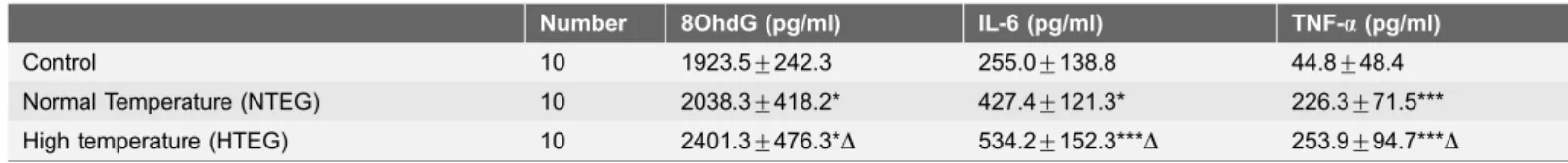
Table 2.Comparison of the changes between groups of 8OhdG in the liver tissue, and serum inflammation factors (IL-6, TNF-a) in the rat.
Number 8OhdG (pg/ml) IL-6 (pg/ml) TNF-a(pg/ml)
Control 10 1923.5¡242.3 255.0¡138.8 44.8¡48.4
Normal Temperature (NTEG) 10 2038.3¡418.2* 427.4¡121.3* 226.3¡71.5*** High temperature (HTEG) 10 2401.3¡476.3*D 534.2¡152.3***D 253.9¡94.7***D
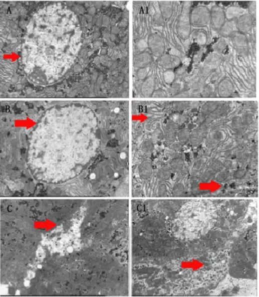
Liver cells lost their structures upon exercises in hot and humid
environment
In order to investigate particularly liver tissue damages, we analyzed liver cell structures with electron microscopy. Cell organelles dissolved and mitochondrial structures as well as normal cell organization disappeared and became incoherent, particular in the HTEG (Fig. 3).
Fig. 2. Histopathological changes in major organs.Microscopical images of H&E stained rat myocardium, brain, lung, liver and kidney tissue sections from control group (CG), normal temperature exercise group NTEG (middle panel) and high temperature exercise group (HTEG) (right panel) (magnification x50).
Discussion
In our rat model study, we induced heat strokes by forced extreme exercises in a hot and humid environment and observed changes of blood biochemistry, organ histology, inflammatory responses as well as enhancements of the oxygen radical indicator 8-OhdG and changed liver cell mitochondrial structures. 8-OhdG is an oxidized derivative of deoxyguanosine and the major product of DNA oxidation under oxidative stress [15]. Our 8-OhdG results are in line with previous literature about oxidative stress in human livers after swimming [16] and in over
Fig. 3. Electron microscope images of rat liver cell changes.Control group: (A) large and round nucleus in the center with smooth nuclear membrane (arrow), visible nucleolus and rich in shallow euchromatin. (A1) The cells had a neat structure with clear and complete mitochondrial organization. NTEG: (B) Nucleus with thickened and dissolved boundaries of the nuclear membrane (arrow). (B1) The cellular structures became unclear and mitochondrial cristae changed their shapes (upper arrow). The endoplasmic reticulum and other organelles became diffuse (lower arrow). HTEG: (C) membrane rupture, scattered organelles outside the cell (arrow), C1) disappearance of normal cell organization and dissolved mitochondrial structures (arrow).
exercised rats [17], which further confirmed that oxidative stress is occurring during exertional heat stroke. Because oxidative damage occurs 3 times more in mitochondria than in the cytosol since the mitochondria are the major source of free oxygen radicals [18] the enhanced 8-OhdG concentrations also reflected in mitochondrial depolarization, indicating liver cell apoptosis in the HTEG (Fig. 1). In our study, liver was the major affected organ of heat stress damage, probably because of its central body location and accelerated metabolic rate at temperature rises [19,20]. It is known that after physical exercises ALT, AST, LDH and CK serum levels are significantly enhanced [16,21,22], which we also detected in both exercise rat groups (Table 1). AST, which serum concentration was higher in the HTEG, is mainly located in the mitochondria and the de Ritis ratio (AST/ALT) as measure for liver damage severity [23] was higher in the HTEG (3.9 vs. 2.8) than in the NTEG, both indicating that liver and particularly mitochondrial damage was more obvious in the HTEG. Lower LDH and CK serum concentrations in the HTEG might reflect the reduced average running time in the HTEG compared to the NTEG (15¡3 vs. 60¡5 min). BUN was higher concentrated in the NTEG, whereas CREA was higher concentrated in sera of the HTEG (P,0.05). We suggest, that in the rats which exercised for an extended time in normal
temperature, increasing protein decomposition occurred and more pronounced elevated BUN serum concentrations developed [24], while in the heat exercise rats, multiple organ failure included acute renal failure causing in turn obviously strong elevated CREA concentrations, which were reported to be mortality indicators for heat stroke patients [25].
Proinflammatory cytokines such as TNF-a and IL-6 in high temperature exercising rats increased significantly compared to the normal temperature exercise and control groups, which is in accordance with previous literature of 30-fold serum IL-6 concentration increases after prolonged ultra-endurance exercises [26]. Exercise related splanchnic hypoperfusion as a cause of intestine damages [27] is similar to heat stroke induced intestinal mucosal injury and hyper-permeability changes, which leads to leakage of endotoxins and increased production of inflammatory cytokines such as TNF- a like during sepsis [8]. A high plasma TNF-a level can be considered as a possible trigger of systemic inflammatory responses via stimulating the release of other cytokines such as IL – 4 and IL –6, which eventually may expand the initial biological effect to systemic progression of the inflammatory response, thus leading to the occurrence of a multiple organ dysfunction syndrome [28–30].
Taken together, our results suggested that exercising in hot and humid environment could further aggravate particularly liver damage and induce hepatic failure. During strenuous exercises liver blood flow decreased leading to liver ischemia/hypoxia and increases of free nitrogen and oxygen radicals, lipid peroxidation and inflammation with high cytokine IL-6 and TNF-a releases caused by activated intrahepatic Kupffer cells finally resulting in complete liver cell damage.
that this conditions induced a systemic inflammatory response syndrome which caused multiple organ damages. At the same time, the mitochondrial membrane potential in liver cells decreased whereas the content of 8-hydroxy deoxyguano-sine acid increased in the heat stress exercise rats, suggesting that lipid
peroxidation and oxidative stress induced by tissue ischemia/hypoxia played an important role in liver injury when extended exercises and heat were combined in our EHS rat model.
Author Contributions
Conceived and designed the experiments: DLL. Performed the experiments: DLL XW. Analyzed the data: DLL XW Z. Zeng YZL B. Liu ZhiYu Zheng B. Li. Contributed reagents/materials/analysis tools: DLL XW YZL ZhiYong Zheng ZongFu Zheng B. Li. Wrote the paper: DLL XW LLL. Other (please specify): designed the study: DLL XW. Statistical analysis: Z. Zeng ZongFu Zheng.
References
1. Kravchenko J, Abernethy AP, Fawzy M, Lyerly HK(2013) Minimization of heatwave morbidity and mortality. Am J Prev Med 44: 274–282.
2. Miyake Y (2013) [Characteristics of elderly heat illness patients in Japan-analysis from Heatstroke STUDY 2010]. Nihon Rinsho 71: 1065–1073.
3. Carter R 3rd, Cheuvront SN, Williams JO, Kolka MA, Stephenson LA, et al.(2005) Epidemiology of hospitalizations and deaths from heat illness in soldiers. Med Sci Sports Exerc 37: 1338–1344.
4. Rohe ST(2012) Exertional heat illness in a Marine training on the endurance course. JAAPA 25: 34, 36– 38.
5. Camus G, Deby-Dupont G, Duchateau J, Deby C, Pincemail J, et al.(1994) Are similar inflammatory factors involved in strenuous exercise and sepsis? Intensive Care Med 20: 602–610.
6. Leon LR, Helwig BG(2010) Heat stroke: role of the systemic inflammatory response. J Appl Physiol 109: 1980–1988.
7. Gathiram P, Wells MT, Brock-Utne JG, Gaffin SL(1987) Antilipopolysaccharide improves survival in primates subjected to heat stroke. Circ Shock 23: 157–164.
8. Bouchama A, Knochel JP(2002) Heat stroke. N Engl J Med 346: 1978–1988.
9. Diesen DL, Kuo PC(2011) Nitric oxide and redox regulation in the liver: part II. Redox biology in pathologic hepatocytes and implications for intervention. J Surg Res 167: 96–112.
10. Thakur V, Pritchard MT, McMullen MR, Wang Q, Nagy LE(2006) Chronic ethanol feeding increases activation of NADPH oxidase by lipopolysaccharide in rat Kupffer cells: role of increased reactive oxygen in LPS-stimulated ERK1/2 activation and TNF-alpha production. J Leukoc Biol 79: 1348–1356.
11. Tukov FF, Luyendyk JP, Ganey PE, Roth RA (2007) The role of tumor necrosis factor alpha in lipopolysaccharide/ranitidine-induced inflammatory liver injury. Toxicol Sci 100: 267–280.
12. Hsieh YC, Chen RF, Yeh YS, Lin MT, Hsieh JH, et al.(2011) Kynurenic acid attenuates multiorgan dysfunction in rats after heatstroke. Acta Pharmacol Sin 32: 167–174.
13. Harmon BV, Corder AM, Collins RJ, Gobe GC, Allen J, et al.(1990) Cell death induced in a murine mastocytoma by 42–47 degrees C heating in vitro: evidence that the form of death changes from apoptosis to necrosis above a critical heat load. Int J Radiat Biol 58: 845–858.
15. Klungland A, Rosewell I, Hollenbach S, Larsen E, Daly G, et al.(1999) Accumulation of premutagenic DNA lesions in mice defective in removal of oxidative base damage. Proc Natl Acad Sci U S A 96: 13300–13305.
16. Ramos D, Martins EG, Viana-Gomes D, Casimiro-Lopes G, Salerno VP (2013) Biomarkers of oxidative stress and tissue damage released by muscle and liver after a single bout of swimming exercise. Appl Physiol Nutr Metab 38: 507–511.
17. Ogonovszky H, Sasvari M, Dosek A, Berkes I, Kaneko T, et al. (2005) The effects of moderate, strenuous, and overtraining on oxidative stress markers and DNA repair in rat liver. Can J Appl Physiol 30: 186–195.
18. de Souza-Pinto NC, Eide L, Hogue BA, Thybo T, Stevnsner T, et al. (2001) Repair of 8-oxodeoxyguanosine lesions in mitochondrial dna depends on the oxoguanine dna glycosylase (OGG1) gene and 8-oxoguanine accumulates in the mitochondrial dna of OGG1-defective mice. Cancer Res 61: 5378–5381.
19. Dickson JA, McKenzie A, McLeod K (1979) Temperature gradients in pigs during whole-body hyperthermia at 42 degrees C. J Appl Physiol Respir Environ Exerc Physiol 47: 712–717.
20. Thrall DE, Page RL, Dewhirst MW, Meyer RE, Hoopes PJ, et al.(1986) Temperature measurements in normal and tumor tissue of dogs undergoing whole body hyperthermia. Cancer Res 46: 6229–6235.
21. Lippi G, Schena F, Montagnana M, Salvagno GL, Banfi G, et al. (2011) Significant variation of traditional markers of liver injury after a half-marathon run. Eur J Intern Med 22: e36–38.
22. Wu HJ, Chen KT, Shee BW, Chang HC, Huang YJ, et al.(2004) Effects of 24 h ultra-marathon on biochemical and hematological parameters. World J Gastroenterol 10: 2711–2714.
23. Hall P, Cash J(2012) What is the real function of the liver ‘function’ tests? Ulster Med J 81: 30–36.
24. Xing JQ, Zhou Y, Fang W, Huang AQ, Li SB, et al.(2013) The effect of pre-competition training on biochemical indices and immune function of volleyball players. Int J Clin Exp Med 6: 712–715.
25. Pease S, Bouadma L, Kermarrec N, Schortgen F, Regnier B, et al.(2009) Early organ dysfunction course, cooling time and outcome in classic heatstroke. Intensive Care Med 35: 1454–1458.
26. Waskiewicz Z, Klapcinska B, Sadowska-Krepa E, Czuba M, Kempa K, et al.(2012) Acute metabolic responses to a 24-h ultra-marathon race in male amateur runners. Eur J Appl Physiol 112: 1679–1688.
27. van Wijck K, Lenaerts K, van Loon LJ, Peters WH, Buurman WA, et al.(2011) Exercise-induced splanchnic hypoperfusion results in gut dysfunction in healthy men. PLoS One 6: e22366.
28. Lim CL, Wilson G, Brown L, Coombes JS, Mackinnon LT (2007) Pre-existing inflammatory state compromises heat tolerance in rats exposed to heat stress. Am J Physiol Regul Integr Comp Physiol 292: R186–194.
29. Shah NG, Tulapurkar ME, Damarla M, Singh IS, Goldblum SE, et al. (2012) Febrile-range hyperthermia augments reversible TNF-alpha-induced hyperpermeability in human microvascular lung endothelial cells. Int J Hyperthermia 28: 627–635.