Volume 44(3):45-53, 2004 www.scielo.br/paz.htm ISSN 0031-1049
Museu de Zoologia da Universidade de São Paulo
1 Departamento de Biologia Geral. Universidade Federal de Viçosa, 36570-000, Viçosa, MG, Brasil. E -mail: jeserrao@ ufv.br. Phone: + 553138991301. Fax: + 553138992549.
2 To whom all correspondence should be addressed.
A C
OMPARATIVES
TUDYOF THEO
VARIE SINS
OMEB
RAZILIANB
E E S(H
YME NOPTE RA; A
POIDE A)
G
USTAVOF
E RRE IRAM
ARTINS1J
OSÉE
DUARDOS
E RRÃO1,2ABSTRACT
The present paper concerns the morphological features of ovaries in 33 species of bees with different social behavior patterns. The ovaries of bees were examined under light microscope. They are polytrophic-meroistic ovaries formed for an anterior germarium and a basal vitellarium. The germarium houses the germ cells and in the vitellarium there are follicles arranged linearly. In general the follicle is constituted by a nutritive chamber (a cluster of nurse cells) and an oocyte chamber, both covered by a single epithelial layer of follicular cells. The number of ovarioles per ovary and the number of mature oocyte per ovary were analyzed. Measurements of ovariole length, oocyte size, oocyte width, follicular epithelial height and the intertegular distance were made to support the comparative study. Statistical analysis showed that representatives of Meliponini and A pini have the largest ovaries. On the other hand, in solitary bees were found the bigger oocytes. Furthermore, our results suggest that there is a tendency for increase in ovary size and ovariole number, with increasing level of sociality.
KEYWORDS: insect morphology, ovary, reproductive tract, social behavior.
oocyte growth occur (Chapman, 1998; Snodgrass, 1935).
Mitotic divisions of germ cells take place within the germarium, whereas the vitellarium contains developing egg chambers in a linear arrangement. Each chamber consists of an oocyte and the nurse cells that are formed by incomplete cytokineses from the same germ cell (Zacaro & Cruz-Landim, 1996; Bili
n
sk et al.,1998; Patrício & Cruz-Landim, 2001). I n the polytrophic ovariole, its own group of nurse cells accompanies each oocyte. In general, this structure is delimited by follicular epithelium constituting the follicles or egg plus nutritive chambers (Bili
n
sk, 1998).INTRODUCTION
Insect ovaries are formed of several functional and elongated units called ovarioles (Bili
n
sk, 1998) and46 MARTINS, G.F. & SERRÃO, J.E.: THEOVARYOFBEES
The number of ovarioles per ovary is variable and shows interspecific differences (Jaglarz, 1998). The ovary morphology and its phylogenetic relationships have been studied by various authors (Simiczyjew et al., 1998; Szklarzewicz, 1998; Jaglarz, 1998; Kubrakiewicz
et al., 1998; Bili
n
sk et al.,1998).Iwata (1955; 1965) studied the polymorphism of the ovaries in bees considering the change that may occur on the ovary structure, observing the number of mature and immature oocytes, the size of mature oocyte and the speed of oocyte maturation, showing that these characteristics have a distinct correlation with the different behavioral patterns found in this insect group. The following study reports on the variation of ovary morphology in bees to support a comparative study, in order to test the hypothesis that structure is indeed related to their sociality.
MATE RIALS AND ME THODS
Nineteen species of Apidae were analyzed with representatives from Apini, Bombini, Meliponini, E uglossini, Centridini, E ucerini, E ricrocidini, E mphorini and X ylocopinae, one species of Andrenidae, ten species of Halictidae and three species of Megachilidae (Table 1).
Bees were collected in the field in Viçosa, MG, Brazil, while for Meliponini and Apini were analyzed physogastric queens obtained from the Central Apiary, Universidade Federal de Viçosa, MG.
The specimens were dissected in insect saline solution and the pieces were removed from mated bees, what was determined by the presence of spermatozoa in their spermathecae. The ovaries were isolated from the dissected reproductive tract and transferred to 4% paraformaldehyde in phosphate buffer 0.1M, pH 7.2. The samples were dehydrated in an ethanol series, embedded in historesin (Leica) and cut at 4
µ
m serial sections, which were stained with Dominici solution.Some sections were submitted to the following histochemical tests: mercury-bromophenol blue for protein, Nile blue for lipids and methyl green-pyronin for cell death. These tests were performed as described by Pearse (1968) with few variations for historesin embedded tissues.
For each bee the following parameters were analyzed: number of ovarioles per ovary, ovariole length, number of mature oocytes per ovary, oocyte length, oocyte width, follicular epithelium thickness and the intertegular distance (as representative of the body size).
Measurements were performed with aid of the software Image Pro-Plus™, 4.0 version for Windows. For determination of the body size of bees, measurements of the intertegular distance were made with the same software. The number of mature oocyte per ovary was determined as proposed for Iwata (1955; 1965).
To determine the degree of dependency of body size and morphological parameters, we used a linear regression procedure following standard statistical tests described by Snedecor & Cochran (1980).
RE SULTS
The general morphology of the ovaries is almost equal to that was described before for some species (Snodgrass, 1956; Cruz-Landim et al., 1998), and therefore only a brief description will be given, emphasizing only the features that have not been detailed before.
All species studied have meroistic polytrophic ovarioles (Figs. 1-3).
A sheath, which is made up of two layers, encloses each ovariole: an outer peritoneal membrane and an inner non-cellular tunica propria. The former is constituted by a network of cells, including muscle cells, fat body cells, and tracheoles that do not penetrate the tunica propria (Figs. 1-3, 7, 9).
The height of the follicular epithelium of egg chamber changes according to the species, with measurements varying from 6.6
µ
m in Plebeia sp. to45
µ
m in X ylocopa frontalis in mature basal follicles. In E picharis flava and E . affinis extensive cell projectionswere found (64.08 ± 8.6
µ
m length) in follicular tissue and these structures penetrate the corion that is very thick (102.0 ± 7.8µ
m length) (Figs. 7, 9).The cytoplasm of nurse and follicular cells was strongly stained by methyl green-pyronin, mercury-bromophenol blue and nile blue, marking the following constituents in the cytoplasm: RNA, proteins and neutral-lipid (Figs. 3, 7). The follicular cells however show different staining tonalities for bromophenol blue in the mid-vitellogenic follicles (Fig. 7).
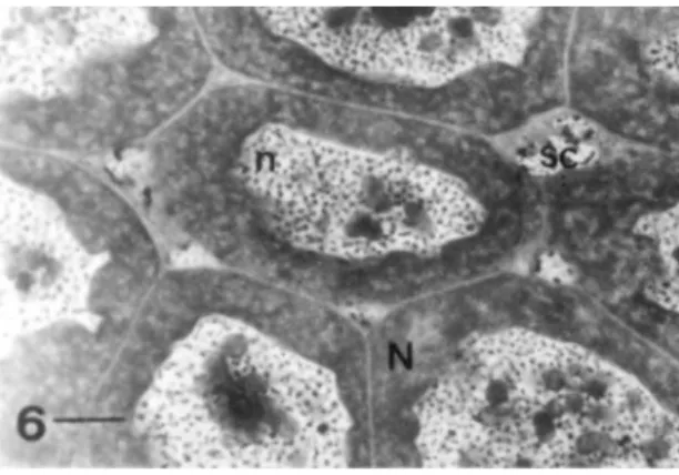
Among nurse cells there is the presence of groups of small cells, which are smaller than nurse cells, the somatic-like cells (Fig. 6).
FIGURE 1. Longitudinal section of the germarium position of
Melipona quadrifasciata. The anterior region have undifferentiated cells and the posterior region have many oocyte-nurse cell complexes; o: young oocyte; on: oocyte nucleus; uc: undifferentiated cells; pm: peritoneal membrane; T: trachea; tp: tunica propria. Bar = 10 µm.
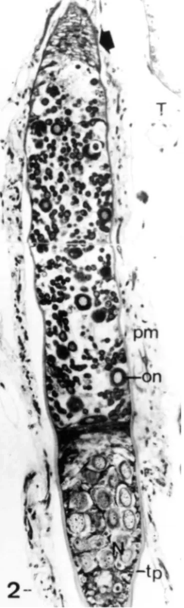
FIGURE 2. Longitudinal section of the anterior region of the ovariole of the X ylocopa frontalis, showing a short terminal filament (arrow) followed by a short germarium with few oocyte-nurse cell complexes followed by a nutritive chamber with the nurse cells (N); on: oocyte nucleus; pm: peritoneal membrane; tp: tunica propria; T: trachea. Bar = 10 µm.
region (Figs. 3-5). In Halictidae this structure was more developed in comparison with other species (Figs. 4, 5).
X ylocopa frontalis had the greatest oocyte (1.1 cm long), occupying almost the entire extension of the ovariole (1.3 cm length) (Table 1).
Apidae species have four ovarioles per ovary, while Andrenidae, Halictidae and Megachilidae have three ovarioles per ovary. These numbers were constant in both ovaries and bee group, except A . mellifera. However, in Apidae the number of mature oocytes per ovariole varied from 1 to many (Table 1).
48 MARTINS, G.F. & SERRÃO, J.E.: THEOVARYOFBEES
FIGURE 3. Longitudinal section of a follicle of E picharis affinis, observed under phase contrast microscope, Methyl Green-Pyronin stained, showing one vitellogenic growing oocyte (o) that have the nucleus (on) and the accessory nuclei (a) placed in the peripheral region of the cytoplasm; E: follicular epithelium covering the oocyte chamber; e: follicular layer of the nutritive chamber; m: muscle; N: nurse cells; pm: peritoneal membrane; T: trachea; tp: tunica propria. Bar = 10 µm.
FIGURE 4. Longitudinal section of vitellogenic growing oocyte of Pseudaugochlora graminea, showing many accessory nuclei (a) surrounding the oocyte nucleus (on). The oocyte is covered by the columnar follicular epithelium (E ). en: follicular epithelial cell nucleus. Bar = 10 µm.
F IGURE 5. Longitudinal section of mature oocyte of
Pseudaugochlora graminea with accessory nuclei placed in the peripheral oocyte region. The follicular cells are flattened; E : follicular epithelium; a: accessory nucleus; en: follicular epithelial cell nucleus. Bar = 10 µm.
Meliponini have the proximal region of the ovarioles with an accumulation of follicles with degenerative nutritive chamber.
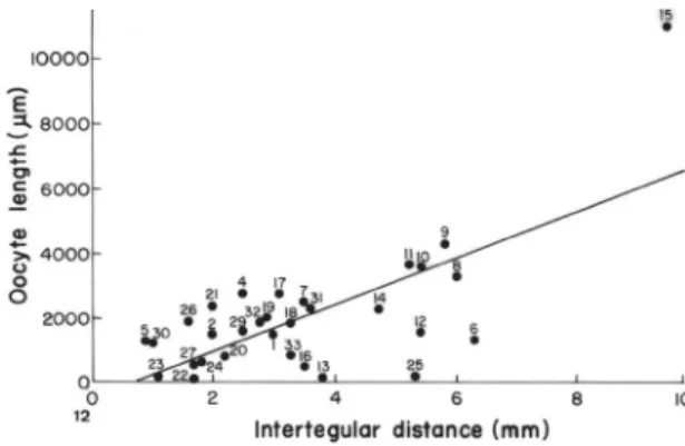
The statistical analysis showed that the ovariole size [R² = 0.010, F (1,31) = 0.030, p < 0.862] have negative correlation with body size (Fig. 10), because Meliponini have the longest ovariole while the others have the largest body size (Table 1). On the other hand, when Meliponini is excluded from the analyses, there is a positive correlation of these features [R² = 0.527, F (1,28) = 27.864, p < 0.00002] (Fig. 11). Furthermore the statistical analysis showed that the oocyte size and oocyte width have a positive correlation with intertegular distance [oocyte size: R² = 0.485, F (1,31) = 27.315, p < 0.0001; oocyte width: R² = 0.418, F (1,31) = 20.835, p < 0.00008] (Figs. 12; 13).
DISCUSSION
The small cells scattered among nurse cells are similar to those found in the beetle Badister bipustulatus, which present characteristics of somatic cells derived from pre follicular cells (Jaglarz, 1998), in spite of their role is unknown.
FIGURE 6. Longitudinal section of the nutritive chamber of
Pseudaugochlora graminea, showing the nurse cells (N) with irregular nucleus (n) and multiple nucleoli. Between the nurse cells there are somatic-like cells (sc). Bar = 10 µm.
FIGURE 7. Longitudinal section of the ovariole of E picharis affinis
obeserved under phase contrast microscope, Bromophenol Blue stained, showing the follicular epithelium (E) of two successive follicles with different developmental stages. Covering the mature oocyte, the follicular projection (p) that penetrate the corion (c). Notice that the epithelium cover the vitellogenic growing oocyte (o) without projections and with cells presenting different staining tonalities (*); en: follicular epithelial cell nucleus; (tp) tunica propria; (pm) peritoneal membrane; en: follicular epithelial cell nucleus. Bar = 10 µm.
FIGURE 8: Oocyte-nurse cell complex in the posterior region of the germarium of Melipona bicolor. Notice the presence of accessory nuclei (a) into the oocyte cytoplasm. The nurse cells present a linear arrangement and they are connected with the oocyte; tp: tunica propria; (o): oocyte; (on): oocyte nucleus; (N): nurse cell; (n): nurse cell nucleus. Bar = 10 µm.
FIGURE 9: Longitudinal section of a mature follicle of E picharis affinis, Methyl Green Pyronin, observed under phase contrast microscope. Notice the follicular cells (E) with extensive cellular projections (p) that penetrate the thick corion (c); en: follicular epithelial cell nucleus; F: fat body cells. Bar = 10 µm.
coriogenesis, follicular cell degenerates in A . mellifera
that is in agreement with the results obtained in this study for others species of bees.
Accessories nuclei have been studied in
E omenacanthus stramineus (Mallophaga) (Bilinsk, 1989) and in the bee M. quadrifasciata anthidioides and in the ant A tta sexdens rubropilosa (Cruz-Landim, 1991) and their formation were very similar among them. It is evident that these
50 MARTINS, G.F. & SERRÃO, J.E.: THEOVARYOFBEES
In Apini, Meliponini and Bombini all ovarioles have mature oocyte suggesting a synchronism in the oocyte production in these ovaries what did not occur in the solitary bees that have only one mature oocyte per ovary. Social bees have an elongated germarium followed by many follicles in a linear arrangement while the solitary ones have short germarium followed by few mature follicles which can be correlated with egg production, because social bees lay many eggs, while solitary bees lay only few eggs.
Number of ovarioles per ovary was found to be a multi-state character. We, Iwatta (1955), Rozen (1986) and Alexander & Rozen (1987) found that Andrenidae, Megachilidae, Halictidae, Colletidae and Melittidae have three ovarioles/ovary, while Apidae presents four or more. T hus, three ovarioles per ovary is the plesiomorphic condition, the others being derived
states. Whether these are ordered or unordered states is not known for sure, but it seems reasonable to believe that an increased feature derived from a smaller one, hardly would evolve to decrease, by the criterion of similarity. It seems more likely that four ovarioles/ovary, five ovarioles/ovary found in Nomadinae (Alexander & Rozen, 1987), six to 18 ovarioles/ovary in parasitic Bombini (Cumber, 1949) and hundreds ovarioles/ ovary derived independently from the three ovarioles/ ovary condition, because they are very discontinuous. Among corbiculate Apinae, Euglossini is the most similar to the ancestor because this tribe presents few mature oocytes, such as found in others non-corbiculate Apinae and bee families, so that orchid bees can be considered as sister-group of other corbiculate Apinae, which corroborates the hypothesis of Roig-Alsina & Michener (1993), based on external morphology, and
FIGURE 10. Relationship between the ovariole size and intertegular distance of bees (Linear regression). Numbers regards to number of the species listed in the Table 1.
FIGURE 12. Relationship between the oocyte size and intertegular distance of bees (Linear regression). Numbers regards to number of the species listed in the Table 1
FIGURE 11. Relationship between the ovariole size and intertegular distance of bees (Linear regression). Data on Meliponini and Apini were excluded. Numbers regards to number of the species listed in the Table 1.
P
A
P
.
A
V
U
L
S
Z
O
O
L
. 4
4(
3)
, 2
00
3
51
Family Subfamily/Tribe Species* ovarioles lenght oocyte length width epithelial heigh distance Behavior*** per ovary (mm) per ovary (µm) (µm) (µm) (mm)
Apidae Apini A pis mellifera (1) 100-180 ** 10.4 Many 1492.6 168.9 18.5 3 Highly eusocial Meliponini Scaptotrigona sp. (2) 4 84 Many 1494.4 500.0 9.4 2 Highly eusocial Melipona bicolor (3) 4 56.2 Many 2143.4 987.8 7.8 - Highly eusocial Melipona quadrifasciata (4) 4 40 Many 2779.0 926.6 12.9 2.5 Highly eusocial Plebeia sp. (5) 4 10 4 1275.8 420.0 6.6 0.9 Highly eusocial Bombini Bombus morio (6) 4 19 4 1317.9 789.5 20.02 6.3 Primitively eusocial Euglossini E uglossa sp. (7) 4 4.7 1 2514.4 396.3 41.93 3.5 Primitively eusocial E ulaema nigrita (8) 4 7.8 1 3303.8 832.2 26.60 6.0 Primitively eusocial Centridini E picharis flava (9) 4 9.5 1 4317.8 813.5 19.52 5.8 Solitary
E picharis affinis (10) 4 6.7 1 3610.0 800 17.74 5.4 Solitary Centris aenea (11) 4 6.5 1 3694.8 471.6 20.20 5.2 Solitary Centris fuscata (12) 4 5.8 1 1589.7 395.1 13.8 5.4 Solitary Centris tarsata (13) 4 5.8 1 127.3 269.5 21.91 3.8 Solitary
Centris sp. (14) 4 6.8 1 2293.1 379.3 23.8 4.7 Solitary
52 MARTINS, G.F. & SERRÃO, J.E.: THEOVARYOFBEES
Serrão (2001) and Peixoto & Serrão (2001) based on digestive tract features, that placed Euglossini in a more basal position in their trees. In this sense, synchronism of egg production represented by accumulation of mature oocytes in the proximal region is a synapomorphy for Apini, Bombini and Meliponini.
Females of the solitary bee, A ndrena erythronii lay approximately 8 diploid eggs in the reproductive lifetime (Michener & Rettenmeyer, 1956), while
Megachile rotundata lay approximately 20 diploid eggs (Gerber & Klostermeyer, 1970). Primitively eusocial
L asioglossum laevissimum lay approximately 75 diploid eggs (Packer, 1992) and L . marginatum lay over 2000 eggs (Plateaux-Quénu, 1960). In the high social bees,
A . mellifera queens lay hundreds of thousands of diploid eggs in the reproductive lifetime (Snodgrass, 1956). In Meliponini, M. compressipes fasciculata lay 25.6 to 30.43 eggs/day, M. quadrifasciata anthidioides lay 10-22 eggs/ day (Kerr, 1949) and P. remota probably produce 60-180 eggs/day (Van Benthem et al., 1995). According to Iwata (1955; 1964), Iwata & Sakagami (1966) and Alexander & Rozen (1987) an increased number of ovarioles functions to increase the reproductive potential of an individual. In this sense, Cruz-Landim
et al. (1998) stated that the achievement of reproductive efficiency in bees is attained through the increase of ovariole numbers and length. In Meliponini it seems that the chosen mechanism is mostly that of the ovarioles length, because there is a negative correlation between body size and ovary length in this bee group (see Figs. 10, 11), while in A pis the achievement of reproductive efficiency is attained through the increase of ovariole number and length (see Table 1).
We suggest that the queen ovary of highly eusocial species are more efficient than that of the solitary and primitively eusocial ones, because in eusocial bees the germarial zone is more developed, housing a higher numbers of germinative cells, ovarioles and mature follicles.
The primitively eusocial Bombus morio have not the same reproductive efficiency in comparison to eusocial species, because its ovary has 4 ovarioles per ovary, one mature oocyte per ovariole, and are shorter than those found in the highly eusocial honey bee and stinglessbees.
We are in agreement with Cruz-Landim et al.
(1998) with the possibility that the number of ovarioles and their length are related to the oviposition rate. Our results showed that the increasing of ovariole number, ovariole size, number of follicles per ovary and size of the germarium, follows the increasing of egg number laying and degree of sociability.
RE SUMO
A morfologia do ovário em 33 espécies de abelhas apre-sentando diferentes graus de sociabilidade foi estudada. Todas as espécies apresentaram ovário do tipo meroístico politrófico for-mado por um germário anterior e um vitelário basal. No germário estão localizadas as células germinativas e o vitelário apresenta folículos arranjados linearmente. Cada folículo é constituído pela câmara nutridora e pela câmara ovocítica, ambas revestidas por uma camada única de células foliculares. O número de ovaríolos/ ovário e de ovócitos maduros/ ovário, o comprimento dos ovaríolos, o tamanho e a largura dos ovócitos, a altura do epitélio folicular e a distância intertegular foram analisadas, mostrando que os representantes das tribos altamente eussociais Meliponini e A pini têm os maiores ovários, enquanto as abelhas solitárias apresen-tam maiores ovócitos. Os resultados obtidos sugerem que há uma tendência para o aumento no tamanho do ovário e número de ovaríolos conforme há um aumento no nível de sociabilidade das abelhas.
PALAVRAS-CHAVE: comportamento social, insetos,
morfologia, ovário, sistema reprodutor.
ACKNOWLE DGME NTS
We are grateful to Dr. F.A. Silveira (Instituto de Ciências Biológicas – ICB/UFMG) and Dr. L.A.O. Campos (Departamento de Biologia Geral, Universi-dade Federal de Viçosa – DBG/UFV) for identification of some bees, to Dr. P. De Marco Jr. (Departamento de Biologia Geral, Universidade Federal de Viçosa – DBG/UFV) and D.C. Resende (Departamento de Bi-ologia Animal – DBA/UFV) for statistical analysis, to anonymous referees for criticism and suggestion to the improvement of the paper and to Brazilian Research Agencies: Fundação de Amparo à Pesquisa do Estado de Minas Gerais (FAPEMIG) and Conselho Nacional de Desenvolvimento Científico e Tecnológico (CNPq) to support this research. G.F. Martins is an undergraduate student from UFV and J.E. Serrão is a staff member of Departamento de Biologia Geral, Universidade Federal de Viçosa and research fellow of Conselho Nacional de Desenvolvimento Científi-co e TecnológiCientífi-co (CNPq).
RE FE RE NCE S
Alexander, B. & Rozen, J.G. 1987. Ovaries, ovarioles and oocytes in parasitic bees (Hymenoptera: Apoidea). Pan Pacific E ntomologist,
Bilinski, S.M. 1989. Formation and function of accessory nuclei in the oocytes of the bird louse, E omenacanthus stamineus (Insecta, Mallophaga) I. Ultrastructural and histochemical studies.
Chromosoma, 97(4):321-326.
Bilinski, S.M. 1998. Introductory remarks. Folia H istochemica Cytobiologica, 36:143-145.
Bilinski, S.M.; Büning, J. & Simiczyjew, B. 1998. Neuropteroidea: different ovary structure in related groups. Folia Histochemica et Cytobiologica, 36:189-195.
Billen, J. 1985. Ultrastructure of the worker ovarioles in Formica ants.
International Journal of Insect Morphololy and E mbryology, 14:21-32. Büning, J. 1994. The Insect Ovary. Ultrastructure, Previtellogenic Growth
and E volution. Chapman & Hall, London.
Camargo-Mathias, M.I. 1993. Histoquímica e ultra-estrutura dos ovários de operárias e rainhas de formigas N eoponera villosa (Hymenoptera: Ponerinae). Tese (D outorado), Instituto de B iociências, Universidade Estadual Paulista, Rio Claro, SP.
Chapman, R.F. 1998. The insects: structure and function. 4.ed. Cambridge University, Cambridge.
Cruz-Landim, C. 1991. Accessory nuclei in Hymenoptera oocytes and the germ plasm, a study of Melipona quadrifasciata anthidioides
Lep. (Hymenoptera, Apidae, Meliponinae). A tta sexdens rubropilosa Forel (Hymenoptera, Formicidae, Attinae). Naturalia, 16:171-182.
Cruz-Landim, C.; Reginato, R.D. & Imperatriz-Fonseca, V.L. 1998. Variation on ovariole number in Meliponinae (Hymenoptera, Apidae) queen’s ovaries, with comments on ovary development and caste differentiation. Papéis A vulsos de Zoologia, 40:289-296. Cumber, R.A. 1949. Bumble-bees and commensals found within a thirty mile radius of London. Proceedings of the Royal E ntomological Society of L ondon, Série A, 24:119-127.
Fleig, R. 1995. Role of the Follicle Cells for Yolk Uptake in Ovarian Follicles of the Honey Bee A pis mellifera L. (Hymenoptera: Apidae). Internalional Journal of Insect Morphology and E mbryology,
24:427-433.
Gerber, H.S. & Klostermeyer, E.C. 1970. Sex control by bees: a voluntary act of egg fertilization during ovopositon. Science,
167:82-84.
Iwata, K . 1955. The comparative anatomy of the ovary in Hymenoptera. Part I. Mushi, 29:17-34.
Iwata, K. 1964. E gg giantism in subsocial Hymenoptera, with ethological discussion on tropical bamboo carpenter bees. Nature L ife Southeast A sia, 3:399-435.
Iwata, K . 1965. The comparative anatomy of the ovary in Hymenoptera. Mushi, 38:101-110.
Iwata, K. & Sakagami, S.F. 1966. Giantism and dwarfism in bee eggs in relation to the modes of life, with notes on the number of ovarioles. Japanese Journal of E cology, 16:4-16.
Jaglarz, M.K . 1998. The number that counts. Phylogenetic implications of the number of nurse cells in ovarian follicles.
Folia Histochemica et Cytobiologica, 36:167-178.
Kerr, W.E. 1949. Algumas comparações entre a abelha européia (A pis mellifera) e as abelhas brasileiras (Meliponini). O Solo, 1:40-47.
K ubrakiewicz, J.; Jedrzejowska, I . & B ilinsk, S.M. 1998. Neuropteroidea: different ovary structure in related groups. Folia Histochemica et Cytobiologica, 36:179-187.
Michener, C.D. & Rettenmeyer, C.W. 1956. The ethology of A ndrena erythronii with comparative data on other species. University of Kansas Science Bulletin, 37:645-684.
Packer, L. 1992. The social organization of L asioglossum laevissimum
(Dialictus) in southern Alberta. Canadian Journal of Zoology,
70:1767-1774.
Patrício, P. & Cruz-Landim, C. 2001. Tipos de ovários presentes nos insetos. Características morfológicas e ultra-estruturais: uma revisão. Naturalia, 26:53-68.
Pearse, A.G.E. 1968. Histochemistry theoretical and applied. Little Brown & Company, Boston.
Peixoto, E.B.M.I. & Serrão, J.E. 2001. A comparative study of the cardia and cardiac valves in corbiculate bees (Hymenoptera, Apinae). Sociobiology, 37:707-721.
Plateaux-Quénu, C. 1960. Nouvelle preuve d’un déterminisme imaginal des castes chez Halictus marginatus Brulle. Compte Rendu de l’A cademie des Sciences, Paris, 250:4465-4466.
Roig-Alsina, A. & Michener, C.D. 1993. Studies of the phylogeny and classification of long-tongued bees (Hymenoptera: Apoidea). University of Kansas Science Bulletin, 55:123-173. Roubik, D.W. 1992. E cology and N atural H istory of Tropical Bees.
Cambridge University Press, New York.
Rozen, J.G. 1986. Survey of the number of ovarioles in various taxa of bees (Hymenoptera: Apoidea). Proceedings of the E ntomological Society of Washington, 88(4):707-710.
Serrão, J.E. 2001. A comparative study of the proventricular structure in corbiculate apinae (Hymenoptera, Apidae). Micron,
32:397-385.
Simiczyjew, B., Ogorzalek, A. & Stys, P. 1998. Heteropteram ovaries: variations on the theme. Folia H istochemica et Cytobiologica,
36:147-156.
Snedecor, G.W. & Cochran, W.G. 1980. Statistical Methods. 7.ed. The Iowa State University Press, Iowa.
Snodgrass, R.F. 1935. Principles of Insect Morphology. McGraw-Hill Book Co., New York.
Snodgrass, R.F. 1956. The A natomy of Honey Bee. Comstock Publ. Assoc., Ithaca.
Szklarzewicz, T. 1998. The ovaries of scale insects (Hemiptera, Coccenea). Morphology and phylogenetic conclusions. Folia Histochemica et Cytobiologica, 36:157-165.
Van Benthem, F.D.J.; Imperatriz-Fonseca, V.L. & Velthuis, H.H.W. 1995. Biology of the stingless bee Plebeia remota (Holmberg), observations on evolutionary implications. Insectes Sociaux,
42:71-87.
Zacaro, A.A. & Cruz-Landim, C. 1996. Ovogênese previtelogênica e diferenciação dos ovaríolos prepostura: considerações ultra-estruturais em A pis mellifera. In: Encontro sobre Abelhas, 2.
A nais. Universidade de São Paulo, Ribeirão Preto, SP, p.94-104.
Recebido em: 13.12.2002 Aceito em: 17.07.2003
Se çã o de P ub lic aç õe s do M us eu d e Z oo lo gi a da U ni ve rs id ad e de S ão P au lo