The role of α2,3- and α2,6-sialylation
in functional aspects of dendritic cells
HÉLIO JOÃO DOS SANTOS CRESPO
Tese para obtenção do grau de Doutor em Ciências da Vida,
na Especialidade em Imunologia
pela Faculdade de Ciências Médicas da Universidade Nova de Lisboa
The role of α2,3- and α2,6-sialylation
in functional aspects of dendritic cells
Hélio João dos Santos Crespo
Orientador: Paula Alexandra Quintela Videira, professora assistente
Co-orientador: Joseph Tien Yuen Lau, professor assistente
Tese para obtenção do grau de Doutor em Ciências da Vida,
na Especialidade em Imunologia
According with Article 8 of the Decreto-Lei n. 388/70, number 2, the author declares that he participated in the conception and execution of the scientific work, as well as in the obtained data treatment and writing of the following original peer-reviewed, published papers, which are part of this dissertation:
Cabral MG, Silva Z, Ligeiro D, Seixas E, Crespo HJ, Carrascal MA, Silva M, Piteira AR, Paixão P, Lau JT, Videira PA (2013),
The phagocytic capacity and immunological potency of human dendritic cells is improved by ɑ2,6-sialic acid deficiency. Immunology. 138(3):235-45. (doi: 10.1111/imm.12025.)
Crespo HJ, Cabral MG, Teixeira AV, Lau JTY, Trindade H. Videira PA (2009),
Effect of sialic acid loss on dendritic cell maturation.Immunology. 128: e621-31 (doi:10.1111/j.1365-2567.2009.03047.x)
Videira PA, Amado I, Crespo HJ, Algueró C, Dall’Olio F, Trindade H (2008), ɑ2,3 and ɑ2,6-sialylated glycans expressed at surface of monocyte derived dendritic cell
and their influence on the endocytosis. Glycoconjugate Journal. 25, 259-68; (doi: 10.1007/s10719-007-9092-6)
Copyright disclaimer
The reproduction of published content and figures was authorized by all editors. All included figures are property of the author of this dissertations, cited authors and/or proprietary institutions.
Funding disclaimer
“I love it when a plan comes together!” (Adoro quando um plano dá certo!) -Col. John ‘Hannibal’ Smith, ‘The A-Team’ TV series, as portrayed by actor George Peppard
“Your reason and your passion are the rudder and the sails of your seafaring soul. If either your sails or your rudder be broken, you can but toss and drift, or else be held at a standstill in mid-seas. For reason, ruling alone, is a force confining; and passion unattended, is a flame that burns to its own destruction. Therefore, let your soul exalt your reason to the heights of passion, that it may sing; And let it direct your passion with reason, so that your passion may live through its own daily resurrection, and like the phoenix rise above its own ashes. […] Among the hills, when you sit in the cool shade of the white poplars, sharing the peace and serenity of distant fields and meadows – then let your heart say in silence, ‘God rests in reason.’” - in The Prophet, Khalil Gibran
Acknowledgements
“We learned about gratitude and humility - that so many people had a hand in our success, from the teachers who inspired us to the janitors who kept our school clean... and we were taught to value everyone's contribution and treat everyone with respect.”
- Michelle Obama
“The discipline of gratitude is the explicit effort to acknowledge that all I am and have is given to me as a gift of love, a gift to be celebrated with joy.” -Henri Nouwen
The people who know me personally know that I don’t follow the “oft-beaten path”
regarding aspects in life that enjoy a certain freedom. The present section is one of them,
I believe.
To Prof. Dr. Paula A. Videira, I am grateful for the seven years’ opportunity that quite
literally changed my scientific (and overall professional) life. After an interregnum of
practically 3 years, I returned to academia. Under her guidance, I grew up scientifically
and personally. Her scientific advice was excellent, her project vision was even better,
but even more outstanding was her patience and (sometimes restrained) kindness in the
face of my mistakes and delays in achieving the desired results. More than a ‘boss’, I
had the pleasure to call her a friend, even if she struggled against that status in the
beginning. In my honest opinion, I believe that our working relation only gained from it
and that’s what I take from this experience, besides the scientific growth.
To Prof. Dr. Joseph T.Y. Lau, I am grateful for all the excellent scientific advice and for
transition between Lisbon and that great city. I am also grateful for your patience and
understanding of any negative disturbance in the Lau Lab routines I might have caused.
To Dr. Guadalupe Cabral and Dr. Zélia Silva, I am grateful for the kind advice and
moments (some better than others, I reckon). You have also taught me to be patient, in
what I think was a two-way street.
To all the M.Sc. interns/trainees that chose to do their thesis work with our group, I am
grateful for the patience in putting up with my work remarks and, sometimes hard, advice.
To the Instituto Militar dos Pupilos do Exército, I am grateful for the lessons in
perseverance, discipline, cooperation, ethical and human values and hard work, learned
continuously in my early years, in a unique environment in the world.
To all the lab, Immunology Department staff at FCM-UNL and the Roswell Park Cancer
Institute, I am grateful for the best support to both teams I could get. I will always
remember you for your competence and kindness.
As for the family and friends’ part…
No man is an island, or so the saying goes, and family always comes first. Everything
that we are is, largely, the result of our ancestor’s decisions and/or work. But none of it
impacts us as much as our parents’. Mom and Dad, it’s true that my life path wasn’t
exactly what you expected it to be, especially ever since my 12th grade. But sometimes,
we have got to stop resisting and let Life lead us to other places than those we wanted
or planned – a lesson that you both recently have learned. That said, I know the great
pride you take at this step of my life. I am, naturally, deeply grateful for your gift of Life,
all the patience and great Love for me, which obviously encompasses all the patience
and trust when I proposed you to start the PhD, with no financial guarantee. By now, you
already know that all your faith – not only on the PhD but on other former academic
training – has paid up. So, all you have to do is to believe. Life will always find a way…
Of course, I have to make the following acknowledgement: I am extremely grateful to my
elementary school teacher, Mrs. Zulmira Infante. She knew how to potentiate the work
begun by my mother in a way that, I believe, will, in a not so far future, be the gold
standard for education: with freedom, solidarity and responsibility. We learned, played
and learned while we played. Testing came later and always by surprise, and you know
what? We didn’t care. Because, whether we succeeded or failed, she would love us all
the same, all four years. And even today, we are still her children. That is her greatest
legacy to us.
To Mylène Carrascal… I had to thank you in this section. You have been more than a
colleague, and, although we shared the lab for 3 or 4 years, it seemed to me we were
colleagues longer than that (for good reasons). The rides to the train station I gave you
were moments of laughter and pure madness, and were the extension of the great lab
time we had. You are a kind, honest, very hard working person, who just need a little bit
more faith in herself – as I told you several times. Time will make you justice, I am
positively sure.
My final word goes to my soul mate, Joana Matias. Quite possibly you have entered my
life at the eye of the storm, softly as a breeze. Together, we endured the hardest part of
the work, sharing with you my countless frustrations. You truly have been my guiding
Light during the writing of this thesis, and everything else, actually. You were there at my
darkest hours, unyielding, and were my strength when I couldn’t walk no further. I
couldn’t ask anything else from you, and yet, you continue to give me everything. Need
I say more?
For all the above, this thesis is dedicated to the four women, my Pillars, who always (or
since we met), and for many reasons (most of them unbeknownst to me, honestly!),
believed in and had complete faith in me: my Mother, my first teacher Zulmira, Paula and
Table of contents
List of Abbreviations ... 25
Abstract ... 31
Resumo ... 35
1 INTRODUCTION ... 41
1.1 Immune system overview ... 45
1.1.1 The innate immune response ... 46
1.1.1.1 Bridging the adaptive immune response ... 46
1.1.1.2 Dendritic cells ... 47
1.1.1.2.1 DC subtypes and their hematopoietic differentiation ... 48
1.1.1.2.2 Endocytosis ... 50
1.1.1.2.3 Maturation ... 52
1.1.1.2.4 Migration ... 52
1.1.1.2.5 Cytokine production ... 54
1.1.2 Pathogen recognition by dendritic cells ... 58
1.1.3 Dendritic cells-based therapy ... 60
1.2 Glycosylation as an immunomodulation target ... 62
1.3 Sialylation and modulation of the immune response ... 67
1.3.1 Sialic acid-recognition in immune processes ... 68
1.4 Dendritic cells and glycosylation ... 70
1.4.1 Sialylation in dendritic cells ... 70
1.4.1.1 Sialic acid-recognizing dendritic cell receptors ... 71
1.4.1.2 Dendritic cell sialylation and endocytosis ... 74
1.4.1.3 Sialylation and dendritic cell migration ... 74
1.4.1.4 Sialic acid in dendritic cell-T cell interactions. ... 77
1.4.2 Dendritic cell glycan-recognition of tumors ... 78
1.5 Concluding remarks ... 81
3 MATERIALS AND METHODS ... 91
3.1 moDC and BMDCs differentiation and maturation ... 93
3.1.1 Human monocytes’ separation ... 93
3.1.2 DC differentiation and maturation ... 94
3.1.3 Flow cytometry analysis of antibody- and lectin-cell staining of moDCs . 95 3.1.4 Animals ... 96
3.1.5 Murine lymphoid tissue-resident and blood DCs extraction ... 96
3.1.6 Bone marrow-derived DCs ... 97
3.1.7 Flow cytometry analysis of murine DC subsets ... 97
3.2 Analysis of sialyltransferases (ST) activity ... 98
3.2.1 Real-time PCR analysis of ST gene expression ... 98
3.2.2 Sialyltransferases enzymatic activity assays ... 99
3.3 Sialidase treatment ... 101
3.4 Endocytosis assay ... 102
3.4.1 Endocytosis by moDCs ... 102
3.4.2 Endocytosis by BMDC ... 102
3.4.3 Bacterial strain and labelling ... 102
3.4.4 Phagocytosis assay ... 103
3.5 T cell activation assay ... 104
3.6 Mouse in vivo studies ... 105
3.6.1 DC adoptive transfer ... 105
3.6.2 Inflammatory DC migration ... 105
3.7 Intracellular signalling pathways studies (MAPK western blot) ... 107
3.8 Microscopy studies ... 109
3.8.1 Confocal laser scanning microscopy ... 109
3.8.2 Nuclear factor-κB translocation ... 109
3.9 Rho GTPases activation assay ... 110
3.10 Determination of α2,6-sialylated proteins ... 111
3.10.1 SNA-affinity protein separation ... 111
3.10.2 SDS-PAGE and lectin western blot ... 111
3.10.3 MALDI TOF/TOF assay ... 112
3.10.4 Protein Identification and Data Mining ... 112
4 RESULTS AND DISCUSSION ... 117
4.1 Sialylation profiling of DCs ... 119
4.1.1 Characterization of sialylation in DCs ... 119
4.1.1.1 Sialyltransferase expression alterations during moDC differentiation 120 4.1.1.1.1 STs involved in α2,6-sialylation of N-linked chains ... 121
4.1.1.1.2 STs involved in α2,3 sialylation of N-linked chains ... 121
4.1.1.1.3 STs involved in α2,3-sialylation of O-linked chains ... 122
4.1.1.1.4 ST involved in α2,6-sialylation of O-linked chains. ... 122
4.1.1.2 Maturation induces changes in the sialylation profile of moDC ... 124
4.1.1.3 Discussion ... 126
4.1.2 DC subtypes and tissue distribution in ST3Gal-1- and ST6Gal-1-deficient mice 130 4.1.2.1 Discussion ... 131
4.1.3 Sialylation-induced DC maturation ... 133
4.1.3.1 Discussion ... 140
4.1.4 Intracellular activation pathways of DCs and potentially involved sialylated receptors ... 142
4.1.4.1 Discussion ... 145
4.2 Sialylation influence in DC endocytosis and phagocytosis ... 149
4.2.1 Endocytosis ... 149
4.2.2 Phagocytosis ... 152
4.2.2.1 Desialylated moDCs and mature moDCs (m-moDCs) exhibit an improved phagocytic capacity for E. coli ... 152
4.2.2.2 Desialylation, moDC cytoskeleton organization and the activation of Rho GTPases upon E. coli stimulation ... 155
4.2.2.3 Enhanced phagocytosis mediation by sialic acid moieties ... 156
4.2.2.4 Phagocytosis of pathogenic E. coli isolates ... 158
4.2.2.5 Improved phagocytic capacity for E. coli of ST6Gal-1-/--mouse BMDCs 159 4.2.3 Discussion ... 160
4.3 α2,6-sialylation is involved in DC homing to lymph nodes ... 167
4.3.1 Discussion ... 171
4.4 Sialylation role in triggering the adaptive response ... 177
4.4.1 Altered expression of cytokines in sialidase-treated DCs ... 177
4.4.2 Increased T cell priming induced by sialidase-treated human DCs ... 179
5 CONCLUSIONS AND FUTURE PERSPECTIVES ... 187
7 ANNEXES ... 197
7.1 Annex I – Results of MALDI TOF/TOF determination of N-α2,6-sialylated proteins, in human moDCs. ... 199
25
List of Abbreviations
A
Ab - Antibody
ACN – acetonitrile
ANOVA – Analisys of Variance APC (cell) – Antigen-presenting cell
APC (fluorochrome) - allophycocyanin
Asn - Asparagine
B
BDCA – Blood Dendritic Cell Antigen
BMDCs – Bone Marrow-derived Dendritic Cells
BSA – Bovine sérum Albumin
CCL – Chemokine (C-C motif) ligand
CCR – Chemokine (C-C motif) receptor
C
CD – cluster of differentiation
Cdc42 – Cell division cycle 42 protein
cDCs – conventional Dendritic Cells
CFSE – carboxyfluorescein succinimidyl ester
CLR – C-type lectin receptor
CMP – cytidine monophosphate
CMP-5-NeuAc – cytidine 5′ -monophospho-N-acetylneuraminic acid
CR3 – complement receptor 3
CXCR – Chemokine (C-X-C motif) receptor
D
DAMP – Danger-associated molecular pattern
DCAR – Dendritic Cell immuno-Activating Receptor
DCIR – Dendritic Cell Immunoreceptor
DCs – Dendritic Cells
DC-SIGN – Dendritic Cell-Specific Inter-cellular adhesion molecule-3-Grabbing Non-integrin
DEC-205 – Dendritic and Epithelial Cells, 205kDa (protein)
DNA – Deoxyribonucleic acid
DNGR-1 – Dendritic Cell Natural Killer lectin Group Receptor 1
E
E. coli – Escherichia coli
ECM – Extracellular matrix
EDTA – Ethylenediamine tetraacetic acid
ERK – Extracellular signal-regulated Kinase
F
26 Fc – Fragment crystallizable (of Ig)
FCS/FBS – Fetal Calf/Bovine Serum
FITC – Fluorescein isothiocyanate
FLT-3 – fms-like Tyrosine Kinase 3
FLT-3L – fms-like Tyrosine Kinase 3 Ligand Fuc – Fucose
G
GADPH – Glyceraldehyde 3-phosphate dehydrogenase
Gal – Galactose
GalNAc – N-acetyl-galactose
GD1a, c – disialic ganglioside Glc – Glucose
GlcNAc – N-acetyl-glucosamine
GM1 – monosialotetrahexosylganglioside
GM-CSF – Granulocyte-macrophage colony-stimulating factor
GTP – guanosine triphosphate
GTPase – guanosine triphosphatase
H
HIV – Human Immunodeficiency Virus
HLA – Human Leukocyte Antigen HRP – horseradish peroxidase
I
ICAMs – Intracellular Adhesion Molecules
iDCs – inflammatory Dendritic Cells IFN – Interferon
Ig – Immunoglobulin
IL – Interleukin
ITAM – Immunoreceptor Tyrosine-based Activation Motif
ITIM – Immunoreceptor Tyrosine-based Inhibition Motif
J
JAM-3 – Junctional adhesion molecule C
JNK – c-Jun protein kinase
KO – knock-out (genetic)
L
LacNAc – N-acetyl-lactosamine Ley – Lewis Y antigen
LFA-1 – Lymphocyte function-associated antigen 1
LN – Lymph node
LP – lamina propria LPS – Lipopolysaccharide
M
27 Mac-1 – Macrophage-1 antigen
MAG – Myelin-associated glycoprotein
MALDI-TOF/TOF – Matrix-assisted Laser Desorption/Ionization – Time Of Flight/Time of Flight
MAPK – Mitogen-activated protein kinases
MFI – mean fluorescent intensity
MGL – Macrophage galactose-binding lectin
MHC – Major histocompatibility complex
moDCs – monocyte-derived Dendritic Cells
MR – Mannose Receptor
mRNA – messenger Ribonucleic acid MS – Mass Spectrometry
MUC1 – Mucin-1
m-moDCs – mature moDCs
N
Neu 1(to 4) – Neuraminidase 1(to 4) Neu5Ac – N-acetylneuraminic acid
NF-κB – Nuclear Factor kappa-light-chain-enhancer of activated B cells, or, Nuclear factor kappa B
NOD – Nucleotide-binding oligomerization domain (receptors)
O
Ova – Ovalbumin OX40-L – OX40 Ligand
P
PAMPs – Pathogen-associated molecular patterns
PBMC – Peripheral Blood Mononuclear Cells
PBS – Phosphate Buffer Saline
PBS-T – Phosphate Buffer Saline with Tween-20 added
PCR – Polymerase chain reaction
pDCs – plasmacytoid Dendritic Cells PE – Phycoerythrin
PerCP – Peridinin-Chlorophyll protein
pERK – Extracellular-signal-regulated kinase, phosphorylated (activated) form
PGE2 – Prostaglandin E2
PHA – Phytohemagglutinin (lectin) PLT – Primary Lymphoid Tissue
PNA – Arachis hypogaea (peanut) agglutinin (lectin)
PRRs – Pattern Recognition Receptors
PSGL-1 – P-selectin glycoprotein ligand-1 PTA – phosphotungstic acid
PVDF – Polyvinylidene difluoride
R
RA – Retinoic Acid
28 RNA – Ribonucleic acid
ROR-γt – Retinoic acid-related Orphan Receptor-γ t
S
SAMP – Self-associated molecular pattern
SDS-PAGE – Sodium dodecyl sulphate-Polyacrylamide gel electrophoresis
Ser – Serine
Siglec – Sialic acid-binding Immuno-globulin-like lectin
sLea– sialyl-Lewis A antigen
sLex– sialyl-Lewis X antigen
SLOs – Secondary Lymphoid Organs
SNA – Sambucus nigra (elderberry) agglutinin (lectin)
ST – sialyltransferase
ST3Gal-1(to -4) – β-galactoside ɑ 2,3-sialyl-transferase 1(to 4)
ST3GalNAc-2(to -6) – ɑ -N-acetylgalactosa-minide ɑ2,6-sialyltransferase 2(to 6)
ST6Gal-1 – β-galactoside ɑ 2,6-sialyltrans-ferase 1
ST8Sia – ɑ2,8-sialyltransferase
STAT3 – Signal transducer and activator of transcription 3
sTn – sialyl-Tn antigen
T
TAC – Tumor-associated antigens
TBS – Tris-buffer Saline
TD – Thoracic duct
TGF – Tumor Growth Factor
Th – T helper (cell)
Thr - Threonine
TLR – Toll-like receptors
TNF – Tumor necrose factor Treg – regulatory T (cell)
TT – tetanus toxoid
V
VCAMs – Vascular Cell Adhesion Molecules
W
WB – western blot
31
Abstract
Glycans decorating cell surface and secreted proteins and lipids occupy the junction
where critical host–host and host-pathogen interactions occur. In spite of the wide
acceptance that glycans are centrally implicated in immunity, exactly how glycans and
their variety and variability contribute to the overall immune response remains poorly
defined.
Glycans, frequently terminated by sialic acid residues, may be modified by external
factors such as pathogens or upon specific physiological cellular events. The terminal,
privileged positions of sialic acid-modified structures makes them key, fundamental
determinants for a number of immune receptors with known involvement in cellular
adhesiveness and cell trafficking, such as Selectins and Siglecs, with known relevant
immune functions. At the time this thesis was initiated, it was established that sialic acids
expressed at cell surface could modulate important mechanisms of the adaptive immune
responses. Given the key role of dendritic cells (DCs) in the transition from innate to the
adaptive immune responses, we anticipated that sialic acids could also modulate
important mechanisms of human DCs. DCs have a relevant role in antigen screening
and uptake, migration to lymph nodes and antigen presentation to lymphocytes,
ultimately triggering the adaptive immune response. Therefore, our primary hypothesis
was that sialic acids may modulate DC functions, such as antigen uptake, maturation,
homing to lymph nodes and antigen presentation to T cells.
To test this hypothesis, we divided our work in four parts.
1) Surface sialylated glycans expressed during differentiation from human monocytes to
DCs (moDCs) were analyzed. Our data showed that α2,3-sialylated O-glycans and α2,6- and α2,3-sialylated N-glycans expression increased during moDC differentiation. Three main sialyltransferases (STs) are committed with this new glycan configuration:
32
contributes for the α2,3-sialylation of O-glycans, especially T antigens; and ST3Gal-4 may contribute for the increased α2,3-sialylated N-glycans. Upon moDC maturation, ST6Gal-1 and ST3Gal-4 are downregulated and ST3Gal-1 is altered in a stimulus
dependent manner.
2) We subsequently analyzed the consequences of the modulation of cell surface sialic
acids in DC functions. We observed that removing surface sialic acid by sialidase
significantly decreased the capacity of moDCs to micropinocytose and
receptor-mediated endocytose. In contrast, treatment with a sialidase significantly improved the
capacity of moDCs to phagocytose Escherichia coli. The improved phagocytosis
mechanism required E. coli sialic acids, indicating a mechanism of host–pathogen
interaction dependent on sialic acid moieties.
Sialidase-treated moDCs have increased expression of MHC and co-stimulatory
molecules, suggesting a more mature phenotype. Experiments using mouse bone
marrow-derived DCs (BMDCs) from ST3Gal-1-/- and ST6Gal-1-/- strains indicated that
endocytosis and maturation are influenced by changes in either α2,3 or α2,6-sialylated glycans. The analysis of α2,6-sialylated, N-glycosylated proteins, strongly suggested the potential involvement of β2 integrins, underlying these mechanisms.
3) The effect of α2,6-sialylation in DC homing to lymph nodes was also analyzed. We observed that BMDCs deficient for ST6Gal-1 have an almost 50% reduction in DC
homing, as assayed by in situ inflammation and adoptive transfer studies. A reduction in
DC homing was also observed when wild type BMDCs were transferred into ST6Gal-1
-/-recipient mice. Further investigations are necessary to identify the molecules involved in
this process.
4) Finally, we also analyzed the impact of sialylation on DCs ability to prime T cells.
Sialidase-33
treated DCs induced a higher proliferative response of T cells with concomitant higher
expression of interferon-γ, suggesting that the clearance of cell surface sialic acids contributes to the development of a T helper type 1 proinflammatory response.
Together, our data strongly support sialic acid’s relevance in DC immune functions.
Alterations of cell surface sialic acid content can alter the endocytosis/phagocytosis,
maturation, migration/homing and the ability for T cell priming in human DCs. Moreover,
sialic acid linkages created by ST6Gal-1 and ST3Gal-1 are functionally relevant. The
engineering of cell surface sialylation, mediated by individual sialidases or
sialyltransferases is a likely possibility to fine tune DC phagocytosis and immunological
35
Resumo
Os glicoconjugados que decoram a superfície celular e os lípidos e proteínas secretados
ocupam o ponto de encontro onde normalmente ocorrem interacções críticas homólogas
(hospedeiro-hospedeiro) e heterólogas (hospedeiro-patogénio). Apesar de ser
largamente aceite que os glicanos são parte integrante do processo de imunidade,
continua a não ser claro qual o papel que os glicanos, em toda a sua diversidade, tomam
no quadro geral da imunidade.
Os glicanos, que são frequentemente terminados por resíduos de ácido siálico, podem
ser alterados por factores externos, tais como patogénios, ou por acontecimentos
fisiológicos celulares específicos. Normalmente em posição terminal, as glico-estruturas
que contêm ácido siálico assumem um papel fundamental numa quantidade substancial
de receptores imunes envolvidos na adesividade e tráfico celular, tal como as Selectinas
e as Siglecs, das quais se sabe apresentarem uma relevante função imune.
À altura do início desta tese, era sabido que os ácidos siálicos expressos à superfície
das células poderiam modular mecanismos importantes nas respostas imunes
adaptativas. Considerando a posição de charneira que as células dendríticas (DCs)
ocupam na transição da resposta imune inata para a adaptativa, antecipámos que os
ácidos siálicos poderiam também modular mecanismos relevantes nas DCs humanas.
As DCs têm uma função muito relevante na verificação e captura antigénica, migração
para os gânglios linfáticos e apresentação antigénica aos linfócitos, uma sequência de
funções que conduz, em ultima instância, à indução da resposta inata adaptativa.
Considerando estas premissas, a nossa hipótese principal foi que os ácidos siálicos
podem influenciar funções relevantes das DCs, tais como captura de antigénios,
maturação, migração para os gânglios linfáticos e apresentação antigénica às células
36
Para testar esta hipótese, dividimos o trabalho em quatro partes:
1) Analisámos os glicanos sialilados de superfície, expressos durante a diferenciação
de monócitos humanos em DCs (moDCs). Os nossos dados mostraram que a
expressão dos glicanos com ligações em O (O-glicanos) e sialilados em α2,3, assim como glicanos com ligações em N (N-glicanos) sialilados em α2,6 e α2,3 aumentou durante o processo de diferenciação das moDCs. Contribuindo para esta nova
configuração glicosídica, três sialiltransferases (STs) poderão estar envolvidas: a
ST6Gal-1 correlaciona-se com a expressão aumentada de N-glicanos sialilados em
α2,6; a ST3Gal-1 contribui para a sialilação em α2,3 de O-glicanos, em especial de
antigénios T; e a ST3Gal-4 poderá ser responsável pelo aumento de N-glicanos
sialilados em α2,3. Após estímulo e consequente maturação das moDCs, ambos os níveis de expressão génica de ST6Gal-1 e ST3Gal-4 são negativamente modificados
sendo, também, que a expressão de ST3Gal-1 varia consoante o estímulo.
2) Estudámos posteriormente as consequências da modulação dos ácidos siálicos de
superfície nas funções das DCs. Observámos que a remoção dos ácidos siálicos de
superfície diminui significativamente a capacidade de macropinocitose e endocitose
mediada por receptores nas moDCs. Em contrapartida, o tratamento com sialidase
aumentou significativamente a capacidade das moDCs para fagocitar Escherichia coli.
Determinou-se também que este mecanismo requer a existência de ácido siálico
presente nas E. coli indicando um mecanismo de interacção hospedeiro-patogénio
dependente de ácido siálico em ambas as partes envolvidas.
As moDCs tratadas com sialidase também apresentam um nível superior de expressão
de moléculas de MHC e moléculas co-estimulatórias, sugerindo um fenótipo celular mais
maduro. Recorrendo ao modelo de ratinho, utilizaram-se DCs derivadas de células da
medula (BMDCs) de ratinhos deficientes em ST3Gal-1 e ST6Gal-1. Estes ensaios
37
nos glicanos sialilados em α2,3 ou α2,6. A detecção e quantificação de proteínas N-glicosiladas e sialiladas em α2,6 apontou para um potencial envolvimento de integrinas
β2 nestes mecanismos.
3) O efeito da sialilação em α2,6 na migração das DCs para os gânglios linfáticos foi também analisado. Observámos que BMDCs deficientes para ST6Gal-1 apresentam
uma redução de cerca de 50% nos níveis de migração das DCs para os gânglios
linfáticos, tal como aferido em ensaios de inflamação in situ e estudos de transferência
adoptiva de células. Uma redução dos níveis deste tipo de migração foi também
observada quando BMDCs nativas foram transferidas para ratinhos receptores
deficientes em ST6Gal-1. São, contudo, necessários mais ensaios de forma a identificar
as moléculas envolvidas neste processo.
4) Por último, analisámos o impacto da sialilação na estimulação antigénica das DCs às
células T. Assim, concluiu-se que moDCs tratadas com sialidase apresentam um nível
de expressão superior de IL-12, TNF-ɑ, IL-6 e IL-10, e activação do factor de transcrição nuclear kappa B (NF-κB). As DCs tratadas com sialidase induziram uma maior proliferação nas células T, com expressão correspondente de interferão-γ. Este dado sugere que a remoção de ácidos siálicos de superfície contribui para o desenvolvimento
de uma resposta pro-inflamatória do tipo 1 por células T auxiliares (resposta Th1).
Considerando estes dados no seu todo, concluímos que o ácido siálico tem um papel
marcante nas funções imunes das DCs. Alterações à concentração de ácido siálico à
superfície das células podem alterar a endocitose/fagocitose, maturação, migração para
os tecidos e gânglios linfáticos e capacidade estimulatória para com as células T.
Complementando estes dados, as ligações glicosídicas de ácidos siálicos criados por
ST6Gal-1 e ST3Gal-1 são funcionalmente relevantes. A modulação programada da
38
possibilidade aceitável para a melhoria da fagocitose por DCs e da sua potência
imunológica. Este facto tem um significado particular para imunoterapias baseadas em
DCs, podendo provar-se decisivo para a sua eficiência e aplicabilidade num futuro muito
41
Section 1
43
Dendritic cells (DCs) are key players of the immune system. As
antigen-presenting cells, they occupy the intersection between innate and adaptive immunity. By
surveying the body microenvironment, they are strategically positioned to capture
antigens and correctly classify collected antigen information, in a ‘self’ or ‘foreign’
category, and to respond accordingly. They carry antigen information from the infection
site to the secondary lymphatic organs, presenting them to T cells, thus promoting an
adaptive immune response against a specific antigen. The immune response against
pathogens greatly depends on the function of DCs. Moreover, DCs also play an
important role in anti-tumor immunity, in which specific cytotoxic T cells are primed by
DCs to respond against tumor cells.
In accordance with the wide variety of immune scenarios that DCs abridge, different
DC subsets have been described. Such classification is made based in their origin,
phenotypic profile, and physiological properties.
Impaired DC functions may lead to several pathologies, such as increased
susceptibility to infection and cancer or, oppositely, to a wide range of autoimmune
diseases (Collin et al., 2011). Investigating the underlying mechanisms of DC-pathogen
or DC-host and -tumor cell interactions may help us to better comprehend the immune
response in physiological and pathological events and to identify new targets for
therapeutic intervention.
Glycosylation is the most frequent post-translational modification of all
secreted and cell surface proteins, as well as of lipids. Specific glycan patterns,
or signatures, are present on the surfaces of DCs. These signatures are altered not
only during differentiation but also in response to cytokine signals and antigen stimuli
(Bax et al., 2007). Sialic acid is a sugar moiety that frequently terminates glycan
structures. Due to its terminal position sialic acid is recognized or modulates ligand
recognition by several immune receptors. Therefore sialic acid might modulate many
antigen-44
presentation (Varki, 1997;2008). The addition of this sugar is mediated by the
sialyltransferases, enzymes that are mainly located in the Golgi apparatus.
Sialyltransferase expression is finely regulated during DC differentiation and maturation,
concurring with the expression of specific sialylated structures.
In diverse immune events, the sialylated glycans will be recognized by receptors, i.e.
lectins, such as Siglecs. While promoting cell recognition by some lectins, the presence
of specific sialic acids can also switch off recognition by other lectins specific for
asialylated glycans, such as galectins. Thus, recognition of sialylated glycans may
impact the DC immunobiological functions. The clarification of sialic acid’s influence in
the DC immunobiology, thus, will potentially lead to a better understanding of the immune
mechanisms mediated by DCs and may reveal novel therapeutic means to modulate
immune responses.
Therefore, the scope of this thesis is to study how glycans modulate
DC-mediated functions, with special attention to the sialic acid-DC-mediated ones and
how they modulate the different DC functions. This chapter includes an introduction
of DCs’ function and glycan-recognition receptors, characterization of DCs’
sialome-related factors, followed by a description of processes known to be modulated by sialic
acid such as endocytosis, migration, priming of adaptive immune response and a very
45
1.1
Immune system overview
Living beings, evolutionarily, developed strategies to shield themselves against the
negative effects of pathogens. One such strategy is the immune system, comprising
specific sets of cells and molecules dedicated to protect our body against the harm
caused by pathogens and toxins. Together, these specific immune components
orchestrate a conceptual state commonly termed immunity.
Immunity, as a concept, can be traced as far as the times of the first great Asian and
Mediterranean empires upon the simple observation that following contraction of a
certain diseases (such as smallpox or some variants of plague), its survivors would no
longer be affected by that illness, even when in direct contact with other affected
individuals. However, immunology as a science would only begin to develop from the
industrial Revolution onwards with small but decisive steps taken by doctors and scientist
such as Robert Koch, Louis Pasteur or Edward Jenner. Also decisive were the works of
Elie Metchnikoff, Erin von Behring and Shibasaburo Kitasato, in the 19th century, whose
observations led to the conclusion that the immune system comprises two different types
of response: non pathogen-specific agents like macrophages (one of the first uncovered
immune cells), existing prior to any infection, and specific pathogen recognizing
molecules such as antibodies, present only after contact with a pathogen. These
observations are at the root of the today widely accepted concept of innate and adaptive
immune responses. Yet, one would need to wait for the 20th century to unveil the majority
of the cellular participants of this immune play and its molecular components, and to
establish one of main principles of the immune system: the relation between the innate
46
1.1.1
The innate immune response
The first line of immune system defense against biological aggression is the
innate immune response. The main function of this phase is to provide an immediate
response in order to contain the infection and to avoid the development of the pathology.
During the innate immune response, antigenic information is collected and presented to
lymphocytes, in order to stimulate a specific, enduring adaptive response (Janeway,
1989).
The innate phase response is characterized by a set of cellular and biochemical
defense mechanisms, namely, 1) physical and chemical barriers, i.e. epithelial tissues
and its secreted anti-pathogenic substances; 2) cells, i.e. phagocytes, granulocytes, and
NK cells; 3) serum proteins, i.e. complement proteins, innate antibodies and
inflammatory mediators; and 4) the coordinating/regulating, leukocyte-messaging
molecules, i.e. cytokines and chemokines (Abbas et al., 2010).
1.1.1.1 Bridging the adaptive immune response
Another important function of the innate immune response is to elicit the longer
duration and more effective adaptive immune response. This function is mediated
by phagocytic cells such as the Dendritic Cells (DCs) that capture the pathogens process
them, and present them to T cells in secondary lymphoid organs. The co-localization of
phagocytes and T cells in secondary lymphoid organs after antigen challenging was first
shown by Sharp et al. (Sharp and Burwell, 1960), followed by evidence of clear
interaction between these cell types (Schoenberg et al., 1964). Later, it became clear
that the activation of T cells requires histocompatibility between the antigen-presenter
47
Complex (MHC) loci were uncovered in 1951 (Snell and Higgins, 1951), the premise that
MHC-restricted antigen presentation was necessary for T cell activation was only
confirmed in 1977 (Thomas and Shevach, 1977), thus emerging the concept of antigen
presentation, currently accepted. Completing this concept, later observations concluded
that different antigen presenting cells (APCs) express different levels of antigen
presentation molecules (De Bruijn et al., 1992;Vidard et al., 1992;Robadey et al., 1996)
and consequently, different stimulation abilities. Among APCs, it soon became clear that
DCs are the most potent inducers of T cell activation (Steinman and Witmer,
1978;Van Voorhis et al., 1982;Setum et al., 1993;Mellman et al., 1998).
1.1.1.2 Dendritic cells
DCs are part of the innate immune response and are essential to boost and/or
regulate the adaptive immune response - therefore, DCs sit at the crossroads of both
innate and adaptive immune responses.
DCs capture antigens in an earlier phase, process them ‘on the go’ while
migrating towards secondary lymphoid organs, such as lymph nodes, where they
present the processed antigens to T cells via major histocompatibility complex
(MHC), the processed antigens to T cells thus enacting an adaptive immune
response (Steinman and Cohn, 1973;1974;Steinman et al., 1974;Steinman et al.,
1975;Steinman and Witmer, 1978;Steinman et al., 1979). DCs can also present antigens
to B cells, although by non-classical (non-MHC) mechanisms (Palucka and Banchereau,
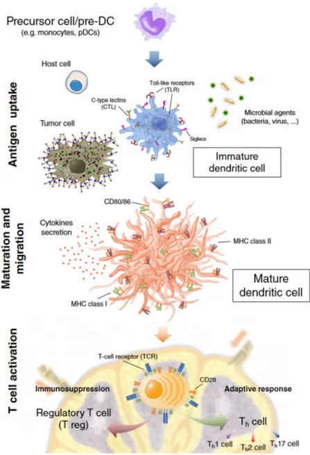
2002;Qi et al., 2006;Harwood and Batista, 2010) (Fig. 1.1).
Morphologically, DCs are relatively large cells with varying numbers of
heterogeneously shaped pseudopods. Given these variety of shapes these cells were
48
Fig. 1.1 – Basic hallmarks and functions of dendritic cells (DCs). InCrespo HJ et al. “Dendritic cells: a spot on sialic acid”,
Frontiers in Immunology (4), 2013.
1.1.1.2.1 DC subtypes and their hematopoietic differentiation
Different DC subsets are widespread throughout the organism, inhabiting different
organs and tissues such as the liver, spleen, thymus, gastrointestinal tract or skin, and
many more, phenotypically adapted to the tissue they reside. DC cell surface present
higher expression levels of MHC class II and co-stimulatory molecules, such as
49
macrophages or B cells. This fact is highlyrelevant for their antigen-presenting function
(van der Valk et al., 1984;Wood et al., 1985;Landry et al., 1988).
DCs can also be phenotypically characterized by the expression of specific markers:
B220, CD103 and CD8 in mouse, and the Dendritic Cell-Specific Intracellular adhesion
molecule 3 (ICAM-3)-Grabbing Nonintegrin (DC-SIGN), CD123, CD1c/Blood Dendritic
Cell Antigen-1 (BDCA-1), CD141/BDCA-2 and BDCA-3 and -4 in human (Dzionek et
al., 2000;MacDonald et al., 2002;Wollenberg et al., 2002;Robbins et al., 2008;Haniffa et
al., 2009;Bachem et al., 2010;Crozat et al., 2010;Jongbloed et al., 2010;Poulin et al.,
2010;Reizis et al., 2011;Haniffa et al., 2012;Langlet et al., 2012;Satpathy et al.,
2012;Lundberg et al., 2013;Plantinga et al., 2013). CD11b and CD11c are also DC
markers widely used on mouse DC phenotyping that are also human DC markers, but
not exclusively.
In the absence of infection and inflammation, DCs are functionally divided in two major
types: migratory and non-migratory, or lymphoid, tissue-resident DCs (reviewed in
(Shortman and Naik, 2007;Collin et al., 2013)). A classic example of the former are
dermal DCs and Langerhans cells that mainly reside in skin tissues. Upon contact with
antigen, they mature and migrate to the draining lymph nodes (Silberberg et al.,
1974;Ross et al., 1994;Ebner et al., 1998). The non-migratory DCs (like spleen or
thymus DCs) reside in secondary lymphoid organs, where they constantly screen
blood or lymph for pathogens (Ardavin, 1997;Henri et al., 2001). Both the canonical
myeloid and lymphoid hematopoietic progenitors contribute to the steady-state DC pool.
As a common trait, the expression of the Fms-like tyrosine kinase 3 (Flt-3) molecules
is characteristic of DC precursors, regardless of the myeloid or lymphoid lineage
and DCs development is driven by Flt3-ligand (Flt-3L) (Manz et al., 2001;D'Amico
and Wu, 2003;Shigematsu et al., 2004;Shortman and Naik, 2007;Watowich and Liu,
50
conventional DCs are marked by the exclusive expression of the DNGR-1 (Schraml et
al., 2013) which could, thus, function as a true lineage DC marker.
Opposed to the steady-state conventional DCs, some DCs are classified as
inflammatory or infection-derived DCs. These populations include the plasmacytoid
DC (pDC) population, a first line of defense against microbial invasion. Functionally
specialized in the detection of viral infections, pDCs develop a fully differentiated DC
phenotype after infection and secrete type I interferon (Grouard et al., 1997;O'Keeffe et
al., 2002). Other inflammatory DCs of interest are the monocyte-derived DCs (moDCs)
(Segura and Amigorena, 2013)). MoDCs are one of the most relevant, well-established
human DC model, especially in inflammation/pathogenic studies (Romani et al., 1996).
This comes as the result of two factors: its relative ease to obtain from monocytes present
in peripheral blood(Chapuis et al., 1997;Caux and Dubois, 2001) and the presence of
all canonical DC phenotypical and functional properties (maturation markers,
endocytosis, allogeneic and/or syngeneic T-cell stimulation) (Akagawa, 1994;Romani et
al., 1996;Palucka et al., 1998;Santin et al., 1999).
In mouse, the main DC model is the Bone Marrow-derived Dendritic Cells
(BMDCs), allowing the reproduction of the differentiation of different DC subsets, by
using different cytokine cocktails (Inaba et al., 1992;Shurin et al., 1997;Zhang et al.,
1997;Yamaguchi, 1998;Lutz et al., 1999;Zhang et al., 1999;Brasel et al., 2000).
In the work here presented, these will also be the two DC models of choice as they
are the best models that generally represent the DC populations and common functions
of all DC subtypes above referred.
1.1.1.2.2 Endocytosis
A fundamental function of DCs is to capture pathogens and then trigger the adaptive
response against them, as previously stated. Thus DCs serve a critical frontline function
51
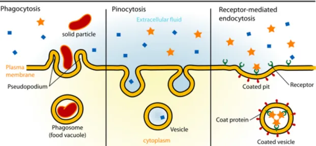
encountered antigens (Sallusto et al., 1995). The three principal means by which DCs
capture antigens are: 1) receptor-mediated endocytosis, in which particles are
endocytosed after a specific cell-surface receptor recognition; 2) macropinocytosis, or
the non-selective endocytosis of solutes, a constitutive process and the major source of
antigens for presentation by DCs (Norbury, 2006); and 3) phagocytosis, the uptake of
large molecules or cells, including virus, bacteria, protein clusters, apoptotic and necrotic
cells, which also involves specific membrane receptors (Fig. 1.2).
The uptake of foreign antigens usually triggers activation signals that will lead DCs
into a mature phenotype that maximizes the potential for antigen presentation and
stimulation of the adaptive response immune cells.
Fig. 1.2 – Types of endocytosis.
Receptor-mediated endocytosis is also fundamental in the maintenance of the
self-tolerance mechanisms. At steady-state, self-antigens are endocytosed and posteriorly
presented by DCs, but the endocytosis of self-antigens does not usually induce
significant maturation changes (Wilson et al., 2003).
Defective maturation contributes to turn DCs tolerogenic and promoting
regulatory T cell responses. Nevertheless, very small foreign and more common
52 1.1.1.2.3 Maturation
DC maturation is the sum of all the phenotypical and functional changes
occurring upon encounter with immune stimuli (i.e., antigens, cytokines, etc.) and it
is crucial to enable DCs to effectively activate T cells. It is characterized by rapid
downregulation of the antigen-uptake process, acidification of lysossomal
compartments, higher expression of MHC II molecules and of CD80 and CD86
co-stimulatory molecules, de novo or up-regulated synthesis of DC-specific
inflammatory cytokines (Thomas and Lipsky, 1994). All these maturation and
migration-changes are necessary hallmarks to enable DCs to perform antigen
presentation and boosting T and B cell responses (Sallusto and Lanzavecchia, 1994). It
is also known that the molecular nature of uptaken antigens, as well as the cytokines to
which DCs are exposed during the uptake process, are responsible for the modulation
of the maturation process. This ultimately influences the differentiation of the DC-pulsed
T cells into functionally distinct subtypes, actively shaping a future active or tolerance
response.
1.1.1.2.4 Migration
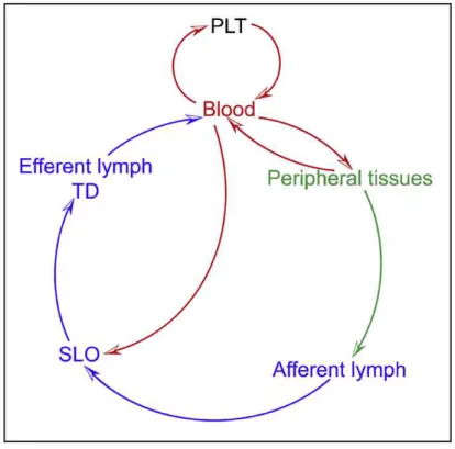
DC migration comprises the whole of the trafficking of DCs: their hematopoiesis on
bone marrow, their entry in circulation via blood towards the peripheral tissues and, from
here or directly from blood, to the secondary lymphoid organs (Fig. 1.3).
The homing of conventional or inflammatory DCs loaded with antigens to T cell niches
(normally, secondary lymphatic organs) is a crucial step for the setting of effective
immune responses. This process is characterized by chemokine-mediated
cell-recruitment to the lymphoid target site and activation of the surrounding tissues
(von Andrian and Mempel, 2003;Bonasio and von Andrian, 2006;Forster et al., 2012).
53
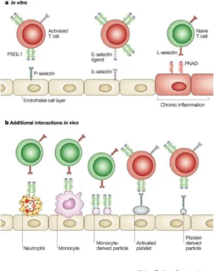
expression of several adhesion molecules, of which integrins and selectins and its
ligands are the most relevant elements.
Fig. 1.3 - Programmatic Outline of DC and DC-Precursor Trafficking Routes. DCs develop from precursors that originate from primary lymphoid tissues (PLT) such as the BM and the thymus. Precursors and committed DCs enter the circulation
and seed peripheral tissues and secondary lymphoid organs (SLOs). From peripheral tissues, they can access afferent
lymph upon receiving a mobilization signal and travel to the draining LN. Leukocytes leave LNs via the efferent lymph and
are collected in the thoracic duct (TD), which eventually guides DCs and their precursors back into the circulation. In
Alvarez D.et al., Mechanisms and Consequences of Dendritic Cell Migration, Immunity, 29, 2008.
DCs also migrate to non-lymphoid tissues, although this form of migration process
and causes are, still, not totally clear. A steady-state setting is an unfavorable
environment for DCs or DC precursors to migrate to non-lymphoid peripheral tissues.
Although the rolling and tethering of DCs are observable in both steady-state and
inflammatory contexts, migration to non-lymphoid tissues requires many of the adhesion
molecules (intracellular adhesion and vascular adhesion molecules (ICAMs and
VCAMs), integrins) normally involved in migration and homing phases that are ordinarily
induced by inflammatory mediators (Robert et al., 1999b;Merad et al., 2002;Pendl et al.,
54
precursors’ migration in the steady state (or inflammation-suppressed pathologies, like
several neoplasias) could prove to be of extreme importance in DC-based therapies,
where this DC attribute needs further improvement.
1.1.1.2.5 Cytokine production
DCs provide the lymph node-based naive T helper (Th) cells with two pathogen-related
signals: the information about the structure of pathogen’s antigens and its pathogenicity.
This comes as the result of the endocytosis and subsequent antigen-processing during
homing towards the lymph nodes. Upon arrival in the lymph nodes, mature DCs can
then present the tissue-derived pathogen information to provide T cells via three
signals: an Ag-specific ‘signal 1’ – MHC:T cell receptor triggering; a ‘signal 2’, resulting
from co-stimulatory stimulus, transmitting information regarding pathogenic potential;
and a ‘signal 3’, comprising cytokine production (Howard et al., 2002;Corthay, 2006).
The latter provides a first, determinant impulse towards one of the Th profiles, namely, T
helper type 1 or 2 (Th1 or Th2) – responsible for the stimulation of a specific cellular and
humoral response (respectively) –, T helper 17 (Th17) – stimulating anti-microbial
mechanisms on epithelial and mucosal barrier tissues –, or regulatory (Treg) cells –
inducing, as the name suggests, immune tolerance towards elicited antigens (Cools et
al., 2007).
In very general terms, IL-12 and IL-4 cytokines are the most relevant cytokines
involved in T cell priming, with IL-12-producing DCs priming Th1 responses, and the non-IL-12-producing DCs priming Th2 responses (Maldonado-Lopez et al., 1999). The
main drivers onto Th1 are inflammatory, monocyte-derived CD11c+ DCs (Macatonia
et al., 1995;Rissoan et al., 1999), and CD11c- cDCs into T
55
The Th1-skewing cytokine IL-12 is the best explored, most relevant
pro-inflammatory third signal in DCs (Trinchieri, 1995), with IFN-γ, IL-4, IL-10, PGE2 and IFN-α activities intimately mediated by this cytokine (Gately et al., 1998;O'Garra, 1998;Wu et al., 1998). Its main inducers are lipopolysaccharide (LPS), artificially
synthesized poly(I)poly(C) or CD40L (Macatonia et al., 1995;Cella et al., 1996;Cella et
al., 1999).
Modulation of IL-12 expression also can be achieved by the action of chemokines,
either positively, such as in the lymphoid-resident DCs in the mouse model (Aliberti et
al., 2000), or negatively, as in the case of human DCs (Braun et al., 2000). DC contact
with most pathogens induces IL-12 production. In contrast, maturation stimuli such as
TNF-α, IL-1, cholera toxin, FasL, fungal hyphae or nematode products seem to have no impact in this cytokine secretion (Braun et al., 1999;d'Ostiani et al., 2000;Gagliardi et al.,
2000;Rescigno et al., 2000;Whelan et al., 2000). IL-12 production can be potently
induced by CD40L, which is expressed at high levels on activated memory T cells (Cella
et al., 1996;Heufler et al., 1996). In terms of activation kinetics, DCs only respond within
8 to 16 hours after challenge with IL-12-inducing stimuli. This activation lag ensures a
regulation of Th1 and Th2 polarization, avoiding unwanted responses (Langenkamp et
al., 2000;Lanzavecchia and Sallusto, 2000;Tanaka et al., 2000).
The Th2-skewing in DCs profile is characterized by secretion of low levels of
IL-1α/β, IL-6, IL-13 or OX40L, but, also importantly, by the absence of IL-12 (Rincon et
al., 1997;Flynn et al., 1998;McKenzie et al., 1998;Rissoan et al., 1999), resulting from
DC contact withlow stimulatory antigens. Recently, it was shown a normally tolerated
antigen became susceptible of a Th2 response after infection with a strong Th1-elliciting
pathogen (Brimnes et al., 2003;Dahl et al., 2004), a fact that further stresses how
56
DCs are also known to actively participate in the induction of Treg cells (Fairchild
and Waldmann, 2000;Steinman et al., 2000). The induction of tolerance, more than the
result of a dedicated, tolerogenic DC lineage or immature DCs, occurs after DC contact
with micro-environmental factors (mostly antigens), inducing DCs to a
tolerogenic-skewing profile. Good examples of these tolerogenic factors, corroborating this
hypothesis, are:
- Non-immunogenic antigens present in respiratory and digestive tracts
(Vermaelen et al., 2001) Antigens from specific pathogens - i.e. Plasmodium
spp. antigens;
- Some self-antigens, such as insulin (Chen et al., 2003);
- Altered “self” peptides (Wildbaum et al., 2002) or self-antigens resulting
from regular cell turnover (Huang et al., 2000).
Tolerance induction may also originate in pro-inflammatory DCs, as well as specialized
subsets/tissue-resident of DCs. For example:
- Activation of DCs that secrete 10 (an immunoregulatory cytokine), but not
IL-12, can direct naïve T cells to a Treg subtype (Akbari et al., 2001;McGuirk et al.,
2002), as in the case of APCs from the liver (Khanna et al., 2000) and Peyer’s
patches (Iwasaki and Kelsall, 1999) secreting high levels of IL-10, selectively
inducing Treg or Th2 cells, respectively;
- In vitro culture of mouse bone marrow progenitor cells in the presence of
GM-CSF, TNF-α and IL-10 induces the differentiation of a distinct subset of dendritic
cells, CD11clow CD45RB+ DCs, similar to a tolerogenic subset
naturally/physiologically present in the spleen and lymph nodes of normal mice
(Wakkach et al., 2003). These DCs were shown to secrete high levels of IL-10
after activation. Functional studies also showed that CD11clow CD45RB+ DCs
57
- The murine CD103+ cDCs, present in hepatic, skin, kidney, intestinal and lung
tissue (Johansson-Lindbom et al., 2005;del Rio et al., 2007;Jaensson et al.,
2008;Monteleone et al., 2008;Ginhoux et al., 2009;Schulz et al., 2009;Desch et
al., 2011;Scott et al., 2011;Murakami et al., 2013;Yu et al., 2013) were proven to
be relevant Treg inducers via a TGF-β and retinoic acid (RA) mechanism
(Coombes et al., 2007). This mouse subset has recently been shown to be the
functional homologue of the human CD141+ DCs (Jongbloed et al.,
2010;Haniffa et al., 2012;Kelly et al., 2013), with similar Treg induction
mechanisms, namely and mainly, via TGF-β with RA also playing a part (Mucida et al., 2007; Nolting et al., 2009).
As an exogenous factor, IL-10 is, thus, the key factor for the induction of
tolerogenic DC in vitro and in vivo, but other cytokines and factors produced in the
peripheral tissues, such as PGE2, TGF-β, RA or endocrine factors should also be taken considered on the generation of tolerogenic DCs (Wilbanks et al., 1992;Hosoi et al.,
1993).
More recently, a newly defined lineage of T cells called T helper 17 (Th17) cells,
was identified with protection functions against some bacterial and fungal
infections (Korn et al., 2009) and with a relevant role in autoimmune disorders, such as
multiple sclerosis (Correale and Farez, 2012).
Th17 cells may be induced by DCs as the result of a combination of IL-6, low
concentration of TGF-β, IL-23, or IL-21 (Acosta-Rodriguez et al., 2007;Korn et al.,
2007;Nurieva et al., 2007;Zhou et al., 2007;Volpe et al., 2008;Yang et al., 2008a;Hu et
al., 2011), with IL-1 also known to enhance this process mediated by the transcription
factor RORγt and the signal transducer and activator of transcription 3 (STAT3) (Bettelli et al., 2006;Mangan et al., 2006;Veldhoen et al., 2006;Manel et al., 2008;Yang et al.,
58
However, IL-6 action may be a lineage dependent factor for Th17 induction. Although
both splenic DCs and intestinal lamina propria DCs induce differentiation of naive T cells
into Th17 cells, splenic DCs do so independent of IL-6, whereas in the presence of LP
and skin CD103+ DCs, IL-6 is required to induce T
h17 lineage cells. This comes as a
result of the necessity of IL-6 to counteract the anti-Th17 differentiation effect of RA and
high concentrations of TGF-β, both known to be produced by skin and intestinal lamina
propria (LP) CD103+ DCs (Coombes et al., 2007;Mucida et al., 2007;Manel et al.,
2008;Nolting et al., 2009) but not by spleen DCs (Hu et al., 2011).
1.1.2
Pathogen recognition by dendritic cells
Pathogen recognition by DCs depends on the identification of distinct microbial
patterns, not present in mammalian cells, but shared by most of the pathogenic microbial,
known as ‘pathogen-associated molecular patterns’ (PAMPs) (Schnare et al.,
2000;Netea et al., 2004). These patterns include bacterial and viral unmethylated CpG
DNA, bacterial flagellin, peptides containing N-formylmethionine residues,
lipoteichoic acids and double-stranded and single-stranded viral RNA. A
substantial part of PAMPs are glycan-containing ones, such as lipopolysaccharide,
N-acetylglucosamine, peptidoglycan, terminal fructose- and mannose-containing
glycans, and glucan-containing cell walls from fungi.
PAMPs are recognized by specific receptors named ‘pattern recognition receptors’
(PRRs), with functions aggregating endocytosis and intracellular signaling. Examples of
PRRs expressed by DCs include Scavenger receptors, NOD-like receptors and
C-type lectins. However, among the most widely studied are the Toll-like receptors
(TLR), a growing family of 12 evolutionary conserved PRRs consisting of type I integral
membrane glycoprotein with relevant role in the microbial response. The outcome of TLR
59
antigen presentation molecules (MHC class II molecules), co-stimulatory molecules
(CD80/86, CD40), inflammatory and/or antiviral cytokines (such as TNF-α, IL-12, IL-23, IFNα/β) and chemokines (i.e., IL-8, RANTES) (Takeda et al., 2003;Dzopalic et al., 2012), thus enacting a powerful response against pathogenic microbes.
C-type lectins (CLRs) are another very relevant family of PRRs expressed by DCs
(Zelensky and Gready, 2005). As lectins, their main function is the recognition of
glycan structures and, in an immunological context, they recognize
pathogen-associated glycans or glycosylated self-antigens. In DCs, some CLRs of note include the
DC-Specific Intracellular adhesion molecule-3 Grabbing Non-integrin (DC-SIGN),
CD207/Langerin, the Selectin family (discussed below), the Macrophage
Galactose/N-acetyl-galactosamine-specific Lectin (MGL1), Mannose Receptor
(MR), DEC205, the Blood DC antigens 2 (BDCA 2), the Dendritic Cell
Immunoreceptor (DCIR), the Dendritic Cell Immunoactivating receptor (DCAR) and
Dectin-1/2/3. In contrast to TLRs, all of these CLRs functionally bind glycan structures
expressed by mammalian cells (except for Dectin-1/2/3 that apparently only recognizes
fungal and/or mycobacterial glycans), a fact that demonstrates its potential role in both
host and pathogen recognition (van Kooyk and Rabinovich, 2008). CLRs can also
recognize and internalize pathogens for presentation without inducing DCs’ maturation.
In fact, the CLR-mediated antigen uptake doesn’t necessarily elicit a factual immune
response, and may instead contribute to induce immunological tolerance (Figdor et al.,
2004). A downside of these phenomena is the potential immune escape of pathogens
recognized via CLRs (van Kooyk et al., 2004;van Gisbergen et al., 2005;van Kooyk,
2008;van Kooyk and Rabinovich, 2008).
Like CLRs, the Sialic acid-binding immunoglobulin-like lectins (Siglecs) can also
recognize pathogens’ glycoproteins and glycolipids thus also contributing to the host’s
innate immune responses. Siglecs specifically recognize sialic acid-containing
60
et al., 2004;Crocker, 2005;Crocker et al., 2007;Kawasaki et al., 2013). The biological and
immunological relevance of CLR and Siglec receptors will be discussed in detail in later
sections.
DCs can also recognize and internalize microbes and its derivate particles by
receptors that bind to opsonins in opsonized (“coated”) microbes. Opsonization of
microbes can occur in two forms: by coating with complement proteins or by binding of
antibodies to antigens expressed on their surface. DC recognition of opsonized microbes
is thus mainly mediated by complement receptors and Fc receptors and assures the
capture of pathogens that might otherwise evade recognition by other DC receptors
(Sedlik et al., 2003;Ben Nasr et al., 2006).
Summarizing, DCs can interact in different ways with microbes, as well as with the
host antigens, through a panoply of receptors. This recognition initiates mechanisms that
will induce or suppress a specific immune response. DC recognition is thus considered
to be of great relevance for the development of a suitable, specific immune outcome,
dictating the balance tolerance/reactivity of the developing host-pathogen response.
1.1.3
Dendritic cells-based therapy
The current knowledge of DC immunobiology allowed several biotechnological and
pharmaceutical companies to develop based immunotherapies. Applications for
DC-based therapy include a plethora of pathologies ranging from infectious and
hypersensitivity diseases to malignancies.
The development of moDC cell culture protocols (Romani et al., 1994;Jonuleit et al.,
1997) allowing its relatively easy obtention, as well as its pro-inflammatory properties,
makes them the preferred DC subset for autologous therapy applications. One strategy
is the ex vivo upload of moDCs with the antigen to turn them able to efficiently develop
61
et al., 2013;Mintern et al., 2013;Phanse et al., 2013;Yao et al., 2013). The best example
of this strategy, with growing media attention, is the vaccination of cancer patients
with DCs loaded with tumor antigens, the Provenge® (sipuleucel-T) vaccine from Dendreon pharmaceutical company, targeting prostate cancer, being the most prominent
example.
Other approaches include the use of specific antibodies targeting DC endocytic
receptors that are used to force the upload of specific antigens towards that receptor.
Antibodies are also used to block specific receptor-ligand interaction and consequent
downstream signaling, counteracting, for instance, the negative immunomodulatory cues
of the tumor microenvironment.
DCs have also been studied as vectors for DNA vaccines encoding for antigens (Cao
et al., 2013). Viral transduction of DCs not only targets antigens to these cells, but also
induces intracellular pathways to modulate the immune response (Liechtenstein et al.,
2013).
All these relatively recent drug-niche that exploits DC unique immune potential is proof
of reconnaissance of DCs’ cornerstone role in the immune system. Nevertheless,
DC-based therapies still face several hindrances until their full application, most notably
having a relatively low overall efficacy (Dougan and Dranoff, 2009;Draube et al., 2011).
The low efficacy is mostly derived from the lack of full knowledge regarding DC biology,
including the pathogenesis/tolerance balance and their migratory mechanisms. The