Unravelling the Evolution of the
Allatostatin-Type A, KISS and Galanin Peptide-Receptor
Gene Families in Bilaterians: Insights from
Anopheles Mosquitoes
Rute C. Felix1, Marlene Trindade1, Isa R. P. Pires2, Vera G. Fonseca1¤, Rute S. Martins1, Henrique Silveira2, Deborah M. Power1, João C. R. Cardoso1*
1Comparative Endocrinology and Integrative Biology, Centre of Marine Sciences, Universidade do Algarve, Campus de Gambelas, 8005–139, Faro, Portugal,2Centro de Malária e outras Doenças Tropicais, UEI Parasitologia Médica, Instituto de Higiene e Medicina Tropical, Universidade Nova de Lisboa, Rua da Junqueira 100, 1349–008, Lisboa, Portugal
¤ Current address: Zoological Research Museum Alexander Koenig (ZFMK), Centre for Molecular Biodiversity Research, Adenauerallee 160, 53113 Bonn, Germany.
*jccardo@ualg.pt
Abstract
Allatostatin type A receptors (AST-ARs) are a group of G-protein coupled receptors acti-vated by members of the FGL-amide (AST-A) peptide family that inhibit food intake and development in arthropods. Despite their physiological importance the evolution of the AST-A system is poorly described and relatively few receptors have been isolated and func-tionally characterised in insects. The present study provides a comprehensive analysis of the origin and comparative evolution of the AST-A system. To determine how evolution and feeding modified the function of AST-AR the duplicate receptors inAnophelesmosquitoes, were characterised. Phylogeny and gene synteny suggested that invertebrate AST-A receptors and peptide genes shared a common evolutionary origin with KISS/GAL recep-tors and ligands. AST-ARs and KISSR emerged from a common gene ancestor after the divergence of GALRs in the bilaterian genome. In arthropods, the AST-A system evolved through lineage-specific events and the maintenance of two receptors in the flies and mos-quitoes (Diptera) was the result of a gene duplication event. Speciation ofAnopheles mos-quitoes affected receptor gene organisation and characterisation of AST-AR duplicates (GPRALS1 and 2) revealed that in common with other insects, the mosquito receptors were activated by insect AST-A peptides and the iCa2+-signalling pathway was stimulated.
GPRALS1and2were expressed mainly in mosquito midgut and ovaries and transcript abundance of both receptors was modified by feeding. A blood meal strongly up-regulated expression of bothGPRALSin the midgut (p<0.05) compared to glucose fed females. Based on the results we hypothesise that the AST-A system in insects shared a common origin with the vertebrate KISS system and may also share a common function as an inte-grator of metabolism and reproduction. Highlights: AST-A and KISS/GAL receptors and ligands shared common ancestry prior to the protostome-deuterostome divergence. OPEN ACCESS
Citation:Felix RC, Trindade M, Pires IRP, Fonseca VG, Martins RS, Silveira H, et al. (2015) Unravelling the Evolution of the Allatostatin-Type A, KISS and Galanin Peptide-Receptor Gene Families in Bilaterians: Insights from Anopheles Mosquitoes. PLoS ONE 10(7): e0130347. doi:10.1371/journal. pone.0130347
Editor:Christian Wegener, University of Würzburg, GERMANY
Received:December 9, 2014
Accepted:May 19, 2015
Published:July 2, 2015
Copyright:© 2015 Felix et al. This is an open access article distributed under the terms of the Creative Commons Attribution License, which permits unrestricted use, distribution, and reproduction in any medium, provided the original author and source are credited.
Data Availability Statement:All relevant data are within the paper and its Supporting Information files.
Funding:This study was co-financed by the Foundation for Science and Technology, Portugal (FCT) project PTDC/BIA-BCM/114395/2009 and the European Regional Development Fund (ERDF) COMPETE - Operational Competitiveness Programme and Portuguese funds through FCT–
Phylogeny and gene synteny revealed thatAST-ARandKISSRemerged afterGALRgene divergence.AST-ARgenes were present in the hemichordates but were lost from the chor-dates. In protostomes,AST-ARspersisted and evolved through lineage-specific events and duplicated in the arthropod radiation. Diptera acquired and maintained functionally diver-gent duplicateAST-ARgenes.
Introduction
Type A allatostatins (AST-As) are a family of insect peptides with a conserved C-terminal FGL-amide motif. They were initially isolated from the cockroachDiploptera punctata[1,2] but are widespread in insects and are mainly detected in the brain and midgut [3–12]. AST-A peptides arise by proteolytic cleavage of a common prohormone precursor and a variable num-ber of peptides of differing lengths have been identified [13–16]. In cockroaches (D.punctata
[1,2],Blattella germanica[17],Periplaneta americana[2]), cricket (Gryllus bimaculatus) [18],
locust (Locust migratoria) [19] and the termite (Reticulitermes flavipes) [20], AST-A peptides inhibit juvenile hormone (JH) secretion by thecorpora allata(CA) but they have numerous other physiological roles including the regulation of food intake in many different insects [13,14,16,21–29]. InB.germanicainjections of AST-A reduce food intake [21]. InDrosophila
melanogastergenetic manipulation of neurons expressing AST-A repress food intake and
responsiveness to sugar [23] and ablation of AST-A and its receptor (DAR-1) significantly reduce larval foraging behaviour in the presence of food [27]. The actions of AST-As on feed-ing are associated with their anti-myotropic actions on insect gut motility and regulation of digestive enzyme activity [29–35].
AST-A peptides activate specific G-protein coupled receptors (GPCRs), the insect allatosta-tin-A receptors (AST-ARs) that are considered orthologues of galanin receptors (GALR) in vertebrates [36–41]. In vertebrates, GALRs have a close evolutionary relationship with kisspep-tin receptors (KISSR) and are activated by galanin (GAL) and spexin (SPX), peptides that are unrelated to insect AST-As [40–42]. AST-A peptide function is relatively well studied but the receptors have only been isolated in a few insect species and their evolution and function is unresolved [11,38,43–46]. In the fruit flyD.melanogastertwo receptors, DAR-1 and DAR-2 have been de-orphanized [36,38,44,47,48] but in most insects only a single receptor gene exists [49,50]. The beetleTribolium castaneumis the exception as it lacks both AST-A and the recep-tors [51–53]. In contrast, in the nematode,Caenorhabditis elegans, an orthologue of the insect AST-ARs was characterised (npr-9) [39], and two putative AST-A peptide encoding genes
(nlp-5andnlp-6) were also identified [41,54,55]. All the studies of AST-A to date suggest that
its role in feeding behaviour emerged early during its evolution and have probably been main-tained during the Ecdysozoa radiation [14,22,25].
Functional specialisation of the AST-A system appears to have occurred in the insects. For example, in larvalD.melanogasterDAR-1 is mainly present in the central nervous system (CNS) and DAR-2 is detected in the gut [56]. Comparison of AST-A activation of DAR-1 and DAR-2 reveals differences in binding and intracellular signalling in the presence of Pertussis toxin (PTX), an inhibitor of Gi-type G-protein activity [47]. In the mosquitoAnopheles
gam-biae(PEST strain) genome, duplicate AST-ARs also exist [50] and microarray data for blood fed females suggests that they are also functionally distinct as only theD.melanogasterDAR-1 orthologue is up-regulated 3 h after a blood meal [57].
Evolution of Allatostatin Type A Receptors and Ligands
and RSM are in receipt of FCT post-doctoral grants SFRH/BPD/89811/2012, SFRH/BPD/80447/2011 and SFRH/BPD/66742/2009, respectively. JCRC is supported by an auxiliary research contract FCT Pluriannual funds attributed to CCMAR under the project PEst-C/MAR/LA0015/2013 and UID/Multi/ 04326/2013.
The present study characterises the origin and evolution of AST-AR and their peptide ligands in arthropods and by isolating and characterizing the duplicate receptors inAnopheles mosquitoes determines if evolution modified receptor function.Anophelesmosquitoes are vec-tors of the malaria parasite and more than 400 species have been identified [58]. The genomes
ofAnophelesspecies are rapidly evolving and they have been used as models of how the
envi-ronment and geographic isolation favour speciation and have modified gene structure and function [59–63]. Phylogeny coupled to gene synteny analysis revealed that the arthropod AST-ARs and AST-A peptides shared a common evolutionary origin with the KISS/GAL systems and that AST-AR and KISSR members probably emerged from the same gene after duplication of the AST-AR/KISSR/GALR ancestor. In arthropods, AST-ARs evolved under lineage-specific pressure and in theAnophelesmosquito speciation affected receptor gene evo-lution. Characterisation of the duplicate AST-ARs fromAnopheles coluzzii, formerly known as
A.gambiae M-form [59], revealed their sequences diverge and their response to a blood meal
differs. The results of the present study are used to develop a model for the evolution of the AST-A system.
Material and Methods
In silico
database mining
AST-ARgenes were identified and retrieved from 21 arthropod genomes available in the
Ensembl metazoan database (http://metazoa.ensembl.org/index.html, January 2014) by con-ducting similarity searches with the deduced mature sequence ofDrosophila melanogaster DAR-1 (FBgn0028961) and DAR-2 (FBgn0039595) and using database gene annotations. The arthropod genomes analysed encompassed three different classes: Insecta, Arachnida and Branchiopoda. The representatives of the Insecta class included members of six orders: Diptera
(D.melanogaster,Megaselia scalaris,Anopheles gambiae,Anopheles darlingi,Aedes aegyptiand
Culex quinquefasciatus); Lepidoptera (Bombyx mori,Heliconius melpomeneandDanaus
plex-ippus); Hymenoptera (Nasonia vitripennis,Apis mellifera,Atta cephalotesandSolenopsis
invicta); Hemiptera (Acyrthosiphon pisumandRhodnius prolixus); Phthiraptera (Pediculus
humanus), and Coleoptera (Tribolium castaneumandDendroctonus ponderosae). Other
arthropod representatives included in this study were members of two Arachnidan orders: Ixo-dida (Ixodes scapularis) and Trombidiformes (Tetranychus urticae)and one representative of the Branchiopoda,Daphnia pulex. Searches for the arthropodAST-Agene were also performed using the deduced mature protein sequence of theD.melanogasterAST-A precursor
(FBgn0015591) and genome annotations.
The reference mosquito genome used forin silicostudies was theA.gambiaePEST strain, which is an M and S chimera [62,64,65]. The assemblies of 20 otherAnophelesgenomes were analysed and included several members of theA.gambiaecomplex (A.arabiensis,A.gambiae S-form,A.merus,A.quadriannulatus,A.melasandA.coluzzii, formerly known asA.gambiae M-form [59]) available from VectorBase (https://www.vectorbase.org/, March 2015).
Sequence comparisons and phylogenetic analysis
Multiple amino acid sequence alignments of receptors were generated using ClustalW (v2) (http://www.genome.jp/tools/clustalw/) software. Conserved sequence motifs were identified and the percentage of receptor amino acid sequence similarities was calculated in GeneDoc [66].
deuterostomes, acorn worm (Saccoglossus kowalevskii), purple sea urchin (Strongylocentratus
purpuratus), amphioxus (Branchiostoma floridae) and the tunicate (Ciona intestinalis).
Deu-terostome KISSR and GALR receptor sequences were obtained from [41] and [67]. Trees were constructed using an alignment of the deduced amino acid sequence from transmembrane (TM) region 1 to 7 (TM1 to 7) including intra and extracellular loops (S1 Table) submitted to ProtTest 2.4 to select the best statistical model to study receptor protein evolution according to the Akaike Information Criterion (AIC) [68].
Phylogenetic trees were constructed using maximum likelihood (ML) and neighbour-join-ing (NJ) methods and bootstrapped to assign measures of accuracy to the clades [69]. Trees were constructed with a total of 128 sequences and rooted with the vertebrate GPR151 receptor cluster (12 sequences). The ML analysis was built in PhyML 3.0 available from ATGC (http:// www.atgc-montpellier.fr/phyml/) [70] using a JTT substitution model with the following parameters: 4 gamma distributed rate categories, a fixed proportion of invariant sites (0.009) and a fixed gamma shape parameter (1.294). Reliability for internal branching was assessed using 100 bootstrap replicates and the aLRT SH-like test [71]. Sequence data was also analysed using the NJ method [72] with 1000 bootstrap replicates in MEGA version 5.1 [73]. The NJ tree was constructed using the pairwise deletion for gaps/missing data treatment option and fixed gamma 4 distributed rate categories (gamma = 1.294) to account for heterogeneity across sites. The consensus trees obtained with ML and NJ analysis shared similar topologies.
Gene synteny analysis
Gene synteny was carried out using the ENSEMBL BioMart comparative tool (http://metazoa. ensembl.org/biomart/martview/). The regions upstream and downstream ofA.gambiae
AST-ARsandAST-Alocus were used to identify homologue genome regions in other insects
(D.melanogasterandT.castaneum) and in the nematodeC.elegansgenomes. Genes in a 10
Mb segment flankingGPRALS1andGPRALS2inA.gambiaechr 2R and a 3 Mb region
flank-ingAST-AinA.gambiaechr 2R were used to searchD.melanogaster,T.castaneumandC.
ele-gansgenomes. Conserved genes flankingAST-ARandAST-Aacross invertebrates were used to establish synteny with humanKISSRandGALRgenome regions. The identity and evolutionary relatedness of the flanking genes identified was confirmed using sequence similarity searches against the human, insect and nematode genome assemblies and confirmed with phylogenetic analysis when necessary. Orthologues of the genes identified in the humanKISSRandKISS/
GAL/SPXparalogon were also mapped in the insect and nematode genomes [42,74].
Ethics statement
All animal experiments were performed at the Centro de Malária e outras Doenças Tropicais, Instituto de Higiene e Medicina Tropical (IHMT, Lisbon). This study was approved by the IHMT committee on the ethics for animal experiments and by the Direção-Geral de Veteri-nária, Ministério da Agricultura do Desenvolvimento Rural e das Pescas, Portugal licences (id approvals: 023351 and 023355). Animal experiments were carried out in strict accordance with Portuguese law and following the guidelines for care and use of laboratory animals. All the authors that performed animal manipulations were licensed to conduct research using labora-tory animals.
Mosquito rearing
Mosquitoes from a laboratory colony ofA.gambiae(Yaoundé strain), recently renamedA.
coluzzii[59] were maintained under standard insectary conditions. Temperature was
maintained at 26 ± 1 °C, humidity at 75% and a 12:12 h light:dark cycle were used in all experi-ments. Adult mosquitoes were allowed to feedad libitumon a 10% glucose solution.
Feeding and tissue collection
A.coluzziifemales were food deprived for 3 h and then fed for 30 min on either a 10% glucose
solution or a blood meal (obtained from anaesthetized 6–8 week old CD1 mice,Mus musculus). Whole mosquito females and dissected tissues (midguts, fat bodies, ovaries and heads) of 20 glucose and/or blood fed mosquitoes from three independent experiments were collected and stored in RNA later at -20°C for RNA extraction.
RNA extractions and cDNA synthesis
Total RNA (tRNA) from wholeA.coluzziior specific tissues was isolated using a kit (total RNA Kit I, Omega, VWR, Portugal) and genomic DNA was eliminated by treating with 1 U DNase (DNA-free Kit, Ambion, UK) for 30 min at 37 °C. DNase treated tRNA (500 ng) was denatured at 65 °C for 5 min, quenched on ice for 5 min and used for cDNA synthesis in a 20μl reaction volume containing 10 ng of pd(N)6 random hexamers (GE Healthcare, UK),
2 mM dNTPs, 100 U of MMLV-RT and 20 U RNasin Plus RNase inhibitor (Promega, Spain). The cDNA was synthesized for 10 min at 25 °C followed by 60 min at 42 °C and 70 °C for 10 min. Due to the small amounts of RNA extracted from the ovaries, cDNA synthesis was per-formed using a single pool of RNA extracted from the 3 experimental groups. The quality of the synthesised cDNA was assessed by PCR amplification of a reference gene, ribosomal pro-tein S7 sub-unit. The thermocycle used was: 95°C for 3 min; 35 cycles of 95°C for 30 s, 60°C for 30 s, 72°C for 30 s, followed by 72°C for 5 min. PCR reactions were carried out in a final reac-tion volume of 10μl and contained 1.5 mM MgCl2(Thermo Scientific, Portugal), 0.2 mM
dNTP’s (GE Healthcare, Spain), 0.25μM of gene specific primer pairs (S2 Table) and 0.5 U of
DreamTaq DNA Polymerase (5 U/μl, Thermo Scientific, Portugal).
Quantitative Polymerase Chain Reaction (q-PCR)
Quantitative real-time PCR (q-PCR) was used to quantify the expression of AST-ARs and of AST-A in femaleA.coluzziimidgut, fat body, head and ovaries when feed with glucose and blood meals. Specific primers were designed using the cloned receptor transcripts (S2 Table). Primers to amplifyAST-Awere designed based on the sequence retrieved fromA.gambiae PEST AGAP003712 that is 100% identical to the EST clone (CR530883) isolated from a head cDNA library of a species from theA.gambiaecomplex. Expression of zinc carboxypeptidase A1 (CP, AGAP009593, a gut specific marker of the digestive process) [75] and the vitellogenin-1 precursor (Vtg, AGAP004203, a protein biomarker of egg production [76]) were also
analysed.
Duplicate q-PCR reactions (<5% variation between replicates) were amplified in a StepOne
Plus Real-Time PCR Detection system (Applied Biosystems, Portugal) for 96-well microplates (Bio-Rad, Portugal). Analysis was performed in a final reaction volume of 10μl that contained
300 nM of forward and reverse primer, SsoFast EvaGreen supermix (Bio-Rad, Portugal) and 2μl of template cDNA (1:5). Optimized cycling conditions consisted of 95°C for 30 s, followed
subunit (S7, AGAP010592) [77,78] and mitochondrial solute carrier family 25 (MC, AGAP001297) [79].
Receptor cloning and mammalian cell transfections
The full-length ofA.coluzzii GPRALS1andGPRALS2were amplified by PCR using proofread-ing DNA polymerase (iProof, BioRad, Portugal) and the specific primers designed usproofread-ing the sequence predicted in ENSEMBL (AGAP003658,GPRALS1and AGAP001773,GPRALS2;S2 Table). Receptors were amplified from cDNA obtained from whole femaleA.coluzziiusingPfu proofreading DNA polymerase (Promega, Spain). The thermocycle used was: 2 min at 95 °C, 35 cycles (95 °C for 1 min, the appropriate annealing temperature (°C) for 45 s, 72 °C for 4 min) and a final extension step of 10 min at 72 °C. PCR products were sequenced to confirm their identity and cloned into pGEM T easy vector (Promega). The purified PCR products were ligated into pcDNA3.1/V5-His TOPO TA expression vector (Invitrogen, USA). The com-pleteD.melanogasterDAR-2-RA (FBtr0085316) was also isolated and amplified from adult cDNAs and cloned into a HindIII/NotI digested pcDNA3.1/V5-His TOPO TA vector.
The amplified insect receptors included the initiation and termination codons but were not cloned in frame with vector tag proteins. The recombinant constructs were used to transfect mammalian CHO cells that had been maintained in complete Dulbecco’s modified Eagle’s medium (DMEM, Sigma, Spain) containing 4.5 g/L glucose, 110 mg/L sodium pyruvate and L-glutamine and supplemented with 10% sterile foetal bovine serum and 0.1% penicillin: strepto-mycin antibiotic mix (10.000 U: 10 mg/ml, Sigma) and 250μg/ml sterile filtered amphotericin
B solution (1:100 dilution, Sigma, Spain). One day prior to transfection, 2–3 x10⁵cells were seeded into 6 well plates (Sarstedt, Portugal) and transfected using Fugene 6 reagent (1: 6 DNA: Fugene, Roche, Germany) according to the manufacturer’s protocol. The efficiency and success of cell transfection was estimated by performing a simultaneous transfection with a plasmid encoding a fluorescent protein. 72 h after incubation, transformed cells were selected by supplementing medium with 800μg/ml of the antibiotic Geneticin (G418 sulphate,
Gib-coBRL, USA) and cell recovery was monitored daily and the medium changed until no cell death was observed. Establishment of AST-AR CHO stable cell lines were confirmed by PCR using receptor specific primers.
Peptides
TheA.gambiaeAST-A1 (SPKYNFGL-NH2) and AST-A2 (LPHYNFGL-NH2) peptides were
chemically synthesized. Peptide sequences were deduced from AGAP003712 by comparing
withD.melanogaster(FBgn0015591) and theA.aegypti(U66841) AST-A precursors [80,81].
A.gambiaepeptides were 100% identical to the deduced peptide sequence ofA.coluzzii
(Yaoundé strain) transcripts and to the gene homologues of other members of theA.gambiae complex and were designated Ano_AST-A1 and Ano_AST-A2. Peptides with a purity>95%
(ChinaPeptides, China) were diluted in 1 × PBS buffer for the cAMP and iCa2+assays. Other peptides used included: the German cockroachBlattella germanicaBLAST-2 peptide (DRLYSFGL-NH2, kindly donated by Dr. Maria Dolors Piulach, CSIC-UPF, Barcelona, Spain
[17]), 1–29 rat galanin (Sigma-Aldrich, Spain) and the vertebrate KISS peptides, sea bass KISS 1–10 (YNLNSFGLRY-NH2) and KISS 2–10 (FNFNPFGLRF-NH2) (kindly donated by Dr Ana
Gomez, CSIC-IATS, Spain [82]).
Receptor signalling
GPRALS1 and GPRALS2 stable CHO cell lines were stimulated with Ano_AST-A1 and Ano_AST-A2 peptides, theB.germanicaBLAST-2 peptide and rat GAL and sea bass KISS
peptides and iCa2+release and cAMP production were measured. TheB.germanicaBLAST-2 peptide shared an identical amino acid (aa) sequence withD.punctataAST-5 that stimulates iCa2+mobilization by bothD.melanogasterreceptors [47].
For iCa2+release (Relative Fluorescent Units, RFU) a Ca2+sensitive fluorescent dye Fluo-4 NW (Molecular Probes, Invitrogen) was used. Approximately 50,000 cells were assayed per well and the variation in fluorescence after addition of increasing peptide concentrations (1 nM to 1μM, diluted in the assay buffer) was measured every 5s over a total of 2 min and
fluorescence was excited using a 485/20 nm filter and captured with a 528/20 nm filter in a Bio-tek Synergy 4 plate reader (BioBio-tek, USA). Background RFU of transfected cells was measured prior to peptide stimulation. The controls consisted of transfected cells exposed to carbachol (100 nM; Sigma, Spain) and non-transfected CHO cells stimulated with 1μM of each peptide
(negative control).
The amount of cAMP produced was determined using a competitive immunoassay with a cryptate labelled anti-cAMP antibody (Cisbio, France) and following the manufactures proto-col. Approximately 15,000 cells were assayed per well and peptide incubations were performed in a final reaction volume of 20μl in white 384 well small Volume HiBase Polystyrene
micro-plates (Greiner, Germany). Prior to the assay, cells were ressuspended in 1 x PBS with 1 mM of 3-isobutyl-1- methylxantine (IBMX, Sigma) and incubated for 5 min at 37°C. Peptides were diluted in 1 x PBS/ 1 mM IBMX and were added to the cells for 30 min at 37°C in a CO2
incu-bator before measuring with 620/10 and 665/8 nm filters in a Biotek Synergy 4 plate reader (Biotek, USA). All assays were performed in triplicate on three independent occasions.
Statistical analysis
Quantitative expression data is presented as mean ± SEM of cDNA from 3 independent experi-ments analysed in duplicate. Significant changes in transcript expression were assessed using a nonparametric Mann-Whitney two-tailed test. Receptor activation is presented as the
mean ± SEM of three independent experiments carried out in triplicate and statistical signifi-cance was assessed using a Kruskal-Wallis test with Dunn’s Multiple Comparison Test. All the analyses were performed in Prism GraphPad version 5 and statistical significance was consid-ered atp<0.05.
Results
AST-AR and peptides in arthropods
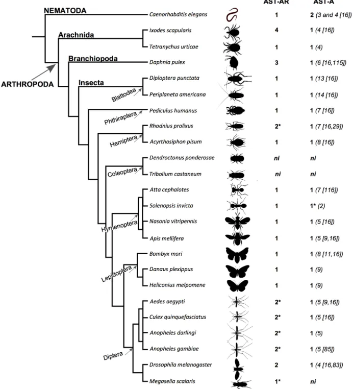
Searches performed in arthropod genomes identified 30 putativeAST-ARand 18 putative
AST-Agenes (Fig 1,S1 Table). The results obtained indicated that receptor gene number across
arthropods was very variable but that the number of genes encoding the peptides was con-served. In common withDrosophila melanogaster, two receptor genes were identified in Culici-dae including the malaria vectorAnopheles gambiaePEST strain, the yellow fever mosquito
Aedes aegyptiand the southern house mosquitoCulex quinquefasciatus. InAnopheles darling
genome two receptors homologues of theA.gambiaeAST-AR genes were identified. In other Diptera representatives (11Drosophilaspecies:D.ananassae,D.erecta,D.grimshawi,D.
moja-vensis,D.pseudoobscura,D.persimilis,D.sechellia,D.simulans,D.virillis,D.willistoniandD.
yakuba) two receptors were also identified (data not shown). The exception was the
identified: silkwormBombyx mori, monarch butterflyDanaus plexippus, postman butterfly
Heliconius melpomene, honey beeApis mellifera, jewel waspNasonia vitripennis, red fire ant
Solenopsis invicta, leaf-cutter antAtta cephalotes, pea aphidAcyrthosiphon pisumand human
Fig 1. Number of predicted AST-AR and peptide genes identified in arthropods andC.elegans.Accession numbers are available inS1 Table. The number of AST-A peptides is indicated within brackets and references are provided. TheT.urticae,D.plexippus,H.melpomene,S.invictaandA.darlingi
AST-A peptides were predicted by comparison with the insect homologues and identification of the C-terminal FGL-amide motif.*indicates species in which a putativeAST-ARpseudogene (orthologue of the third CulicidaeAST-ARgene) was identified. Data fromD.pulexandA.cephalotesobtained from [115,
116].
doi:10.1371/journal.pone.0130347.g001
licePediculus humanus. The exception was the Coleopterans; noAST-ARgenes were retrieved from the genome of the red flour beetleTribolium castaneumor the mountain pine beetle
Den-droctonus ponderosae. In the branchiopodDaphnia pulex, 3 genes were recovered. In the
ara-chnidanIxodes scapularis4AST-ARgenes were identified and in the red spider mite
Tetranychus urticaeonly a single receptor gene was identified (Fig 1).
In the genomes ofA.gambiae(AGAP001774) andA.aegypti(AAEL006077) a third
AST-ARgene that mapped close to, and was more likeGPRALS2but had a different orientation
(antisense) was found. Orthologues were identified inA.darlingi(deduced from Scaffold_ 1464) andC.quinquefasciatus(CPIJ011118) and also in the genomes ofM.scalaris (MESCA004796) andR.prolixus(RPRC004705) (S1 Table). The predicted insect receptor sequences encoded 3 or less TM domains and were excluded from the analysis. Despite strenu-ous efforts it was not possible to identify full-length genes and the sequences may represent pseudogenes arising from species-specific genome events (S1 Table).
In arthropods a singleAST-Agene was identified in the genomes of all species analysed with the exception of the two beetle genomes that lacked the genes (Fig 1). The number of mature AST-A peptides was highly variable across insects. Cockroaches had the most numerous AST-A (13 inDiploptera punctataand 14 inPeriplaneta americana[16]) and the Diptera and Arachnida had the fewest AST-A (4 peptidesin D.melanogaster[16,83] andIxodes scapularis [16] and 5 peptides in mosquitoes [9,16]) (Fig 2).
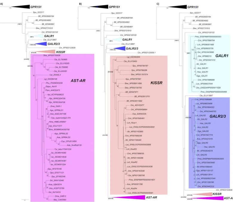
Phylogeny of AST-AR
Phylogenetic analysis suggested that in arthropods gene duplications and deletions affected AST-AR evolution. Orthologues ofD.melanogasterDAR-1 in other Diptera were highly con-served but the duplicate receptors were highly divergent (Fig 3). A cluster of receptors that included DAR-1 and mosquito orthologues was identified but no equivalent cluster existed for DAR-2. In contrast, species-specific expansion of AST-ARs gene number occurred inR.
pro-lixus(2 receptors),D.pulex(3 receptors) andI.scapularis(4 receptors) (Fig 3A). The tree
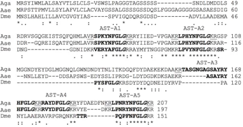
topology of arthropod AST-ARs with homologues in other metazoans including the nematode Fig 2. AST-A peptide precursor inA.gambiae.The deduced sequence of AST-A inA.gambiae(Aga, PEST) was obtained from the AGAP003712 gene and confirmed using EST data. TheA.aegypti(Aae, AAEL015251,[81]) andD.melanogaster(Dme, FBgn0015591,[48]) orthologues were used for comparisons. The predicted mature peptides are highlighted in bold and the Gly residues processed to the C-terminal amide in mature AST-A’s are indicated in italics.
Caenorhabditis elegans, the annelid,Capitella teleta, the mollusc,Lottia giganteaand the early deuterostomeSaccoglossus kowalevskiisuggested that they all shared a common ancestor.
The arthropod and other invertebrate AST-ARs tended to cluster in the phylogenetic trees with the protostome and deuterostome KISSR group rather than the GALRs (Fig 3). Paradoxi-cally, the dipteran receptors (D.melanogasterandA.gambiae) shared slightly higher sequence identity/similarity with human GALR1 compared to KISSR1 (Table 1). The KISSR (Fig 3B) Fig 3. Phylogeny of the AST-AR with the KISSR and GALR.Phylogenetic analysis was performed using the ML method and three subsets of the same phylogenetic tree showing the expansion of the different family members (A, B and C) are represented to facilitate interpretation. Only bootstrap support values above 50% are indicated. In the most important receptor family nodes statistical support was established using the aLRT SH-like test and is indicated (bootstrap method/ aLRT SH-like test). The deducedA.darlingi(Scaffold_325) was not used, as the receptor sequence was very incomplete and only 3 TM domains were predicted. The phylogenetic tree was rooted with the vertebrate GPR151 cluster (12 sequences). Species names and accession numbers of the receptor genes are available inS1 Table.
doi:10.1371/journal.pone.0130347.g003
and the GALR (Fig 3C) clusters contained representatives from several vertebrate and inverte-brate lineages including annelids, mollusc and early deuterostomes.
AST-AR and peptide gene synteny
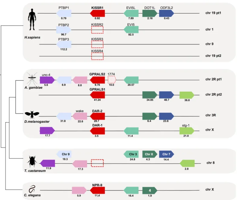
The gene environment of insect receptor and peptide genes was compared withC.elegansand human (Figs4and5). The genes in linkage withAST-ARinA.gambiaeandD.melanogaster were compared to the homologue genomic regions of humanGALR (GALR1, chr 18;GALR2, chr 17 andGALR3, chr 22), humanKISSR1(chr 19) andC.elegans npr-9(chr X) (Fig 4,S3 Table). InA.gambiae GPRALS1andGPRALS2genes were localised on chr 2R, while in theD.
melanogasterthey mapped to chr X (DAR-1) and chr 3R (DAR-2), although gene synteny was
retained. The genome arrangement ofA.gambiaeandD.melanogasterchromosome regions containingAST-ARssuggested that they underwent distinct evolutionary pressure after gene duplication. The conserved gene linkage between the insect and the nematodeC.elegans ortho-logue regions suggested that duplication ofAST-ARoccurred after the divergence and radiation of the nematodes.
None of the genes flanking insectAST-ARswere identified in the humanGALRs loci. In contrast, neighbouring genes that flanked protostomeAST-ARgenes mapped to the human
KISSR1chromosome paralogon (Fig 4). Members of 4 gene families (Polypyrimidine tract
binding protein,PTBP; ecotropic viral integration site 5 proteins,EVI5; DOT1-like histone H3K79 methyltransferase proteins,DOT1L; and outer dense fiber of sperm tails 3 protein,
ODF3L) flanked the humanKISSR1gene on chromosome 19,A.gambiae AST-ARson chr 2R,
andD.melanogaster DAR-1on chr X andDAR-2on chr 3R. TheAST-ARgenome region in
the nematodeC.eleganscontained members of 3 gene families linked to humanKISSR1and insectAST-AR(Fig 4). The conserved gene environment that flankedAST-ARsinC.elegans,A.
gambiae,D.melanogasterandKISSR1in human was absent from theT.castaneumgenome
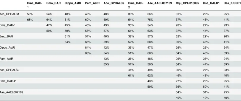
Table 1. Sequence identity and similarity of insect AST-ARs with human GALR1 and KISSR1.
Dme_DAR-1
Bmo_BAR Dippu_AstR Pam_AstR Aco_GPRALS2 Dme_DAR-2
Aae_AAEL007169 Cqu_CPIJ013095 Hsa_GALR1 Hsa_KISSR1
Aco_GPRALS1 59% 54% 48% 49% 48% 39% 66% 31% 29% 25%
68% 64% 61% 60% 59% 54% 75% 37% 46% 41%
Dme_DAR-1 47% 45% 45% 43% 35% 54% 28% 27% 23%
59% 59% 58% 57% 51% 63% 37% 44% 37%
Bmo_BAR 51% 51% 46% 38% 57% 32% 29% 26%
64% 65% 59% 52% 68% 39% 46% 41%
Dippu_AstR 84% 42% 35% 47% 26% 26% 24%
88% 54% 51% 60% 34% 45% 39%
Pam_AstR 43% 36% 48% 26% 26% 24%
55% 51% 59% 34% 44% 39%
Aco_GPRALS2 44% 49% 39% 27% 23%
61% 62% 46% 48% 40%
Dme_DAR-2 43% 27% 29% 25%
59% 36% 50% 41%
Aae_AAEL007169 34% 31% 25%
40% 49% 40%
Percentages were calculated using the full-length amino acid sequence of the receptors.
(Fig 4). The genes in linkage withAST-ARin Diptera were distributed between chromosome 8, 9, 7, 3 and X inT.castaneumthat lacks theAST-ARgenes. The conservation on the beetle chr 8 of a greater number of genes from the dipteranAST-ARbearing chromosomes suggests it may be the homologue chromosome.
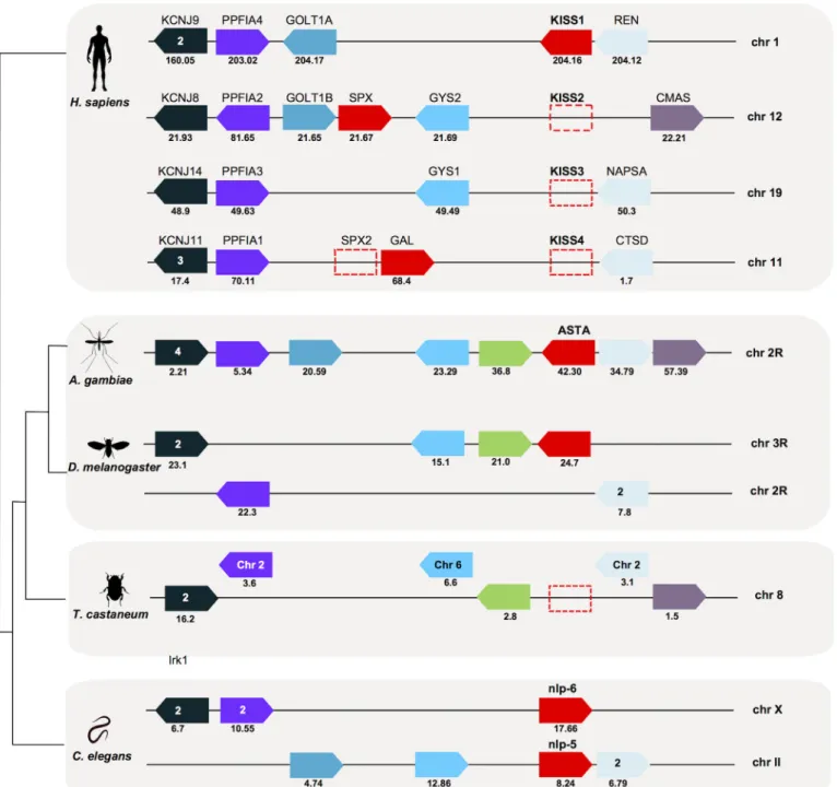
The genome region ofAST-Agenes inA.gambiaeandD.melanogasterwas also compared with the humanKISS/GAL/SPXparalogon (chr 1, 11, 12 and 19) [42,74] and with theC.elegans chromosome that contained thenpl-5(chr II) andnpl-6(chr X) genes (Fig 5,S4 Table). InA. Fig 4. Conserved gene synteny of theA.gambiae,D.melanogasterandC.elegans AST-ARgenome regions with the humanKISSR1
chromosomes.Conservation forT.castaneumis also shown. Horizontal lines represent chromosome fragments and block arrows indicate genes and orientation in the genome. Orthologue genes are represented in the same colour and their position (Mb) is indicated. An arrow with red stripes represents the putativeAST-ARpseudogene (AGAP001774) localized nearGPRALS2. Dotted boxes represent the absent humanKISSRgenes (that emerged during early vertebrate tetraploidizations) [67,74] and theT.castaneum AST-ARgene. Note that the mosquito 2R and human ch19 have been divided into two parts (pt1 and pt2) to facilitate visualization. Only shared genes are represented. The number of family members that map to the same chromosome is indicated and the closest toAST-ARandKISSR1is represented. A full description of gene families and names and accession numbers is given inS3 Table.
doi:10.1371/journal.pone.0130347.g004
gambiae,AST-Ashared the same chromosome localisation as theGPRALSs(chr 2R) and mapped closest toGPRALS1. A similar situation occurred inD.melanogasterandC.elegans and the gene encoding the peptide was also localised on the same chromosome as the receptors.
InD.melanogaster,AST-Amapped nearDAR-2on chr 3R and inC.elegansnearnpr-9on chr
Fig 5. Conserved gene synteny of the genome regions containingAST-Agene inA.gambiae,D.melanogasterandC.elegans(npl-5andnpl-6) compared to the humanKISS/GAL/SPXchromosomes.The gene homologues inT.castaneumare also represented. Horizontal lines indicate chromosome fragments and coloured arrow identify genes and their orientation in the genome. Orthologue genes are indicated in the same colour and their positions are indicated below (Mb). Dotted boxes represent the absent humanKISSandSPX2genes (that emerged during early vertebrate
tetraploidizations) [42,74] and theT.castaneum AST-Agene. Only shared gene family members are represented. The number of family members that map to the same chromosome is indicated and those closest toAST-AandKISS/GAL/SPXare represented. A full description of gene families and names and accession numbers is given inS4 Table.
X. Members of metazoan gene families (inward rectifying potassium channel superfamily, KCNJ; LAR protein-tyrosine phosphatase-interacting protein,PPFIA,PTPRFandLiprin; Golgi TransportGOLT; glycogen synthase,GYS; aspartic protease family,REN,NAPSA,CTSD,
CathD; and members of the N-acylneuraminate cytidylyltransferase,CMAS) were conserved in
the region flanking theAST-Agene inA.gambiae,D.melanogasterand the humanKISS/GAL/ SPXparalogon. Representatives of five genes families that were in linkage with humanKISS/
GAL/SPXand insectAST-Awere split between chr X that containednpl-6and chr II that
con-tainednpl-5inC.elegans. Conservation of genes that flankedAST-AandKISS/GAL/SPXgenes suggests that they shared a common evolutionary origin (Fig 5). InT.castaneumthese genes mapped to different chromosomes including chr 8.
AST-ARs
and
AST-A
genes in
Anopheles
mosquitoes
GPRALS1(1137 bp) andGPRALS2(1080 bp) were isolated fromA.coluzziiwhole female
cDNA and the deduced proteins shared 48% aa identity.A.coluzziiGPRALS1 shared greatest aa sequence identity withD.melanogasterDAR-1 (59%) and with the orthologues from other arthropods such asBombyx mori(54%),Diploptera punctata(48%) andPeriplaneta americana (49%). The deduced protein sequence ofA.gambiaeGPRALS2 shared 43% and 44% aa identity
withD.melanogasterDAR-1 and DAR-2, respectively (Table 1).
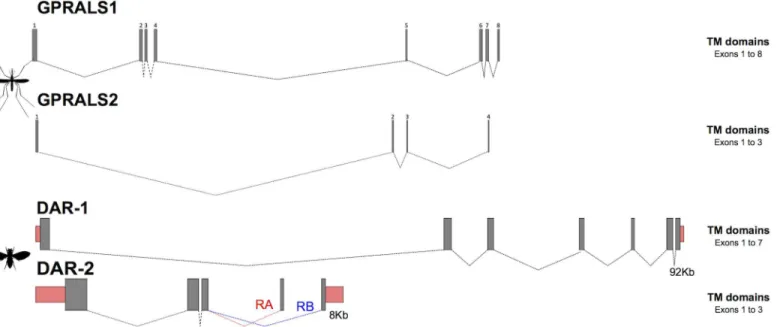
In theA.gambiaegenome assembly, the twoAST-ARgenes had a different gene organisa-tion and number of predicted transcripts. Two alternative transcripts of the same length
(GPRALS1-RAandGPRALS1-RB) were predicted that shared 7 common exons but had a
dif-ferent exon 1 (S5 Table). The predictedGPRALS2gene structure was more complex and com-posed of 10 exons and alternative splicing was predicted to generate three almost identical transcripts:GPRALS2-RA;GPRALS2-RBandGPRALS2-RC. The transcripts shared the first exon but alternative splicing of 3 consecutive tandem duplicated clusters of three exons gener-ated three predicted proteins that shared 96–99% aa identity. To confirm gene predictions ESTs forA.gambiaewere analysed and a partial clone (BX620556) was identified that was identical toGPRALS2-RC. Other ESTs identified were very incomplete and the existence of multiple transcripts remains to be confirmed.
Characterization ofGPRALS1in the genomes of otherAnophelesmosquitoes revealed that receptor gene structure was conserved and thatGPRALS1was composed of 8 exons and
GPRALS2of 4 exons (S5andS6Tables,Fig 6). The exceptions were theGPRALS2genes in two
other species of theA.gambiaecomplex:A.arabiensis(Dongola strain) that had duplicated exons 2 and 3 andA.quadriannulatus(SANGQUA strain) in which exon 2 was duplicated. Furthermore, in common with theA.gambiaePEST, putativeGPRALS2-like pseudogenes that map close toGPRALS2but had a different orientation (antisense) were identified inA.
arabien-sisandA.quadriannulatusgenomes (S6 Table). This seems to be indicative of paracentric
inversions, a characteristic of chr 2R evolution [63,65,84].
The amplifiedA.coluzzii GPRALS1shared 98% and 99% nucleotide identity, respectively with the predictedA.gambiaePEST GPRALS1-RA and RB. The nucleotide sequence of theA.
coluzzii GPRALS2was 95% identical to theA.gambiae GPRALS2-RAand 99% identical to
GPRALS2-RBandGPRALS2-RC. In common withAnophelesmosquitoes, the duplicate
recep-tors inD.melanogasteralso had a different gene organisation (Fig 6). TheDAR-1gene had a higher number of exons thanDAR-2and the latter receptor gene generated two distinct tran-scripts via alternative splicing of the last exon [36,38,44].
A single gene that encoded 5 putative AST-A peptides was identified in allAnopheles mos-quito genomes (data not shown). The AGAP003712 deduced mature AST-A peptides all had a conserved C-terminal FGL-amide motif and were of different lengths: Ano_AST-A1 and
Ano_AST-A2 were 8 aa’s; Ano_AST-A3 was 17 aa’s; Ano_AST-A4 was 7 aa’s and Ano_ AST-A5 was 9 aa’s (Fig 2) [85]. TheD.melanogasterAST-A precursor (151 aa) generated four peptides (Dme-AST-1 to Dme-AST-4) [16,83,86] and was shorter than theA.gambiaeAST-A precursor (207 aa). The Ano_AST-A1 and Ano_AST-A2 shared 50% aa identity with Dme_ AST-A1 and 75% and 87% aa identity with Dme_AST-A2, respectively. Ano_AST-A2 only dif-fered at a single amino acid position (His3) from Dme_AST-A2 peptide (Val3) (Fig 2). The remaining peptides Ano_AST-A3, Ano_AST-A4 and Ano_AST-A5 shared little similarity with the otherD.melanogasterAST-A peptides.
Comparisons of the dipteran AST-ARs and AST-A with vertebrate
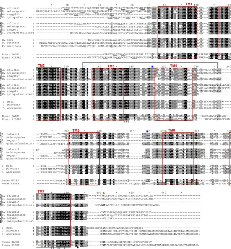
orthologues
Sequence comparisons of the dipteran AST-ARs were performed with other insects to identify motifs characteristic of the paralogue receptors and similarities with the human orthologues. The duplicate insect AST-ARs shared highly conserved TM domains and divergent N- and C-terminal domains. Both dipteran AST-AR paralogues and other insect AST-ARs shared con-served structural and functional motifs with the vertebrate KISSR1 and GALR1 (Fig 7). This included the seven transmembrane regions and several conserved motifs [87,88]: i) a short N-terminal domain, ii) a DRY/F motif between TM3 and intracellular loop 2, and iii) a
NSxxNPxxY motif within TM7. Putative N-glycosylation (N-x-T/S) motifs important for the structure of the N-terminus were also predicted. In addition, the characteristic conserved cyste-ine residues of the vertebrate KISS/GAL receptors that may form a disulphide bond between ECL1 (between TM2 and TM3) and ECL2 (between TM4 and TM5) were also conserved in insect AST-ARs.
The amino acid motifs important for peptide affinity in GALR, His264in TM6 and His267, Glu271and Phe282before TM7, were not conserved in the insect AST-ARs with the exception Fig 6. Gene organisation of the AST-A receptors inAnophelesandD.melanogaster.The structure of theAnophelesreceptor genes was deduced from the consensus organisation obtained from several mosquito genomes (S5andS6Tables). TheD.melanogaster AST-ARsgene organizations were obtained from ENSEMBL. In theA.gambiaePEST genome duplicated exons highly similar in sequence toGPRALS1(exon 1) andGPRALS2(exon 2, 3 and 4) are predicted and are not represented. Closed boxes represent exons and dashed lines introns. Mosquito exons are numbered and exons encoding the UTR are represented by pink boxes. Gene structures were built using FancyGene 1.4 software. The figure is not drawn to scale.
Fig 7. Sequence conservation of the duplicate dipteran AST-ARs with the insect and human orthologues.The predicted seven transmembrane domains are boxed in red and numbered. Potential sites for N-glycosylation are underlined in the N-terminal region and two conserved motifs D-R-Y/F localized after TM3 and NSxxNPxxY within TM7 are annotated with asterisks [87,88]. Two conserved cysteine residues that may form a disulphide bond were identified are connected by a line; predicted residues involved in protein kinase C phosphorylation are indicated by a blue square and potential protein A phosphorylation sites are annotated by a green diamond; C-terminal cysteine residues for potential palmitoylation after TM7 are denoted in italics and indicated with an orange pentagon. Amino acids important for binding of human galanin to GALR1 are indicated in red. The arginine residue important for the
of His264that was preserved in DAR-1 [89]. However, the Arginine (Arg) residue, localized in the C-terminal region after TM7, that has been linked with the role of the human KISSR1 receptor in precocious puberty, was conserved across all insects [90]. Consensus amino acid signalling motifs responsible for protein kinase C phosphorylation (T/SxR/L), protein kinase A phosphorylation (RxS/T) and the potential palmitoylation cysteine located shortly after TM7, were also conserved between insect AST-ARs and the human orthologues (Fig 7).
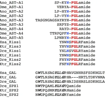
The dipteran AST-A peptides shared a C- terminal FGL-amide motif with the vertebrate KISS family and this region is essential for peptide binding and activation of the vertebrate KISSR (Fig 8, [91]). In addition, a conserved Asparagine (Asn) was also found in all insect AST-As and was shared by vertebrate KISS. The vertebrate GAL and SPX peptides shared no sequence conservation with insect AST-A peptides (Fig 8, [42]).
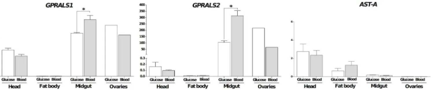
Tissue expression and effect of blood feeding in the female
A
.
coluzzii
GPRALS1andGPRALS2transcripts had an overlapping tissue distribution inA.coluzziiand
were most abundant in the midgut. The relative abundance ofGPRALS2was approximately
>1000 times higher thanGPRALS1suggesting that receptors may intervene in different
func-tions (Fig 9). Transcripts of both receptors were of very low abundance in the fat body and in function of human KISSR1 that is proximate to the end of TM7 is indicated in bold and circled. Shading denotes amino acid conservation and dark grey means 80% and black 100% conservation. Shading after TM4 was manually edited and did not considered the incomplete receptor regions*indicate incomplete mosquito receptor sequences. Accession numbers of receptor genes are available inS1 Table.
doi:10.1371/journal.pone.0130347.g007
Fig 8. Amino acid sequence alignment of the dipteran AST-A mature peptides with the vertebrate KISS, GAL and SPX family members.The highly conserved FGL motif between AST-A and KISS peptides is indicated in bold and red and conserved N residues in bold and blue. Sequence conservation of GAL and SPX is indicated in italics and totally conserved are in italics and bold. The vertebrate predicted peptide sequences were obtained from [74] and [42] and theXenopus laevismature galanin peptide deduced from EU446417.1.
the head and of higher abundance in the ovary. Expression ofA.coluzzii AST-Atranscripts were also characterized and were most abundant in the head compared to fat body, midgut and ovaries (Fig 9).
After blood feeding, tissue abundance ofAST-ARswas modified but no difference in abun-dance ofAST-Atranscripts was detected (Fig 9).GPRALS1andGPRALS2were both signifi-cantly up-regulated in the midgut (p<0.05) and down-regulated in the head and ovaries
(statistical analysis in the latter tissue could not be performed as only a single biological repli-cate of pooled ovaries was used). Expression of carboxypeptidase (CP) (p<0.01) and
vitello-genin (Vtg), well-established markers of physiological events triggered by blood feeding, were up-regulated in whole females 3h after a blood meal compared to glucose fed female mosqui-toes (S1 Fig).
Functional characterisation of the duplicate
A
.
coluzzii
receptors
The capacity of AST-A peptides to stimulate GPRALS1 and GPRALS2 was assessed by measur-ing the cAMP and iCa2+signalling response toAnophelesAno_AST-A1 and Ano_AST-A2 and
B.germanicaBLAST-2 peptides. None of the insect peptides induced increased intracellular
cAMP but there was a dose dependent increase in iCa2+mobilization (Fig 10). The twoA.
coluzziireceptors were activated byB.germanicaBLAST-2 and also by the homologue
AST-A peptides, suggesting that they are functional insect AST-A receptors. The potency of Fig 9. Tissue distribution and effect of a blood meal on the expression of the paralogueGPRALSandAST-Atranscripts in the femaleA.coluzzii. Expression was analysed in the head, fat body, midgut and ovaries 3 hours after a glucose (white bars) or blood (grey bars) meals. Receptor expression levels were normalized using the geometric mean of two reference genes (S7andMC). The results are represented as mean±SEM of three biological replicates with the exception of ovaries where only a single biological replicate was analysed (~60 ovaries). Prism GraphPad v5 software was used to assess the significance of differences between experimental groups using the Mann-Whitney (two-tailed) test (*p<0.05).
doi:10.1371/journal.pone.0130347.g009
Fig 10. Capacity of the insect AST-A peptides to activate theA.coluzziiGPRALS.The mosquito Ano_AST-A1 and Ano_AST-A2 peptides and the cockroach BLAST-2 peptide were tested at several different concentrations and the response of the receptor monitored by measuring concentrations of intracellular calcium (RFU). TheD.melanogasterDAR-2-RA receptor was used as a positive control: A) Response to BLAST-2 peptide (0.5μM to 0.005μM);
B) Receptor response to the presence of decreasing concentrations of Ano_AST-A1 and Ano_AST-A2 peptides (1μM to 1nM). A Kruskal-Wallis test with a
Dunn’s Multiple Comparison test was performed using Prism GraphPad version 5 software. No significant differences were found.
doi:10.1371/journal.pone.0130347.g010
Ano_AST-A2 (EC50= 1.47x10⁻⁸M) was greater than Ano_AST-A1 (EC50= 1.37x10⁻⁷M) for
GPRALS2. Both peptides also activated GPRALS1 however, since saturation of the peptide response was not achieved, accurate determination of peptide potency was not possible. Verte-brate GAL (rat 1–29 GAL) and KISS peptides (sea bass KISS 1–10 aa and 2–10 aa) failed to
acti-vateA.coluzziiGPRALS1 and GPRALS2.
Discussion
AST-As are a functionally important peptide family that regulates development, reproduction and feeding in insects [13,16,25]. In the present study putative AST-ARs were retrieved from the genomes of several arthropods and the origin and evolution of the receptors and their pep-tide ligands was analysed. The involvement of the duplicate AST-ARs in blood feeding was characterised inA.coluzzii. Comparative bioinformatics analysis revealed that AST-AR evolu-tion in arthropods was lineage-specific and that the receptors and peptide ligands emerged early in evolution and evolved in parallel with the KISS and GAL family members. Receptor gene duplication occurred in the ancestral bilaterian genome and the invertebrate AST-ARs share the same ancestral gene precursor that originated the KISSR members in lophotrochozo-ans, early deuterostomes and vertebrate genomes. In dipterlophotrochozo-ans, two AST-AR genes exist and characterization of the AST-AR duplicates revealed that their sequences diverged presumably as a result of different evolutionary pressures. The tissue distribution and abundance of AST-ARs in femaleA.coluzziiindicates that they probably acquired different functions but that together they probably integrate feeding and reproduction in common with what occurs in the vertebrate KISS system. We hypothesise that the regulatory function of the ancestral gene has been retained by the AST-A system in protostomes and the KISS system in vertebrates.
Evolution of AST-AR and AST-A in arthropods was lineage specific
In arthropods, a variable number ofAST-ARgenes and deduced AST-A peptides derived from a unique gene were identified suggesting that both receptors and peptides have evolved by line-age specific events. In the arachnidanI.scapularis, the branchiopodD.pulexand the insectR.
prolixusmultiple receptors exist and the paralogues are highly related in sequence as the result
of species-specific gene duplication. In the other arthropods a singleAST-ARgene was found. The exceptions were the genomes ofT.castaneumandD.ponderosaethat lack the AST-A sys-tem [51,52] and the dipteran genomes where two highly distinct AST-ARs co-exist. The origin of the two Diptera AST-ARs is intriguing and phylogeny and gene structure analysis suggests that after gene duplication the two receptors evolved under distinct evolutionary pressures. The divergence between the dipteran paralogues may be because they arose from a gene dupli-cation event early in the radiation of the insects or that AST-AR gene duplidupli-cation only occurred in Diptera and suffered considerable modifications in flies and mosquitoes after their diver-gence (>200 MYA, [46]).
In arthropods, adaptation to different ecological niches has modulated genome evolution and led to differential gene retention [63,84,92–95]. Deletions ofT.castaneum AST-ARand
AST-Agenes may be the result of a species-specific genome rearrangement. The factors
Analysis of several differentAnophelesgenomes suggests that in some species, theAST-AR gene structure was modified and that exon tandem duplication and exon inversions occurred during the radiation ofAnopheles. Modification inAST-ARgene structure has mostly affected
GPRALS2suggesting that speciation modified the evolution of this gene duplicate. Similar
mechanisms of gene evolution have also been described for other GPCRs involved in the regu-lation of development, feeding and reproduction in insects [60]. It was not possible to obtain DNA for theA.gambiaePEST strain to confirm experimentally the gene structure of AST-ARs and it remains to be established if heterozygosity of the original DNA source led to assembly artefacts that generated additional transcript copies.
The variable number of putative AST-A peptides encoded in each insect gene and their spe-cies-specific characteristics has functional implications that remain to be explained [16,24]. For example, in cockroaches the AST-A peptides identified have different potencies for inhibition of JH production by the CA and for stimulation of gut contraction [96,97]. In species with mul-tiple receptor encoding genes, theAST-Agene encodes fewer peptides and the inverse is also true. It is tempting to speculate that retention of multiple receptors or peptide encoding genes may be a mechanism to guarantee functional divergence but further studies are required to test this hypothesis.
AST-A system shared ancestry with KISS and GAL systems
Recently, based upon sequence and gene structure resemblance, eight bilaterian peptidergic signalling systems were identified [41]. Here, using a combination of molecular phylogeny and gene synteny analysis we identified a further peptidergic system and suggest that the inverte-brate AST-A and the KISS and GAL family shared a common origin prior to the protostome-deuterostome divergence (Fig 11).
To date the insect AST-ARs and nematode AST-AR-like were considered to be the homo-logues of the vertebrate GALRs [36,38,39,43]. In fact, sequence similarity searches revealed that AST-AR’s share highest sequence similarity with GALR and the role in feeding and energy metabolism of the AST-A system in nematodes and insects and the GAL system in mammals have been taken as evidence of their functional homology [14,23,36,37,39,98]. The results of the present study suggest thatAST-ARshared a common evolutionary origin with both
GALRsandKISSRbut the phylogenetic analysis and gene synteny analysis suggests that
AST-ARs are more related to KISSRs. Identification ofAST-AR,KISSRandGALRgenes in pro-tostome and deuterostome genomes implies that prior to their divergence the genes emerged. The non-identification of putativeAST-ARgenes in chordates indicates that they were subse-quently eliminated from the genome. We propose a new evolutionary model in which the ancestralAST-AR/KISSR/GALRgene duplicated and originated the ancestral gene precursor of
AST-ARandKISSRand the ancestralGALRgene in the bilaterian genome (Fig 11).
and its importance for AST-AR and KISSR activation [46,47,91] also suggests they are closely related.
In vertebrates, KISSR and the respective peptides are key players in reproduction and, in mammals this system acts upstream in the gonadotropic axis mediating gonadotropin secre-tion and regulasecre-tion of the onset of puberty [100,101]. KISS peptides are involved in the regula-tion of the metabolic control of fertility [102,103], regulation of food intake and fat mass production and food restriction increases sex hormone induced KISS1 expression in the adi-pose tissue of rats [104,105]. The abundance of AST-AR transcripts in the mosquito midgut and ovaries and the changes provoked by a blood meal may indicate that, in addition to molec-ular conservation with the KISS system, both insect and vertebrate systems may also share con-served physiological roles as integrators of metazoan metabolism and reproduction.
Fig 11. Proposed model for the origin and evolution of theAST-ARgenes.Circles with different colours represent the AST-AR (light blue), KISSR (green) and GALR (pink) family members. The tetraploidization events basal to vertebrate radiation (1R, 2R) and the teleost specific genome duplication (3R) are indicated. The circle with a cross indicates gene loss during evolution. Numbers within the circles indicate predicted gene numbers of each family. Gene number from early deuterostome and lophotrochozoa representatives were obtained from [41]. For simplicity, lineage-specific duplications are not indicated and the time line is not drawn to scale. Receptor mapping for the vertebrate ancestral chromosomes (VAC) was obtained from [42].
Functional conservation of the AST-A system across insects
TheA.coluzziiAST-ARs share a similar pharmacological profile with the other insect receptors
and activation by cockroach BLAST-2 peptide, that shares the conserved C-terminal FGL-amide, suggests this motif is essential for receptor activation. Despite the limitations of using heterologous expressions systems that inevitably express the receptors out of context our studies revealed that insect AST-A peptides are able to trigger activation of the mosquito recep-tor and stimulate iCa2+signalling but not cAMP. In contrast in thecorpus cardiacumofL.
migratoria,D.punctataAST-2 induced an increase in cAMP [19]. In fact functional
characteri-sation of the AST-AR in several insects revealed that the peptides trigger multiple signalling pathways and that the insect receptors can associate with different members of the G-protein complex [46,47,106]. For full functional characterisation of AST-ARs inAnophelesit will be important to also take into consideration the signalling pathways activated by different ligands.
The insect AST-ARs activate similar intracellular signalling pathways to the vertebrate KISSR and GALR homologues but if promiscuous interaction and activation of mosquito GPRALS with human peptides occursin vivois not yet known. In our study, incubation of GPRALS transfected CHO cells with the vertebrate KISS and GAL peptides (the cognate pep-tides of the homologue deuterostome receptors) failed to elicit activation of cAMP or iCa2+. Nonetheless, before a final conclusion can be reached about GPRALS activation the involve-ment of other intracellular signalling pathways (eg: PKC-MAPK/ERK) remains to be characterized.
The overall tissue distribution of the duplicate mosquitoGPRALSand ofAST-Ais similar to other insects and suggests that the AST-A system may play a key role in the regulation of food intake and reproduction [13,14,25]. InD.melanogasterboth peptide and receptors share over-lapping tissue distribution and are highly abundant in brain and midgut [11,14,21,86]. InD.
punctata,AST-ARis mainly expressed in the brain but it is also expressed in the ovaries and in
B.moriit is mostly detected in the midgut and is of low abundance in the head [11,45,107]. In
R.prolixusAST-A is abundant in the CNS and the receptor is detected in the CNS and in
dif-ferent gut regions [29,46]. The different expression levels ofGPRALS1andGPRALS2in the midgut and ovaries ofA.coluzziitend to support the notion that they have divergent functions.
InD.melanogaster, DAR-2 was mainly associated with gut function while DAR-1 was brain
specific [36,48,108]. In mosquito the relative importance of the duplicate receptors in physiol-ogy remains to be determined. Comparison of the AST-A system between male and female mosquitoes was not carried out in the present study but available data suggests that males express higher levels of the AST-A peptide precursor than females but no differences in recep-tor expression were found [109]. Future studies will be directed at defining the specific function of the duplicate receptors in male and female mosquitoes.
Mosquito receptors are responsive to a blood meal
A unique characteristic of theA.gambiaereceptors, in comparison to other insects, is their responsiveness to a blood meal suggesting that participation of the AST-A system in mosquito feeding and reproduction may be triggered by blood feeding.Anophelesmosquitoes are anau-togenous (feed on blood to reproduce) and during the first hours after a blood meal, the mos-quitoes undergo profound physiological, morphological and hormonal changes and significant up-regulation (p<0.05) of AST-ARs occurs. The change in receptor expression levels in
tis-sues involved in blood digestion (midgut,p<0.05) and reproduction (ovaries) suggests that
these receptors may be important mediators of the crosstalk between protein digestion and egg maturation in mosquitoes. InR.prolixus(hematophagous insect) AST-A was proposed to par-ticipate in blood digestion as transcript expression was modified by a blood meal and this
affected the amount of peptide available for release [29]. In contrast, a blood meal did not mod-ify transcript abundance ofA.coluzzii AST-Atranscripts, although it was not possible to assess if peptide levels changed or if feeding modified the ratio of the 5 predicted mosquito AST-A’s. It will be important to carry out further studies that assess peptide levels and the relative importance of the different forms in males and females.
In mosquito females, a blood meal stimulates the secretion of several proteolytic enzymes including carboxypeptidases (CP) from the midgut, which in female mosquitoes is at a higher concentration than in males [110,111]. The nutrient rich (proteins, lipids and carbohydrates) mammalian blood triggers vitellogenesis and restores energy levels for egg development [76,112,113]. Transcription ofVtg-1, an egg yolk precursor protein, is triggered by the blood meal and reaches a maximum 24 hours post-feeding [111]. In our study, 3 h post blood feeding, an increase inCPoccurred relative to glucose fed females (p<0.01) andVtg-1was
up-regu-lated, but to understand the physiological involvement of AST-AR and AST-A in blood protein digestion or egg maturation will require further studies.
Conclusion
The protostomeAST-ARandAST-Agenes emerged prior to the protostome-deuterostome divergence and their similar evolutionary trajectory is suggestive of co-evolution of receptors and their peptides in common with what has been suggested for the metazoan GnRH/AKH, CCK/SK, NMU/PK and Orexin/AT GPCRs and their peptide ligands [40,41,114]. The results of the present study indicate that they evolved in parallel with the KISS and GAL receptor and peptide family members. Evidence is presented that reveals thatAST-AR,GALRsandKISSR emerged from a common ancestral gene. Moreover, the analysis performed raises an alterna-tive hypothesis to that which proposes protostome AST-AR as the orthologue of vertebrate GALR. Instead the data indicates that the ancestral gene that originatedAST-ARalso gave rise
toKISSRand that this occurred after the divergence of aGALR-likegene. Duplication of
AST-ARoccurred during the arthropod radiation but in Diptera their divergence in sequence
and function indicates they underwent distinct evolutionary pressure. InA.coluzzii, both receptor transcripts were affected by a blood meal and, it remains to be established if in com-mon with the KISS system in vertebrates they regulate and integrate the link between metabo-lism and reproduction in insects.
Supporting Information
S1 Fig. Quantitative expression analysis of carboxypeptidase A and vitellogenin1 precur-sors in glucose fed and blood fed femaleA.coluzzii.The results are presented as
mean ± SEM of 3 experiments analysed in duplicate. Expression was normalized using the geo-metric mean of two reference genes (S7andMC). A Mann-Whitney (two-tailed) test was per-formed using Prism GraphPad version 5 software to evaluate if differences between groups were significant. Statistical significance is represented with(p<0.01).
(PDF)
S1 Table. Accession numbers of the predicted AST-AR and AST-A mature proteins and KISSR and GALR used in the study.scaffolds where genes were deduced; + putativeAST-AR
pseudogenes; # not used for phylogeny.1obtained from [45],2obtained from [67]. The annelid
(C.teleta), mollusc (L.gigantea), acorn worm (S.kowalevskii), purple sea urchin (S.
S2 Table. List of primers used for receptor cloning and real-time PCR expression analysis.
(PDF)
S3 Table. Human (H.sapiens),D.melanogaster,T.castaneumandC.elegansgenes ortholo-gues of theA.gambiae GPRALS1andGPRALS2gene environment on chr 2R.Accession numbers, chromosome positions, symbol and initial gene positions (base pair) are given. The data was extracted using Ensembl Biomart software and confirmed using sequence similarity searches. (PDF)
S4 Table. Human (H.sapiens),D.melanogaster,T.castaneumandC.elegansgene ortholo-gues of theA.gambiae AST-Agene environment on chr 2R.Accession numbers, chromo-some position, symbols and initial gene positions (base pair) are given. The data was extracted using Ensembl Biomart software and confirmed using sequence similarity searches.
(PDF)
S5 Table. Gene organisation of theGPRALS1inAnophelesmosquitoes.Mosquito genomes were accessed in VectorBase (https://www.vectorbase.org/, March 2015) and receptor gene structure was deduced by homology with theGPRALS1transcript. The number and approxi-mate size of the deduced exons (E) and introns (I) are given in base pairs (bp). E5 ofA.gambiae PEST is not predicted in the reference genome and was deduced by homology. E1 is duplicated
inA.gambiaePEST.Anophelesspecies in which sequence hits were found to short genome
scaffolds are not represented. ni—not identified,incomplete sequences.
(PDF)
S6 Table. Gene organisation of theGPRALS2inAnophelesmosquitoes.Mosquito genomes were accessed in VectorBase (https://www.vectorbase.org/, March 2015) and receptors gene structures were deduced by homology with theGPRALS2transcript. The predicted transcripts based on genome annotation are also indicated. The number and approximate size of the exons (E) and introns (I) are given in base pairs (bp). Duplicate exons and inverted exons were found forAnophelesspecies (A.gambiaePEST strain,A.arabiensisDongola strain andA.
quadriannulatusSANGQUA strain) suggesting that alternative receptor transcripts may exist.
Anophelesspecies in which sequence hits were found to short scaffolds are not represented.
The predicted gene structure ofA.coluzzii(Yaoundé strain) gene is presented and was deduced by homology with theA.coluzzii(MALI-NIH strain) and I2 size was estimated based on PCR of genomic DNA (data not shown). ni—not identified.incomplete sequences. Transcripts
that cover similar exon regions are in italics. (PDF)
Acknowledgments
The authors would like to acknowledge Dr. Maria Dolors Piulach (CSIC-UPF, Barcelona, Spain) for donating German cockroachBlattella germanicaBLAST-2 peptide and Dr Ana Gomez (CSIC-IATS, Torre de la Sal, Spain) for donating the sea bass KISS peptides (KISS 1–10 and KISS 2–10). This study was co-financed by the Foundation for Science and Technology, Portugal (FCT) project PTDC/BIA-BCM/114395/2009 and the European Regional Develop-ment Fund (ERDF) COMPETE—Operational Competitiveness Programme and Portuguese funds through FCT–Foundation for Science and Technology, under the project“PEst-C/ MAR/LA0015/2013”and PEst-OE/SAU/LA0018/2013. RCF, VGF and RSM are in receipt of FCT post-doctoral grants SFRH/BPD/89811/2012, SFRH/BPD/80447/2011 and SFRH/BPD/ 66742/2009, respectively. JCRC is supported by an auxiliary research contract FCT Pluriannual funds attributed to CCMAR under the project“PEst-C/MAR/LA0015/2013”.
Author Contributions
Conceived and designed the experiments: JCRC HCS DMP. Performed the experiments: JCRC RCF MT IRPP VGF RSM. Analyzed the data: JCRC RCF VGF DMP. Contributed reagents/ materials/analysis tools: JCRC HCS DMP. Wrote the paper: RCF DMP JCRC.
References
1. Pratt GE, Farnsworth DE, Siegel NR, Fok KF, Feyereisen R (1989) Identification of an allatostatin from adult Diploptera punctata. Biochem Biophys Res Commun 163: 1243–1247. PMID:2783135
2. Woodhead AP, Stay B, Seidel SL, Khan MA, Tobe SS (1989) Primary structure of four allatostatins: neuropeptide inhibitors of juvenile hormone synthesis. Proc Natl Acad Sci U S A 86: 5997–6001. PMID:2762309
3. Audsley N, Down RE, Isaac RE (2014) Genomic and peptidomic analyses of the neuropeptides from the emerging pest, Drosophila suzukii. Peptides.
4. Baggerman G, Boonen K, Verleyen P, De Loof A, Schoofs L (2005) Peptidomic analysis of the larval Drosophila melanogaster central nervous system by two-dimensional capillary liquid chromatography quadrupole time-of-flight mass spectrometry. J Mass Spectrom 40: 250–260. PMID:15706625
5. Davey M, Duve H, Thorpe A, East P (1999) Characterisation of the helicostatin peptide precursor gene from Helicoverpa armigera (Lepidoptera: Noctuidae). Insect Biochem Mol Biol 29: 1119–1127. PMID:10612045
6. Ding Q, Donly BC, Tobe SS, Bendena WG (1995) Comparison of the allatostatin neuropeptide precur-sors in the distantly related cockroaches Periplaneta americana and Diploptera punctata. Eur J Bio-chem 234: 737–746. PMID:8575430
7. Hernandez-Martinez S, Li Y, Lanz-Mendoza H, Rodriguez MH, Noriega FG (2005) Immunostaining for allatotropin and allatostatin-A and-C in the mosquitoes Aedes aegypti and Anopheles albimanus. Cell Tissue Res 321: 105–113. PMID:15909164
8. Maestro JL, Belles X, Piulachs MD, Thorpe A, Duve H (1998) Localization of allatostatin-immunoreac-tive material in the central nervous system, stomatogastric nervous system, and gut of the cockroach Blattella germanica. Arch Insect Biochem Physiol 37: 269–282. PMID:9543710
9. Predel R, Neupert S, Garczynski SF, Crim JW, Brown MR, Kahnt J, et al. (2010) Neuropeptidomics of the mosquito Aedes aegypti. J Proteome Res 9: 2006–2015. doi:10.1021/pr901187pPMID:
20163154
10. Reichwald K, Unnithan GC, Davis NT, Agricola H, Feyereisen R (1994) Expression of the allatostatin gene in endocrine cells of the cockroach midgut. Proc Natl Acad Sci U S A 91: 11894–11898. PMID:
7991553
11. Secher T, Lenz C, Cazzamali G, Sorensen G, Williamson M, Hansen GN, et al. (2001) Molecular clon-ing of a functional allatostatin gut/brain receptor and an allatostatin preprohormone from the silkworm Bombyx mori. J Biol Chem 276: 47052–47060. PMID:11590150
12. Veenstra JA (2009) Peptidergic paracrine and endocrine cells in the midgut of the fruit fly maggot. Cell Tissue Res 336: 309–323. doi:10.1007/s00441-009-0769-yPMID:19319573
13. Stay B, Tobe SS (2007) The role of allatostatins in juvenile hormone synthesis in insects and crusta-ceans. Annu Rev Entomol 52: 277–299. PMID:16968202
14. Bendena WG, Donly BC, Tobe SS (1999) Allatostatins: a growing family of neuropeptides with struc-tural and functional diversity. Ann N Y Acad Sci 897: 311–329. PMID:10676459
15. Belles X, Graham LA, Bendena WG, Ding Q, Edwards JP, Weaver RJ, et al. (1999) The molecular evolution of the allatostatin precursor in cockroaches. Peptides 20: 11–22. PMID:10098619
16. Bendena WG, Tobe SS (2012) Families of allatoregulator sequences: a 2011 perspective. Canadian Journal of Zoology-Revue Canadienne De Zoologie 90: 521–544.
17. Belles X, Maestro JL, Piulachs MD, Johnsen AH, Duve H, Thorpe A. (1994) Allatostatic neuropeptides from the cockroach Blattella germanica (L.) (Dictyoptera, Blattellidae). Identification, immunolocaliza-tion and activity. Regul Pept 53: 237–247. PMID:7846299
18. Lorenz MW, Kellner R, Hoffmann KH (1995) Identification of two allatostatins from the cricket, Gryllus bimaculatus de Geer (Ensifera, Gryllidae): additional members of a family of neuropeptides inhibiting juvenile hormone biosynthesis. Regul Pept 57: 227–236. PMID:7480872