Validation and reliability of a modified
sphygmomanometer for the assessment of handgrip
strength in Parkinson’s disease
Soraia M. Silva1, Fernanda I. Corrêa1 ,Paula F. C. Silva1, Daniela F. T. Silva2, Paulo R. G. Lucareli1,João C. F. Corrêa1
ABSTRACT | Background: Handgrip strength is currently considered a predictor of overall muscle strength and functional capacity. Therefore, it is important to ind reliable and affordable instruments for this analysis, such as the modiied sphygmomanometer test (MST). Objectives: To assess the concurrent criterion validity of the MST, to compare the MST with the Jamar dynamometer, and to analyze the reproducibility (i.e. reliability and agreement) of the MST in individuals with Parkinson’s disease (PD). Method: The authors recruited 50 subjects, 24 with PD (65.5±6.2 years of age) and 26
healthy elderly subjects (63.4±7.2 years of age). The handgrip strength was measured using the Jamar dynamometer and modiied sphygmomanometer. The concurrent criterion validity was analyzed using Pearson’s correlation coeficient and a simple linear regression test. The reproducibility of the MST was evaluated with the coeficient of intra-class correlation (ICC2,1), the standard error of measurement (SEM), the minimal detectable change (MDC), and the Bland-Altman plot. For all of the analyses, α≤0.05 was considered a risk. Results: There was a signiicant correlation of moderate magnitude
(r≥0.45) between the MST and the Jamar dynamometer. The MST had excellent reliability (ICC2,1≥0.7). The SEM and
the MDC were adequate; however, the Bland-Altman plot indicated an unsatisfactory interrater agreement. Conclusions:
The MST exhibited adequate validity and excellent reliability and is, therefore, suitable for monitoring the handgrip strength in PD. However, if the goal is to compare the measurements between examiners, the authors recommend that the data be interpreted with caution.
Keywords: Parkinson’s disease; muscle strength dynamometer; reproducibility of results; aged.
HOW TO CITE THIS ARTICLE
Silva SM, Corrêa FI, Silva PFC, Silva DFT, Lucareli PRG, Corrêa JCF. Validation and reliability of a modified sphygmomanometer for the assessment of handgrip strength in Parkinson’s disease. Braz J Phys Ther. 2015 Mar-Apr; 19(2):137-145. http://dx.doi.org/10.1590/bjpt-rbf.2014.0081
1Programa de Pós-graduação em Ciências da Reabilitação, Universidade Nove de Julho (UNINOVE), São Paulo, SP, Brazil 2Programa de Pós-graduação em Biofotônica Aplicada às Ciências da Saúde, UNINOVE, São Paulo, SP, Brazil
Received: June 10, 2014 Revised: Oct. 13, 2014 Accepted: Nov. 18, 2014
Introduction
Parkinson’s disease (PD) is a chronic disorder of
the central nervous system (CNS) characterized by
the degeneration of dopaminergic neurons located in
the compact part of the substantia nigra, which leads
to a decreased production of dopamine, the main neurotransmitter of the nigrostriatal pathway1. It is
the second most common neurodegenerative disease
in individuals >60 years of age, and the prevalence of PD worldwide is estimated to be approximately 100 to 300 cases per 100,000 inhabitants2.
The decreased function of the dopaminergic neurons leads to a decrease in spontaneous movements and is
responsible for the primary motor symptoms related
to PD, including the following: resting tremor, which
affects primarily the upper limbs and extends to the neck and face; bradykinesia, characterized by a slowness
of voluntary motor activity; muscle stiffness, which
results from an ineficient inhibition of the antagonist muscles; postural instability caused by the loss of postural relexes; and muscle weakness1,3.
The motor sequelae of PD, particularly the gradual loss of muscle strength4-7, cause serious functional limitations and interfere with the performance of
activities of daily living (ADLs) and outside tasks. In
this sense, the evaluation of muscle strength is essential for the functional evaluation of these individuals and is used in clinical practice for several purposes, including as a functional diagnosis for the assessment of clinical outcomes over time and as a predictive or prognostic indicator8 of the occurrence of falls and
Speciically, functional impairment of the upper limbs (ULs) plays an important role in the degree of disability of individuals with PD, and slow muscle contraction and deicits in UL relaxation have been
reported7. Therefore, the assessment of handgrip
strength (HGS) is an important measure because,
in addition to evaluating the strength of the upper
extremity, HGS has been considered a predictor of
overall muscle strength and functional capacity13.
In clinical practice, HGS can be evaluated using a portable Jamar dynamometer, which yields objective,
valid, accurate, and sensitive HGS measurements14.
However, the Jamar dynamometer is costly. An
alternative method for measuring muscle strength in
the clinical setting is the modiied sphygmomanometer test (MST) because this test assumes the functions of the portable dynamometer15-17 and is low cost.
The MST involves the use of an aneroid
sphygmomanometer, which is a low-cost, portable, readily available device that is commonly acquired by health professionals to measure blood pressure. In addition, the MST can be easily performed by
following procedures similar to those adopted in
the use of the portable dynamometer and provides objective measurements that can be correlated with the
measures of muscle strength16,18,19. Some measurement
properties, such as validity and reliability, have been
investigated for the MST in some populations with positive results15-24.
However, to date, no studies are available regarding
the validity of the MST in PD. Therefore, the present study aimed to assess whether the MST had adequate
measurement properties that could be applied to PD patients, thereby providing a new method for evaluating HGS in this population. Speciically, the present study
aimed to assess the concurrent criterion validity of
the modiied sphygmomanometer, to compare the
MST with the Jamar dynamometer, and to evaluate
the reproducibility (i.e. reliability and agreement)
of the MST.
Method
Participants
A total of 50 individuals were enrolled in the study. Of these, 24 were recruited from the Brazilian Parkinson Association and formed the group with PD, with mild to moderate motor impairment classiied
according to the Hoehn and Yahr scale25. The control
group consisted of 26 healthy older individuals
recruited from the Physical Therapy Clinic of the
Universidade Nove de Julho (UNINOVE) in the state of São Paulo, Brazil.
Eligibility criteria
For the individuals with PD, the following inclusion criteria were used: preserved cognitive functions
assessed with the Mini Mental State Examination; a minimum HGS of 2, based on the assessment by
Kendall et al.26; the absence of pain in the upper
limbs that might have limited the performance of the test; a level ≤3 on the Hoehn and Yahr scale25 and being in the “on” period at the time of evaluation. The exclusion criteria included PD patients with
deformities or limitations in the range of motion of the
wrist and ingers that could prevent the correct use of
the measuring devices, having undergone any upper
limb surgery in the last 12 months, and the presence
of decreased tactile somatosensory sensitivity in the
hands and ingers. For the evaluation of sensitivity, a small brush was brushed on the skin. The volunteer subjects closed their eyes during the procedure, and those who did not report tactile sensation were excluded.
The control group, made up of healthy older individuals, was also evaluated with the same
inclusion criteria, except for the use of the Hoehn
and Yahr scale25.
Ethical aspects
This study followed the principles of the Helsinki Declaration and the Guidelines and Rules for research
involving humans that were formulated by the
National Health Council of the Ministry of Health
and established in Brazil in October 1996.
All of the participants signed an informed consent
form and were informed that they could discontinue the study at any stage without penalty. This study
was reviewed and approved by the Research Ethics Committee of UNINOVE under protocol no. 477900/11.
Instruments
Evaluation of HGS using a Jamar dynamometer
The HGS was measured bilaterally using a Jamar®
dynamometer (Fabrication Enterprises Inc., Irvington, New York, USA) set at the second handle position14,27. To perform the test, the subject remained in the sitting
position in a chair without armrests, with the shoulder
between 0° and 30°) and in neutral deviation14. Three
measurements were recorded for the calculation of the arithmetic mean14,27-29, with a rest period of 20 seconds
between each measurement on the same hand14,27. This evaluation procedure is recommended by the American Society of Hand Therapists27 and has been reproduced in studies using Brazilian subjects28,29.
After a 3-minute interval, the same procedure
was repeated on the other hand, restarting the test
using the next device. The order of application of the instruments was determined by drawing by lot performed by the subjects.
Evaluation of HGS using the modified
sphygmomanometer test
The modiications made to the sphygmomanometer were based on previously described methods17,18,30,31
and were adapted according to the dimensions and shape of the Jamar dynamometer. For this purpose, the dimensions of the Jamar dynamometer were measured with the handle set at the second position,
and a metal bar with the same size (10x5x2 cm) was
covered with a paste made of cornstarch and white
glue. When dry, this paste became solid and did not
deform under handgrip pressure.
For the sphygmomemnometer test instrument,
a Premium brand (Fabrication Accumed LTDA., Rio de Janeiro, Rio de Janeiro, Brazil) aneroid sphygmomanometer was used. The modiication
involved the removal of the outer cloth cuff and Velcro
from the device; only the inner cuff (i.e. the bladder) was used because, according to Souza et al.32, the inner cuff could be more easily adapted for training and exhibited better stabilization compared to other adaptations. The device made with the metal bar and paste was wrapped with the cuff and ixed longitudinally
with adhesive tape. The device was then sealed with
clear polyvinyl chloride (PVC) ilm and secured with
tape (Figure 1).
The MST was performed with the sphygmomanometer
pre-inlated to 80 mm Hg17; the subject remained in the
sitting position in a chair without an armrest, with the
shoulder in adduction and neutral rotation, the elbow lexed to 90°, and the forearm in neutral rotation, and the wrist in a neutral deviation and slightly extended (between 0° and 30°), and then, at a simple and precise command of the examiner, the subject performed the handgrip test. The subject was asked to hold each
contraction for 5 seconds, and then a rest period of
20 seconds was allowed between measurements of the
same arm14,27. The MST was performed bilaterally four
times, with the irst measurement being performed to familiarize the subject with the device. The arithmetic
mean of the last three measurements was used as the study outcome14,27-29.
To obtain an exact measurement, the sphygmomanometer was calibrated periodically (once a year). Considering
the need to safeguard the health of the patient and ensure
reliable measurements, we followed the Metrological Technical Regulation, which establishes the conditions
that the mechanical aneroid sphygmomanometers
should meet. According to this regulation, users must submit their devices yearly to metrological control executed by specialized professionals.
Validation of the concurrent criterion
The concurrent criterion validity is evaluated
when the measure to be validated and the criterion measure are obtained at the same time33,34. Therefore,
to assess the concurrent criterion validity, the HGS was
obtained for both the Jamar portable dynamometer and modiied sphygmomanometer.
Assessment of reproducibility
To evaluate the interrater reproducibility, two examiners performed the MST independently to prevent the exchange of information.
Figure 1. (A) sphygmomanometer in the original format; (B) inner sphygmomanometer cuff; (C) modiied device (10X5X2 cm);
To evaluate the intra-rater reproducibility, one of the examiners performed the MST on two different occasions, with a maximum period of 7 days between
each test. The order of application of the instruments in the second evaluation was the same as that adopted
in the irst evaluation.
In the period between the tests, those individuals
who reported information that could change the HGS
test results, such as injuries or pain in the upper limbs, were automatically excluded from the study to avoid interference with the measurement of reproducibility. Patients with PD who were not medicated (in the “on” period) were also excluded.
Statistical analysis
For the sample characterization and distribution of the measurements obtained, descriptive statistics were
performed using the means and standard deviations
for the quantitative variables and frequencies for the categorical variables. To compare the HGS values between the control and PD groups, an unpaired
Student’s t test was used.
To analyze the concurrent criterion validity, the
correlation between the MST and the portable Jamar
dynamometer was assessed. For this purpose, the
Pearson correlation coeficient (r) was used, considering the strength or magnitude of the correlation between variables, based on the following criteria: weak (correlation coeficient between 0.1 and 0.3), moderate (a value between 0.4 and 0.6), and strong (a value between 0.7 and 0.9)35. In addition, a simple linear
regression was used as a measure of validity. For this purpose, HGS evaluated with the MST was considered
the independent variable, whereas HGS evaluated with the portable dynamometer was considered the dependent variable. It was thus possible to formulate
a mathematical equation to predict HGS.
To analyze the reproducibility of the MST, the reliability and the agreement between measures were evaluated at three different periods. To assess reliability, the intraclass correlation coeficient (ICC, type 2.1)36 and the respective 95% conidence interval (CI) for the ICC were used (ICC of 0.80–0.99 = excellent; ICC of 0.60–0.79 = good, and ICC <0.60 = weak)37.
To analyze the intra- and interrater agreement,
two measures were used—the Standard Error of
Measurement (SEM) and the Minimum Detectable
Change (MDC)38. The SEM relects the instrument
error and was calculated by dividing the standard deviation (SD) of the mean difference by the square
root of 2 (SD of the differences/√2). The MDC is the minimum change of the measurement that can be
interpreted as real change and was calculated using
the formula MDC = 1.96 x√2 x SEM 38.
The interrater agreement was measured using the
Bland-Altman plot. Using this test, scatter plots were
constructed, which revealed the individual differences
(y-axis) according to the means observed in both evaluations (x-axis)39.
The Bland-Altman plots were made using the
MedCalc statistical software, whereas the remaining analyses were performed using SPSS for Windows
(SPSS. Inc., Chicago, IL, USA). For all the analyses, a risk of α≤0.05 was considered signiicant.
Results
A total of 36 subjects with PD were recruited, but 9 of these were excluded because of positive cutoff values during the screening for a cognitive deicit, and 3 had pain in the upper limbs; therefore, the sample consisted of 24 individuals with PD. For the control group, 27 healthy subjects were recruited, and, of these, only 1 was excluded for having had orthopedic surgery in the right upper limb within the last 12 months; therefore, 26 older subjects formed
the control group.
The inal sample consisted of 50 subjects, whose
clinical and demographic characteristics are presented in Table 1. In addition, no signiicant difference
(p>0.05) was observed in the assessment of the HGS between the control and PD groups, demonstrating
that the groups were homogeneous.
A moderate correlation was observed between the measurements obtained with the MST and the Jamar
dynamometer in the groups evaluated (Table 2). The simple linear regression test indicated moderate
predictive values, except for the HGS in the left arm of subjects with PD, whose predictive values
were low. Table 2 shows the regression equation that predicted HGS.
With regard to reproducibility, adequate, good, and excellent degrees of reliability were observed in both groups (Table 3). For agreement, the SEM
varied between 2.29 in the control group and 2.67 in the PD group, whereas MDC varied between 6.34 in the control group and 7.40 in the PD group. Table 3 indicates that the values in both groups
Table 1. Demographic and clinical characteristics of the study subjects.
Variable CG elderly (n=26) Parkinsonian (n=24)
Men 11 (42%) 10 (42%)
Women 15 (58%) 14 (58%)
Age (years) 63.4 (7.2) 65.5 (6.2)
BMI (kg/m2) 22.5 (3.9) 24.9 (2.0)
Right UL dominant 19 (73%) 18 (75%)
Left UL dominant 7 (27%) 6 (25%)
Hoehn and Yahr Classiication - - 2 (1/3)
Time since PD diagnosed (years) - - 7.2 (4.1)
MST Right (mm Hg)
First evaluation 55.95 (21.28) 59.35 (20.34)
Second evaluation 56.25 (21.02) 67.43 (21.25)
Third evaluation 60.22 (20.43) 65.71 (14.64)
MST Left (mm Hg)
First evaluation 57.15 (16.73) 60.66 (18.35)
Second evaluation 55.38 (15.97) 70.91 (23.21)
Third evaluation 66.15 (25.24) 72.96 (17.66)
CG: control group; PD: Parkinson’s disease; BMI: body mass index; UL: upper limb; MST: modiied sphygmomanometer test. The data are expressed as the frequency (percentage), median (interquartile range), or mean (standard deviation).
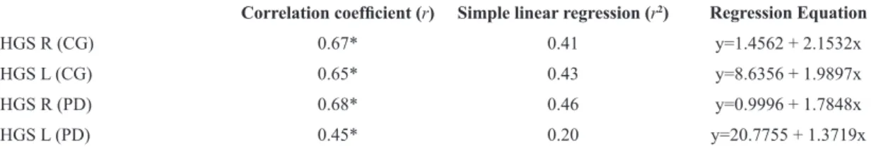
Table 2. Pearson correlation coeficient and simple linear regression analysis between the modiied sphygmomanometer test and the
portable Jamar dynamometer.
Correlation coeficient (r) Simple linear regression (r2) Regression Equation
HGS R (CG) 0.67* 0.41 y=1.4562 + 2.1532x
HGS L (CG) 0.65* 0.43 y=8.6356 + 1.9897x
HGS R (PD) 0.68* 0.46 y=0.9996 + 1.7848x
HGS L (PD) 0.45* 0.20 y=20.7755 + 1.3719x
CG: control group; PD: Parkinson’s disease; HGS: handgrip strength; R: right; L: left. * (P<0.05).
Table 3. Reproducibility (reliability and agreement) of the modiied sphygmomanometer test (MST). Reliability
ICC2,1 (IC 95%)
Agreement SEM
Agreement MDC
MST R (CG) 0.79 (0.55-0.95) 2.56 7.09
MST L (CG) 0.88 (0.75-0.95) 2.29 6.34
MST R (PD) 0.89 (0.62-0.96) 2.55 7.06
MST L (PD) 0.83 (0.50-0.95) 2.67 7.40
MST: modiied sphygmomanometer test; R: right; L: left. CG: control group; PD: Parkinson’s disease; ICC: intraclass correlation coeficient; SEM: standard error of measurement; MDC: minimum detectable change.
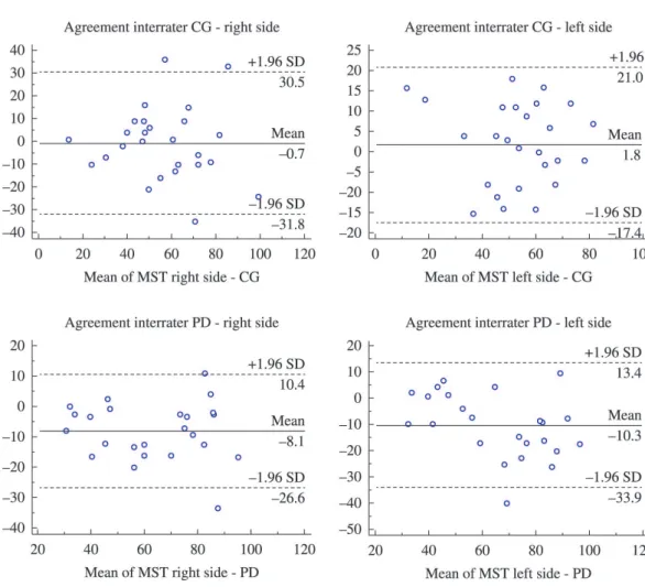
Figure 2 illustrates the interrater agreement in both
groups. When the mean difference of the measurements
obtained by different examiners was compared, a symmetrical distribution was observed around the
mean. However, wide limits of agreement and a high
bias were observed, particularly in the PD group.
Discussion
The HGS is often affected in subjects with PD because of motor changes during disease progression,
and these changes negatively impact the performance
degeneration that occurs in PD patients, HGS must
be constantly monitored by therapists using reliable and easily accessible devices.
Accordingly, the purpose of this study was to analyze the concurrent criterion validity and reproducibility
of the MST for the assessment of HGS in individuals with PD. The results indicate a moderate correlation
between the MST and the portable dynamometer, and the reproducibility of the MST was considered adequate, good, or excellent.
The concurrent criterion validity indicates the adequacy of the instrument using distinct data,
including those obtained from the gold-standard
measurements. In this sense, a positive and moderate
correlation was observed between the MST and the
Jamar dynamometer, which is considered the gold standard for evaluating HGS27. Furthermore, the MST values could moderately predict the HGS assessed
with the Jamar dynamometer, except on the left side,
which exhibited a low predictive value. Therefore, it can be inferred that the measurements assessed by both instruments were similar.
Additionally, the present study aimed to evaluate the reproducibility of the MST measurements, deined as the ability of an instrument to yield reliable results even when used by different examiners or during
different periods40. The reliability of the MST has been tested on different populations (adults and healthy older individuals with rheumatoid arthritis and lower
back pain), and the measurements obtained were
adequate15-24. However, to date, the reliability of the
MST had not been tested in individuals with PD41. Reproducibility studies (i.e. reliability and agreement) are crucial in assessing the variability
of a method or instrument and, consequently, in
avoiding the misinterpretation of variables before and after interventions. Regarding reliability, adequate, good, and excellent intra- and interrater ICC values
Figure 2. Interrater agreement according to the Bland-Altman method. CG: control group; PD: Parkinson’s disease; MST: Modiied
were observed in both groups. Therefore, the MST is a valid and reliable method for measuring HGS in
individuals with PD.
With regard to the intra- and interrater agreement
assessed using the SEM and MDC42, a small SEM
was obtained, and therefore, it is expected that the
measurements made in the same individual at different
times would have a variation of 2.67 mmHg, which is
related to the measurement error and not to changes in the clinical status of the patient. The MDC values
found indicate that a change >7.40 mmHg has a <5% probability of occurring due to random variation
or a random error in the measurement.
Of note, the mean difference between the control and PD groups on the left side exceeded the values established by the SEM and MDC. This variation may be attributed to the non-dominance of the left hand43, considering that most subjects were right-handed.
Although the interrater agreement was assessed using the Bland-Altman plot, no satisfactory results were obtained. The plots showed a high bias and wide
limits of agreement, particularly in the PD group.
The Bland-Altman plot has been used in various reliability studies44. However, it was not possible to compare the results obtained herein with those of other studies because no previous studies used this method to analyze the reliability of the MST in this
particular population.
One of the limitations of this study was related to the use of a sample composed of individuals with mild to moderate PD. In this respect, previous studies have shown that individuals with more severe signs
and symptoms of PD tend to have cognitive deicits
that interfere with or even prevent the adequate performance of the HGS test45. Therefore, in this study,
individuals classiied as levels 4 and 5 in the Hoehn and Yahr scale were excluded. However, further studies should be conducted to verify whether the results presented herein are observed in subjects with more
severe impairments and whether the severity of motor symptoms and postural changes, which are frequent in patients in the advanced stages of PD, interfere with the performance of this analysis.
In summary, it can be concluded that, despite the above limitations, the results reported herein are relevant to the ield of physical therapy and the rehabilitation of patients with PD because the results corroborate the adequate validity and reliability of
the MST. However, if the goal is to compare the
measurements made by distinct examiners, the data should be interpreted with caution. Therefore, HGS,
which is considered a predictor of overall muscle strength13, can be assessed more adequately in the
future. By doing so, the planning of treatment strategies and the progression of PD can be monitored more adequately by the therapist and at a low cost.
References
1. Wirdefeldt K, Adami HO, Cole P, Trichopoulos D, Mandel J. Epidemiology and etiology of Parkinson’s disease: a review of the evidence. Eur J Epidemiol. 2011;26(Suppl 1):S1-58. http://dx.doi.org/10.1007/s10654-011-9581-6.
PMid:21626386
2. de Lau LM, Breteler MM. Epidemiology of Parkinson’s disease. Lancet Neurol. 2006;5(6):525-35. http://dx.doi.
org/10.1016/S1474-4422(06)70471-9. PMid:16713924 3. Jankovic J. Parkinson’s disease: clinical features and
diagnosis. J Neurol Neurosurg Psychiatry. 2008;79(4):368-76.
http://dx.doi.org/10.1136/jnnp.2007.131045. PMid:18344392
4. Cano-de-la-Cuerda R, Pérez-de-Heredia M, Miangolarra-Page JC, Muñoz-Hellín E, Fernández-de-Las-Peñas C. Is there muscular weakness in Parkinson’s disease? Am J Phys
Med Rehabil. 2010;89(1):70-6. http://dx.doi.org/10.1097/
PHM.0b013e3181a9ed9b. PMid:19487924
5. Friedman JH, AbrantesAM. Self perceived weakness in Parkinson’s disease. Parkinsonism Relat Disord. 2012;18(7):
887-9. http://dx.doi.org/10.1016/j.parkreldis.2012.03.023.
PMid:22522072
6. Moreno Catalá M, Woitalla D, ArampatzisA. Central factors
explain muscle weakness in young fallers with Parkinson’s
disease. Neurorehabil Neural Repair. 2013;27(8):753-9. http://
dx.doi.org/10.1177/1545968313491011. PMid:23774123 7. Neely KA, Planetta PJ, Prodoehl J, Corcos DM, Comella CL,
Goetz CG, et al. Force control deficits in individuals with Parkinson’s disease, multiple systems atrophy, and progressive supranuclear palsy. PLoS ONE. 2013;8(3):e58403. http://
dx.doi.org/10.1371/journal.pone.0058403. PMid:23505500
8. Attivissimo F, Cavone G, Lanzolla AML, Savino M. Application
of hand grip signals for an objective evaluation of Parkinson disease: Analysis and comparison with standard functional
clinical tests. Measurement. 2009;42(8):1123-30. http://
dx.doi.org/10.1016/j.measurement.2009.05.001.
9. Gazibara T, Pekmezovic T, Tepavcevic DK, Tomic A, Stankovic I, Kostic VS, et al. Circumstances of falls and fall-related
injuries among patients with Parkinson’s disease in an
outpatient setting. Geriatr Nurs. 2014;35(5):364-9. http://
dx.doi.org/10.1016/j.gerinurse.2014.05.001. PMid:24916437 10. Mak MK, Pang MY, Mok V. Gait difficulty, postural
instability, and muscle weakness are associated with fear of
falling in people with Parkinson’s disease. Parkinsons Dis.
2012;2012:901721. http://dx.doi.org/10.1155/2012/901721.
PMid:22007344
11. Rudzińska M, Bukowczan S, Stożek J, Zajdel K, Mirek E, Chwała W, et al. Causes and consequences of falls in Parkinson disease patients in a prospective study. Neurol Neurochir Pol. 2013;47(5):423-30. http://dx.doi.org/10.5114/
12. Mak MK, Pang MY. Parkinsonian single fallers versus recurrent fallers: different fall characteristics and clinical features. J Neurol. 2010;257(9):1543-51. http://dx.doi.
org/10.1007/s00415-010-5573-9. PMid:20449601 13. Curb JD, Ceria-Ulep CD, Rodriguez BL, Grove J, Guralnik J,
WillcoxBJ, et al. Performance-based measures of physical
function for high-function populations.J Am Geriatr
Soc. 2006;54(5):737-42.
http://dx.doi.org/10.1111/j.1532-5415.2006.00700.x. PMid:16696737
14. Figueiredo I, Sampaio RF, Mancini MC, Silva FCM, Souza
MAP. Teste de força de preensão utilizando o dinamômetro Jamar. Acta Fisiátrica. 2007;14(2):104-10.
15. Helewa A, Goldsmith CH, Smythe HA. Patient, observer and instrument variation in the measurement of strength
of shoulder abductor muscles in patients with rheumatoid
arthritis using a modified sphygmomanometer. J Rheumatol.
1986;13(6):1044-9. PMid:3560090.
16. Helewa A, Goldsmith CH, Smythe HA. Measuring abdominal
muscle weakness in patients with low back pain and
matched controls: a comparison of 3 devices. J Rheumatol.
1993;20(9):1539-43. PMid:8164211.
17. Helewa A, Goldsmith CH, Smythe HA. The modified
sphygmomanometer-an instrument to measure muscle strength:
a validation study. J Chronic Dis. 1981;34(7):353-61. http://
dx.doi.org/10.1016/0021-9681(81)90073-4. PMid:7251815
18. Lucareli PRG, Lima MO, Lima FPS, Gimenes RO, Lucareli
JGA, Junior SAG, et al. Comparação dos métodos de
mensuração da força muscular dos flexores dos dedos das mãos através da dinamometria manual e esfigmomanômetro
modificado. Rev Einstein. 2010;8(2):205-8.
19. Rice CL, Cunningham DA, Paterson DH, Rechnitzer PA. Strength in an elderly population. Arch Phys Med Rehabil.
1989;70(5):391-7. PMid:2719543.
20. Bohannon RW, Lusardi MM. Modified sphygmomanometer
versus strain gauge hand-held dynamometer.Arch Phys Med Rehabil. 1991;72(11):911-4.
http://dx.doi.org/10.1016/0003-9993(91)90010-G. PMid:1929810
21. BalogunJA, Akomolafe CT, Amusa LO. Reproducibility and
criterion-related validity of the modified sphygmomanometer
for isometric testing of grip strength. Physiother Can.
1990;42(6):290-5.
22. Hamilton GF, McDonald C, Chenier TC. Measurement of grip
strength: validity and reliability of the sphygmomanometer and jamar grip dynamometer. J Orthop Sports Phys Ther.
1992;16(5):215-9. http://dx.doi.org/10.2519/jospt.1992.16.5.215.
PMid:18796752
23. Perossa DR, Dziak M, Vernon HT, Hayashita K. The
intra-examiner reliability of manual muscle testing of the hip
and shoulder with a modified sphygmomanometer: a
preliminary study of normal subjects.J Can Chiropr Assoc. 1998;42(2):73-82.
24. Isherwood L, Lew L, Dean E. Indirect evidence for eccentric muscle contraction during isometric muscle testing performed with a modified sphygmomanometer. Physiother Can. 1989;41(3):138-42.
25. Goetz CG, Poewe W, Rascol O, Sampaio C, Stebbins GT, Counsell C, et al. Movement Disorder Society Task Force report on the Hoehn and Yahr staging scale: status and
recommendations. Mov Disord. 2004;19(9):1020-8. http://
dx.doi.org/10.1002/mds.20213. PMid:15372591
26. Kendall FP, McCreary EK, Provance PG. Músculos, provas
e funções. 5th ed. São Paulo: Manole; 2007.
27. Fess EE. Grip strength. Clinical assessement recomendations.
2nd ed. Chicago: American Society of Hand Therapists; 1992.
28. Reis MM, Arantes PMM. Medida da força de preensão
manual- validade e confiabilidade do dinamômetro saehan.
Fisioter Pesqui. 2011;18(2):176-81. http://dx.doi.org/10.1590/
S1809-29502011000200013.
29. Geraldes AAR, Oliveira ARM, AlbuquerqueRB, Carvalho JM, Farinatti PTV. A força de preensão manual é boa preditora do desempenho funcional de idosos frágeis: um estudo
correlacional múltiplo.Rev Bras Med Esporte. 2008;14(1):
12-6. http://dx.doi.org/10.1590/S1517-86922008000100002. 30. Delgado C, Fernandes Filho JF, Barbosa FP, Oliveira HB.
Utilização do esfigmomanômetro na avaliação da força dos músculos extensores e flexores da articulação do joelho em
militares. Rev Bras Med Esporte.2004;10(5):362-6. http://
dx.doi.org/10.1590/S1517-86922004000500003.
31. Kaegi C, Thibault MC, Giroux F, Bourbonnais D. The
interrater reliability of force measurements using a modified sphygmomanometer in elderly subjects. Phys Ther. 1998;78(10):1095-103. PMid:9781703.
32. Souza LAC, Martins JC, Moura JB, Teixeira-Salmela LF, De Paula FVR, Faria CDCM. Assessment of muscular strength with the modified sphygmomanometer test: what
is the best method and source of outcome values?Braz J
Phys Ther. 2014;18(2):191-200. http://dx.doi.org/10.1590/
S1413-35552012005000149. PMid:24839045
33. Portney LG, Watkins MP. Foundations of clinical research: applications to practice. 2nd ed. New Jersey: Prentice-Hall;
2000.
34. Sim J, Arnell P. Measurement validity in physical therapy research. Phys Ther. 1993;73(2):102-10, discussion 110-5.
PMid:8421716.
35. Dancey CP, Reidy J. Estatística sem matemática para psicologia: usando SPSS para Windows. Porto Alegre: Artmed; 2006.
36. Krebs DE. Declare your ICC type. Phys Ther. 1986;66(9):1431.
PMid:3749277.
37. McGraw KO, Wong SP. Forming inferences about some intraclass correlation coefficiens. Psychol Methods.
1996;1(1):30-46. http://dx.doi.org/10.1037/1082-989X.1.1.30.
38. Terwee CB, Bot SD, de Boer MR, van der Windt DA, Knol DL, Dekker J, et al. Quality criteria were proposed for measurement properties of health status questionnaires. J Clin Epidemiol. 2007;60(1):34-42. http://dx.doi.org/10.1016/j.
jclinepi.2006.03.012. PMid:17161752
39. Bland JM, Altman DG. Statistical methods for assessing
agreement between two methods of clinical measurement.
Lancet. 1986;1(8476):307-10. http://dx.doi.org/10.1016/
S0140-6736(86)90837-8. PMid:2868172
40. de Vet HC, Terwee CB, Knol DL, Bouter LM. When
to use agreement versus reliability measures. J Clin Epidemiol. 2006;59(10):1033-9. http://dx.doi.org/10.1016/j.
jclinepi.2005.10.015. PMid:16980142
41. Souza LAC, Martins JC, Teixeira-Salmela LF, Godoy MR,
Aguiar LT, Faria CDCM. Avaliação da força muscular pelo
literatura. Fisioter Mov. 2013;26(2):437-52. http://dx.doi.
org/10.1590/S0103-51502013000200021.
42. Magalhães MO, Costa LOP, Ferreira ML, Machado LAC. Clinimetric testing of two instruments that measure attitudes
and beliefs of health care providers about chronic low back
pain. Rev Bras Fisioter. 2011;15(3):249-56. http://dx.doi. org/10.1590/S1413-35552011000300012. PMid:21829990.
43. Incel NA, Ceceli E, Durukan PB, Erdem HR, Yorgancioglu ZR. Grip strength: effect of hand dominance. Singapore Med J. 2002;43(5):234-7. PMid:12188074.
44. AndradeJA, Figueiredo LC, Santos TRT, Paula AC, Bittencourt NF, Fonseca ST. Reliability of transverse plane pelvic
alignment measurement during the bridge test with unilateral knee extension.Rev Bras Fisioter. 2012;16(4):268-74.
http://dx.doi.org/10.1590/S1413-35552012000400007.
PMid:22899181
45. Zhu K, van Hilten JJ, Marinus J. Predictors of dementia in
Parkinson’s disease; findings from a 5-year prospective study using the SCOPA-COG. Parkinsonism Relat Disord. 2014;20(9):980-5. http://dx.doi.org/10.1016/j.
parkreldis.2014.06.006. PMid:25024059
Correspondence Soraia Micaela Silva